DEF Helps Bring Vision-Saving Treatment To Patients With Retinitis Pigmentosa

Director of the Stem Cell and Retinal Regeneration Program at the  Gavin Herbert Eye Institute (GHEI), Henry Klassen, MD, Ph.D., leads life-changing research that will affect and influence vision-saving treatments for years to come.

Watch below to hear from Dr. Klassen on his recent stem cell research:

February is AMD/Low Vision Awareness Month

Low vision affects millions of Americans — including many older adults. People with low vision aren’t blind, but because of their vision loss, they may not be able to do everyday tasks like driving or reading even with glasses.

What is Low Vision?

When your eyesight is impaired to the degree that you struggle with daily tasks like reading or cooking – or that you have difficulty recognizing faces, you may have a medical condition referred to as low vision. Someone with low vision can’t simply put on a pair of glasses or contacts and see well; this condition is beyond the typical loss of vision that occurs with aging.

Individuals experiencing low vision can struggle with maintaining independence; hobbies, reading and even socializing may become challenging as vision loss progresses. Because of the impact low vision can have on your life, it is important to have your eyes checked by a low vision specialist if you have any concerns. Since it is unusual to be able to restore vision once it is lost, screenings can help preserve the vision you have and help you access adaptive lenses and devices if you need them. While there is not a cure for low vision,  a low vision specialist can help you adapt and if needed, create a vision rehabilitation program designed to meet your specific needs.

If you are experiencing trouble seeing even with prescription eye glasses, ask your optometrist or ophthalmologist for a low vision evaluation. If your practitioner does not perform this evaluation, request a referral to a low vision specialist in a private practice, at a Braille Institute or a University Eye Care Institute. 

Low vision is often caused by one of these conditions:

  • Age-related macular degeneration (AMD) –AMD is a progressive eye condition affecting as many as 15 million Americans. The disease attacks the macula of the eye, where our sharpest central vision occurs, affecting reading, driving, identifying faces, watching television, safely navigating stairs and performing other daily tasks. Although it rarely results in complete blindness, it robs the individual of all but the outermost, peripheral vision, leaving only dim images or black holes at the center of vision. Read more

  • Diabetes Diabetic retinopathy is the most common diabetic eye disease and a leading cause of blindness in American adults caused by changes in the blood vessels of the retina.

In some people with diabetic retinopathy, blood vessels may swell and leak fluid or blood inside the eye. In other people, abnormal new blood vessels grow on the surface of the retina. The retina is the light-sensitive tissue at the back of the eye. A healthy retina is necessary for good vision.  Read More 

  • Glaucoma Glaucoma is a disease that causes damage to the major nerve of the eye called the optic nerve, a part of the central nervous system that carries visual information from the eye to the brain

The eye experiences a gradual increase of intraocular pressure (IOP) due to an imbalance of the fluid produced in the eye and the amount of fluid drained. Over time, elevated IOP can cause vision loss. The most common form of glaucoma is primary open angle glaucoma which affects about 3 million Americans. However, there are other types including narrow angle, congenital, normal tension, and secondary glaucoma. Read more

Reducing your risk for Low Vision

Practicing healthy daily lifestyle habits can reduce your risk for low vision. A healthy diet, not smoking and maintaining healthy blood pressure are important first steps. Other healthy habits that can lower the risk include:

  • Eat a diet with plenty of green, leafy vegetables such as kale, spinach, and collard greens, and fresh fruit.
  • Fish that is high in omega-3 fatty acids is good for eye health. Try to include it in your diet at least once or twice a week. Types of fish that are high in omega-3 fatty acids include salmon, sardines, mackerel, herring, and albacore tuna.
  • It’s important to keep your body healthy. Maintain a healthy weight, exercise regularly, maintain healthy blood pressure and cholesterol levels. The healthier your body, the more it can foster good eye health. Visit our website for eye healthy recipes Eye Cook.
  • Protect your eyes. Wear sunglasses and a hat with a visor in bright sunlight to protect your eyes from potentially harmful ultra-violet (UV) light and blue light.

Low Vision Reading Apps on Your Smartphone

Reading often is one of the most difficult challenges for visually impaired people. Many people with low vision give up reading altogether, because what used to be an enjoyable, effortless activity now requires thought, preparation and a lot of adjustment. In short, reading is just no fun anymore. 

The American Academy of Ophthalmology (AAO) has listed some of the top apps, devices and resources for people with low vision, below are a few listed for reading app, for more apps visit American Academy of Ophthalmology.

Reading

  • KNFB Reader (Android and iOS, $99.99) – This voice-to-text tool lets you take a picture of any text—books, recipes, product labels—and have it read back to you or converted to braille.
  • Audible (Android and iOS, $14.95 per month plus downloads) – Provides downloadable audiobooks, periodicals, newspapers and more. Their collection currently includes 200,000+ books.
  • Kindle app (Android, iOS, PC and Mac, free) – An e-reader app that allows you to download books. Books can be purchased and directly downloaded from Amazon, or downloaded from your library app into the Kindle app. The free Kindle app can be downloaded onto many iOS and Android devices.
  • Bookshare (Android and iOS, $50 per year) – An online library for people with low vision. Currently, the Bookshare collection contains nearly 850,000 titles, downloadable in a choice of formats including ebooks, audio, braille, and large font. Membership requires verification of your print disability, and includes free downloads.
  • BARD Mobile (Android and iOS, free) – A talking book library that offers access to tens of thousands of titles. You must first enroll in the National Library Service (NLS) for the Blind and Print Disabled at the Library of Congress. Note that this program may only be available in the United States.


Regular eye exams are essential for all adults; it is recommended that adults over the age of 60 have eye exams each year. If your vision can not be improved to the point that you are able to see the things you need to see or read, your eye care professional can refer you to a low vision specialist. This specialist differs from a conventional optometrist and is trained to evaluate your low vision problem and offer solutions to help you retain or regain independence and the ability to do things you enjoy again.


More Low Vision resources

January is Glaucoma Awareness Month


Glaucoma is a leading cause of vision loss and blindness in the United States. Glaucoma has no early symptoms — that’s why half of people with glaucoma don’t know they have it. 

The only way to find out if you have glaucoma is to get a comprehensive dilated eye exam. There’s no cure for glaucoma, but early treatment can often stop the damage and protect your vision.   

Anyone can get glaucoma, but those at higher risk include: 

  • Everyone over age 60, especially Hispanics/Latinos 
  • African Americans over age 40 
  • People with a family history of glaucoma 

National Glaucoma Awareness month is important because:

  1. It’s a silent disease – Open-angle glaucoma, the most common type, has no symptoms. Without treatment, those affected will slowly lose their peripheral vision. If glaucoma remains untreated, people may miss objects to the side and out of the corner of their eye.
     
  2. It gives others a voice – National Glaucoma Awareness Month helps patients cope. It’s a time to let them share their stories.
     
  3. It’s important to spread the word – The main objective of this month is to keep people in the know about this disease. Not everyone is aware of how easily they can be affected. Eye care organizations use this month to address the risks and provide treatment tips.


Questions for your doctor

If you or a loved one has been diagnosed with glaucoma, you may have lots of questions. Starting treatment early is the best way to stop the damage and protect your vision. Talk to your eye doctor about your symptoms and treatment options.  

Here are a few questions to ask your doctor:

  • What type of glaucoma do I have?
  • How often do I need to get a checkup for my glaucoma?
  • What are my treatment options?
  • What will happen if I don’t get treated for my glaucoma?
  • How will I know if my treatment is working?
  • What are the possible dangers or side effects from treatment?
  • Is there anything I can do to prevent my glaucoma from worsening?
  • Does glaucoma put me at risk for other eye diseases?
  • What can I expect for my vision — now and in the future?
  • Are there any activities I should avoid?

Visiting the doctor can be stressful, especially when you’re dealing with a new diagnosis. It helps to have questions written down ahead of time. 


Discovery Eye Foundation is currently funding research to find better treatments for glaucoma. Gifts of any size can make a difference.

Donate Today!


Glaucoma simulation video courtesy of NEI


DEF-Funded Retinitis Pigmentosa(RP) and Age-Related Macular Degeneration(AMD) Projects Converge with Promise

Two hallmark DEF-funded projects are converging, providing great hope for those facing vision loss from retinitis pigmentosa (RP) or age-related macular degeneration (AMD).

The first project, headed by UC Irvine researchers Drs. Henry Klassen and Jing Yang, concentrates on putting human retinal progenitor cells into the eyes of those with RP in order to rescue damaged retinal cells. That project is currently in Phase II clinical trials, progressing toward FDA approval.

                                                      Tissue-culture model

According to DEF Research Director Dr. Cristina Kenney, if the project is approved by the FDA for use with RP, the next question is: What other diseases might these retinal progenitor cells be used for? That’s where a second DEF-funded project comes in.

Kenney is working on a “personalized” cybrid cell model to screen agents that specifically target the mitochondria in AMD cells. To date, the researchers have different cybrid cell lines representing 60 different individuals with eye diseases. They are looking for novel mechanisms to protect AMD cells from dying.

Yang and Kenney are now working together to determine whether the retinal progenitor cells can be the agent that rescues AMD cybrids. “When we take the mitochondria from AMD patients and put them into healthy retinal cells, which makes cybrids, we have shown that these AMD cybrid cells will start to die. So we used that model to ask the question: How do we rescue them?” Kenney says.

Kenney and Yang developed a tissue culture model, where the retinal progenitor cells are grown in one part of a chamber, and the AMD cybrids are grown in another part of chamber, surrounded by culture medium. There is a porous separator between the two chambers through which the cells can communicate.

 “We are finding that the retinal progenitor cells produce a factor that protects the AMD cybrids,” Kenney says. “This provides promising evidence that these proprietary retinal progenitor cells that are being tested for treating RP also may be helpful in AMD patients.”

“DEF has been supporting both these retina-related projects for quite some time, and it’s very exciting to see them coming together to potentially treat both RP and AMD.”

To help support this sight-saving research, please donate to the Discovery Eye Foundation by clicking button below.

Cancer in the EYE: Unexpected Thought

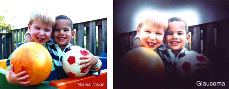
When you think of cancer, most of us do not think about the eye or vision. Though rare, cancer can start inside or outside of the eye. If cancer starts inside the eyeball it’s called intraocular and if it starts outside the eye (eyelid or in the eye socket) then it’s called extraocular tumor. It can occur in both children and adults. Most major eye centers have specialists who are trained in the diagnosis and treatment of eye cancers.

Here are a few types of cancer in eye:

  • Eye Cancer in Children: The most common cancer seen in the eye of children is retinoblastoma. This occurs in children at very early age so these are the youngest cancer patients. This cancer starts inside the eye and affects the retinal cells. This is a cancer that presents very quietly, the child has no pain, no complaints and plays happily without any problem until one day parents notice that the pupil of the eye has some abnormal ‘White Glow’ (leukocoria) rather than the usual ‘red-eye’ reflection seen in a photo—that can be the first sign of retinoblastoma. So, it requires prompt evaluation by an ophthalmologist. Retinoblastoma is a curable cancer but if it is not treated on time, it can grow quickly and fill the eyeball. It can lead to loss of vision and life-threatening problems.

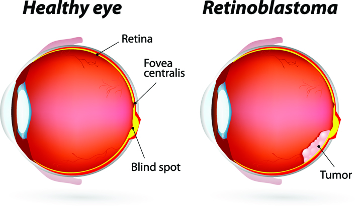
At the later stage of this cancer, the only one way to survive is to remove the eyeball (enucleation). Like many of other types of cancer, retinoblastoma has a genetic component so genetic testing needs to be done. The tumor begins with the RB1 gene mutation that stimulates retinal cells to develop into a tumor called a retinoblastoma. The RB1 mutation can be inherited from the parents, but in some cases it is sporadic and not inherited. There are various treatments such as surgery, chemotherapy, radiotherapy etc. to cure retinoblastoma cancer. Rarely it can spread beyond the eye. 
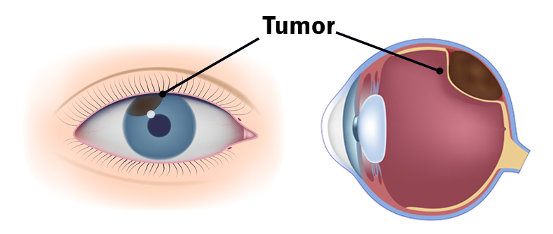
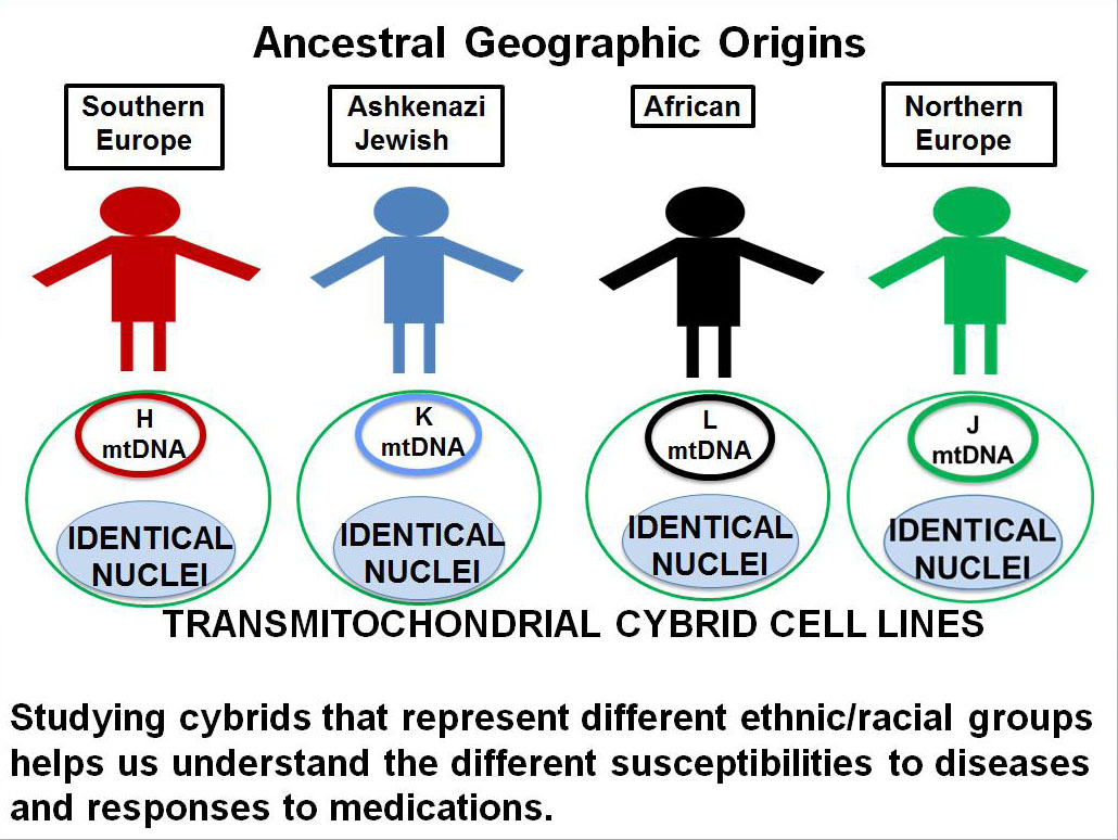
  • Eye Cancer in Adults: The development of a tumor in the back of the eye in adults can be from a metastatic cancer from elsewhere in the body or can arise in the eye itself. The most common primary eye cancer is called uveal melanoma and is a cluster of rapidly growing cells underneath the retina, which can lead to vision loss. The tumor can also appear as a dark black spot on the iris. Ideally the uveal melanoma can be treated when the tumor is still only in the eye.However, unfortunately, in approximately 50% of patients the uveal melanoma spreads (metastasis) to other part of the body, making it the most dangerous eye cancer in adults. Depending upon its location the uveal melanoma may not cause early symptoms but the patient may experience blurred vision or large numbers of floaters. The uveal melanoma can be diagnosed only when an ophthalmologist or eye care specialist examines your eye. What is the main cause of uveal melanoma? How it starts, grow and spread? These questions are still unanswered. The risk of this cancer increases in persons having fair skin (white), light eye color and inability to tan. There are certain changes in the genes linked to uveal melanoma. Currently the most common are GNAQ and BAP-1 gene mutations that are associated with greater risk of metastasis, where it spreads to other parts of the body. The standard treatment of this tumor is fine-needle aspiration biopsy (FNAB), brachytherapy (radioactive material inside a small capsule placed next to the tumor), radiation therapy and possibly enucleation. 
  • Metastasis to Eye: Finally, the other types of cancers found in the eyes are because of the spreading of a primary cancer (from a distant site such as the breast, lung or liver) to the eye.  Sometimes an eye exam can identify a metastatic tumor before the primary tumor is recognized elsewhere in the body.  Examples of cancers that spread to the eye are breast cancer in women and lung cancer in men. Less commonly, the prostate, kidney, gastrointestinal and blood cancers (leukemia and lymphomas) can spread to the eye.  Treatment depends on the type of cancer involved. 
  • Eye Damage from Chemotherapy: Eye problems can also develop through the side effects of chemotherapy or hormone therapies given for tumors outside the eye. When a person has any type of cancer, they often must undergo treatments with chemotherapeutic drugs.  A commonly used cancer drug is called cisplatin.  When treated with cisplatin, there can be damage to the retinal pigment epithelial cells in the inner part of the retina. Hemorrhages or bleeding can occur within the retina itself and vision can decrease temporarily. If a person is undergoing cancer treatments, it is a good idea to have their eyes checked, especially if they are having any vision problems. The chemotherapy does not actually cause cancers but only side effects.

Therefore, if you are having any decreased vision, it is always makes sense to have a good eye examination to identify any problems at early stage.

Age-Related Macular Degeneration Month

Have you ever noticed how many people, places, things, and even a rather unattractive groundhog are designated and honored with a day, a month, or a year?  Now, I am not putting down Punxsutawney Phil because I do want him to see his shadow and promise us an early spring.  In our designation of things, we also should pay homage to values and causes that are important in our history and our health.  I am glad that I am reminded every November of our Veterans on a special day or the Presidents who have served our nation.  Certainly Dr. King’s birthday and Black History Month place an appropriate focus on issues that society is still working to solve.

So, I was delighted when I heard that February is Age-Related Macular Degeneration Month.  More than any other eye disease AMD affects the largest number of people over 65 of European lineage.  In fact, as I write this piece, over 15 million Americans are struggling within its dry form, currently an incurable condition.  It’s projected that if we are not able to solve the riddle of AMD by the year 2050, the number of senior Americans with AMD will be more than 30 million.

The Discovery Eye Foundation is committed to finding the answer that will preserve the vision of millions of people.  Right now research supported by DEF at the Gavin Herbert Eye Institute, University of California Irvine, Medical School is on the cutting edge of promising breakthroughs that could make the difference in the lives of so many.  

Please join me in supporting this most worthy effort by contributing whatever you can to DEF and its important work.  Our vision may depend on your generosity.


Tom Sullivan
DEF’s Ambassador of Vision
sullivanvision.com

Mitochondrial Revolution: New Hope for AMD and Aging Diseases

How many times have we heard: “This field has been thoroughly studied, and we know all there is to know; there is no reason to continue investigating, because there is nothing more to find out on the subject”? This has been the attitude of some researchers with respect to the importance of mitochondria and diseases. For more than 50 years, we have known that mitochondria, which are the “batteries” of the cell, are critical for energy production. But many believed they did not have other major roles in the health of the cell, so when it came to developing drugs against diseases, mitochondria were overlooked.

That idea has now been turned on its head.

Mitochondria

Led by Discovery Eye Foundation (DEF) Research Director Dr. Cristina Kenney, the Mitochondria Research Group believed that, to really discover something new, you have to look in novel areas. This group has done just that. Using the transmitochondrial cybrid model, which are cell lines with identical nuclei, but with each line containing mitochondria from a different person, they have shown that the mitochondria have major regulation powers over cell behavior and expression of disease-related pathways. This is significant, because the mitochondria then become a target for therapies to combat diseases.

Kenney’s group is investigating various drugs and substances that will keep the mitochondria healthy and, ultimately, improve the health of the retinal cells in age-related macular degeneration (AMD). But it does not stop there. This same approach to developing mitochondria-targeting drugs is being pursued for drugs to treat Parkinson’s disease, Alzheimer’s disease, leukemia, various cancers, heart failure, thrombosis, stroke, diabetic retinopathy, Leber hereditary optic neuropathy, and even liver toxicity from acetaminophen.

The continued support from DEF, especially in the early stages of the mitochondria research, has fostered a new area to be opened up, specifically for AMD and diabetic retinopathy. In turn, this has allowed new collaborations among Kenney and researchers from the eye field, and laboratories studying the brain and neurodegeneration, cardiology, cancer therapies and methods to reduce side effects from cancer drugs. Kenney’s discoveries using cybrids have revolutionized the field of mitochondrial research, showing that mitochondria have wide-ranging biological effects never imagined and opening up the field of mitochondrial therapy to careful investigation.

You can help make a difference by supporting DEF’s sight saving research. Help our researchers advance AMD research by donating today! 

DonateNow


Lauren HauptmanLauren Hauptman
Lauren Hauptman Ink

A New Commitment to Vision

Meet Tom Sullivan – DEF’s Ambassador of Vision

Over the last 40 years, I’ve been committed to working on behalf of blind children and their families.  My involvement has spanned the entire gamut of participation – from direct involvement in the classroom and counseling parents to hosting organized 10K races and celebrity golf tournaments that carried my name.  In that time, my wife and I raised just over $8 million thanks to the generosity of so many people.  Though my commitment to this cause has not changed, I’ve chosen to take on a new challenge that has in every way re-energized my passion. 

I’ve recently become the Ambassador of Vision for the Discovery Eye Foundation (DEF), a remarkable organization that funds cutting edge research that I believe someday will eliminate many forms of blindness.  The principle reason for my enthusiastic commitment is largely due to the fact that DEF directly funds researchers and avoids institutional restrictions.

As of this blog, DEF is engaged in ongoing efforts to understand 5 main eye diseases – Retinitis Pigmentosa, Macular Degeneration, Keratoconus, Diabetic Retinopathy, and Glaucoma.  I can honestly tell you that breakthroughs are not only on the horizon, but in many cases they are imminent.  Over the weeks and months I’ll be telling you much more, specifically about our individual research projects.  

Any help you may choose to give on behalf of people struggling with vision loss will be deeply appreciated. I look forward to having many of you join my fight for sight. Your help can make a difference! Click here to donate.


Tom Sullivan
DEF’s Ambassador of Vision
sullivanvision.com


Creating Cybrids to Study Age-Related Diseases

DEF Research Director Dr. M. Cristina Kenney’s research has shown that the mitochondrial DNA from different ethnic/racial populations may play a key role in determining that population’s resistance or susceptibility to disease (see previous article on 11/12/15 – Mitochondria and Age-Related Macular Degeneration). In order to study these effects, Kenney has developed the cybrid model using mitochondria from subjects of different ethnic/racial groups (Figure 1). The comparison of an individual’s mitochondria with that from other ethnic/racial groups (African, European, Asian or Ashkenazi Jewish) allows us to determine if their mitochondria determine that population’s susceptibility or resistance to disease and to response to drugs.

cybrids to study age-related disease
Figure 1 – Cybrids are cell lines with identical nuclei but the mitochondrial DNA from individuals of different ethnic/racial groups.

Personalized cybrids
Kenney’s cybrids are made with mitochondria from the blood taken from individual living donors. Looked at individually they are all really “personalized cybrids” because each cybrid test system has the mitochondria from the original donor and reflects the responses of that donor.

Using Cybrids to Study Age-Related Diseases

How is Kenney using these personalized cybrids?
Kenney is partnering with Dr. Pinchas Cohen, dean of the University of Southern California, Leonard Davis School of Gerontology, to explore how novel, small proteins produced from mitochondria might be used to treat a variety of age-related diseases such as age-related macular degeneration, Alzheimer’s, Parkinson’s, stroke and cholesterol. Cohen’s laboratory has discovered and characterized many of these new, small proteins called “mitochondrial derived peptides” (MDPs). His work has shown that these MDPs can protect brain cells from damage and early death, such as occurs in Alzheimer’s disease. Cohen and Kenney are now testing these MDPs in the K and H cybrids to assess their protective effects to stop retinal cell death, such as seen in AMD.

Kenney explains her approach:
“Our cybrid system represents a very powerful technique. We are now using the Ashkenazi Jewish population as an excellent model to learn how the mitochondria, with their unique mtDNA, influence the risk factors for AMD. We plan to extend the study to investigate Ashkenazi Jewish people’s susceptibility to Alzheimer’s disease, heart disease and stroke. Eventually, we believe the findings for the K haplogroup mitochondrial DNA will be applicable to other groups, as well.”

11/17/15


Anthony B. Nesburn, MD FACSAnthony B. Nesburn, MD, FASC
President/Medical Director
Discovery Eye Foundation

Treatments for Dry Eye Disease

This is the third and final installation of the comprehensive series we have presented on dry eye disease. Dr. Wade first discussed the symptoms you might experience if you have dry eye and the Dr. Garg explained the process of diagnosing they type of dry eye disease you might have. In this article Dr. Farid reviews treatment options based on your diagnosis of dry eye disease.treatments for dry eye

Treatments for Dry Eye Disease

As our understanding of dry eye disease expands, so do treatment options. We now know that dry eye disease is a multifactorial disease. There is no one cause so there is no one magic cure. Treatments aim to improve tear composition, reduce eye surface inflammation, and target eyelid margin disease. Here, we will review many treatment options, but the treatment combination or “cocktail” that is appropriate for you will depend on your specific type of dry eye disease. This is usually determined after some testing by your eye care provider.

Environmental, Dietary, and Medication Adjustments
Before going into specific dry eye treatments, there are modifiable causes and preventative methods to improve dry eyes. Simple changes in the environment, diet, and medications can be easy ways to improve symptoms.

Environmental Changes
As expected, a dry environment will worsen dry eyes. Humidifiers and moisture goggles have been shown to help alleviate these symptoms. Furthermore, situations that cause decreased blinking, such as prolonged use of computer screens, can worsen dry eyes. Patients should take frequent breaks from computer screens and reading, allowing their eyes to rest and resume normal blinking. When in windy, smoky, or dusty situations, sunglasses can act as a barrier to the eyes, reducing dry eye symptoms. Avoiding wind, fans, or any source of air blowing into the eyes may also help.

Dietary Changes
Drinking adequate water keeps patients hydrated and reduces exacerbations of dry eye symptoms. Omega-3 fatty acids, particularly eicosapentaenoic acid (EPA) and docosahexaenoic acid (DHA), have been shown in many clinical studies to improve dry eyes. It is believed that these fatty acids help inhibit inflammatory mediators. Essential fatty acids cannot be synthesized and must be ingested through diet. Foods rich in omega-3 fatty acids include fish, other seafood, and flaxseed oil. Supplements are also available.

Adjusting Medications
Many medications are associated with dry eyes, and patients may benefit from adjusting doses or finding alternative treatments. You can work with your physician to weigh the costs and benefits about modifying your current medications if they are suspected to worsen dry eyes. Some of these medications include: hypertensive drugs, antihistamines, decongestants, antidepressants, acne medications, birth control, and hormone replacement therapy. Eye drops with preservatives, such as glaucoma medications, can also worsen dry eyes.

Lubricating Treatments
For patients with decreased tear production, supplementation of tears or reduction of tear drainage will improve symptoms.

Artificial tears, gels, and ointments
Artificial tears, gels, and ointments are readily available over the counter. Artificial tears are eye drops that are used throughout the day as needed, up to eight times for dry eyes. You must make sure to get “lubricating” drops. There are multiple available brands that are excellent and equivalent in providing artificial moisture. Avoid “redness relief” brands as they are focused on reducing appearance of the vessels in the eyes as opposed to actual dry eye relief. Gels and ointments are thicker and very effective but can blur vision. Non-commercial comparison testing between brands has not been done. Because the ingredients, preservatives, and consistency vary from brand to brand, it is recommended for patients to choose the best option that works well for them based on trial and error.

Preservatives are used in many eye drops to help them to last longer. Unfortunately, they can be irritating to the eyes, especially when used often (more than 4 drops/day) or with other drops containing preservatives (ie. glaucoma medications). Patients who frequently use eye drops are recommended to use preservative-free (PF) drops. Unfortunately, preservative-free drops are more expensive because they come in “single use” containers. You may be able to extend the life of each single use container by spreading out its contents over multiple uses throughout the day.

Lipid-containing lubricants, such as those with castor oil, attempt to mimic the oils found in the tears, reducing evaporative dry eyes, but more research is necessary to study their efficacy.

Punctal Plugs
Your ophthalmologist can place small plugs at a clinic visit into the punctum, a small hole at the upper and inner lids that drain tears from the eyes. One plug can be added initially, and if more tear retention is required, another plug can be added. There are absorbable and non-absorbable types. Absorbable types are made of collagen and last 1 week to 6 months. Once placed, they are not visible or removable. Non-absorbable plugs are usually silicone, and permanent. They are easily placed by your ophthalmologists and remain visible in follow-up exams. Some patients feel the plugs, and there may be mechanical rubbing, especially when the plug is not the proper size. Some patients may also experience excessive tearing with plugs. If problematic, patients can wait for the plugs to dissolve if absorbable or they can be removed with forceps if non-absorbable.

Autologous Serum
Serum is extracted from a patient’s blood and turned into an eye drop. The growth factors, vitamins, and antibodies present in serum are the same as those in natural tears. Evidence is showing significant promise in alleviating symptoms and signs of chronic dry eye disease. A good collaboration between a phlebotomy lab and compounding pharmacy is necessary to make the products. The products must be kept refrigerated or cold between uses. They can be frozen for long-term storage. Each blood draw provides a supply of drops that can last 3-6 months.

Hydroxypropyl Cellulose Ophthalmic Inserts (Lacrisert)
An insert is available for people who find regular artificial tear use to be difficult. A physician can order the inserts through a pharmacy. The patient places the insert in the inferior fornix of the eye, the area between the lower lid and sclera of the eye. It slowly dissolves over 24 hours, giving constant lubrication.

Anti-inflammatory Treatments
Inflammation is now being recognized as a major underlying cause of chronic and worsening dry eye disease. Many patients who have been suffering from chronic dry eyes, particularly those with autoimmune diseases like Sjogren’s Syndrome, will do well on treatments that reduce the amount of inflammation in the tear film and ocular surface. Your physician can order these medications for you if needed.

Cyclosporine A (Restasis)
The FDA approved Restasis for dry eyes in 2002. This is an immunosuppressive and anti-inflammatory eye drop medication. Relief is not instant, and may take 6-8 weeks of sustained use for improvement in dry eye symptoms. Less than 20% of patients may experience a burning sensation with the drops, but the safety and tolerability profile is otherwise excellent.

Antibiotics
Doxycycline and minocycline are used for inflammatory ocular surface and eyelid disease. The antibiotics have a dual effect: they act as anti-inflammatories and anti-microbials. As an anti-microbial, these medications can improve meibomian gland function. They can decrease lid bacterial flora, reducing a cause of meibomian lipid breakdown. A low-dose (doxycycline 20 mg BID) regimen for 1-2 months has been shown to be effective. Side effects are usual mild but include stomach upset, yeast infections, and photosensitivity.

Steroids
Steroids act as anti-inflammatories. Because steroids are associated with complications in long-term use, they are mainly used in short pulses either at the initiation of treatment or as rescue during exacerbations. A short 4-6 week course is generally well tolerated as a “rescue treatment,” or an urgent treatment, to relieve intolerable symptoms quickly before resorting to other treatment options.

Meibomian Gland Treatments
Meibomian glands produce oils that are crucial in preventing our tears from evaporating too quickly. Treatments that target the glands can help patients with meibomian gland dysfunction or lid margin disease.

Warm compresses and lid scrubs
Warm compresses provide heat that warms the oils in the glands, unclogging the glands and improving oil flow. Warm washcloths, small rice bags heated in the microwave, or commercial hydrogel pads are all effective, and no studies have been done to compare the different methods. The heat. The compress should be placed over the eyes for 5-10 minute. Gentle circular or rolling massage of the eyelids can help express the oils from the glands.

Lid scrubs are useful particularly in cases of blepharitis, or mild inflammation of the lids, that cause them to become crusted. There are excellent over the counter commercial lid soap formulations that work well to clean the lid margins. Alternatively, baby shampoo and a washcloth gently applied to the lids can work as well.

Thermal pulsation (Lipiflow)
Lipiflow is an FDA approved in-office treatment for meibomian gland dysfunction and dry eyes. During the 12-minute procedure, a device is placed over the eye and eyelids that provides localized warmth and pressure on the lids (Figure 1).

treatments for dry eye
Image 1 – Lipiflow treatment

The procedure is 100% safe and very effective at clearing out the trapped oil glands and allowing smooth flow to be re-established. After approximately one month, the consistency of the oils in the tear flow will have improved remarkably with associated improvement in dry eye symptoms. With one procedure, the effects last between 12-24 months.

Intense Pulsed Light (IPL)
Originally approved for acne and rosacea dermatologic disease, IPL uses bursts of light to minimize blood vessel size. It can be used off-label for ocular rosacea and meibomian gland dysfunction, but results have not been reported.

Summary
There are many treatment options for dry eye disease. Generally, conservative over-the-counter treatments should be tried first. Commonly, environmental changes, dietary changes, artificial tears, and warm compresses will improve the majority of dry eye symptoms to tolerable levels. However, when these options are exhausted, there are many additional options for patients. Work closely with a trusted health professional to determine the optimal treatment combination as each patient is different.

8/25/15


treatments for dry eyeMarjan Farid, MD
Director of Cornea, Cataract, and Refractive Surgery
Vice-Chair of Ophthalmic Faculty
Director of the Cornea Fellowship Program
Associate Professor of Ophthalmology
Gavin Herbert Eye Institute, University of California, Irvine


treatments for dry eyePriscilla Q. Vu, MS
Medical Student
University of California, Irvine School of Medicine