February is AMD/Low Vision Awareness Month

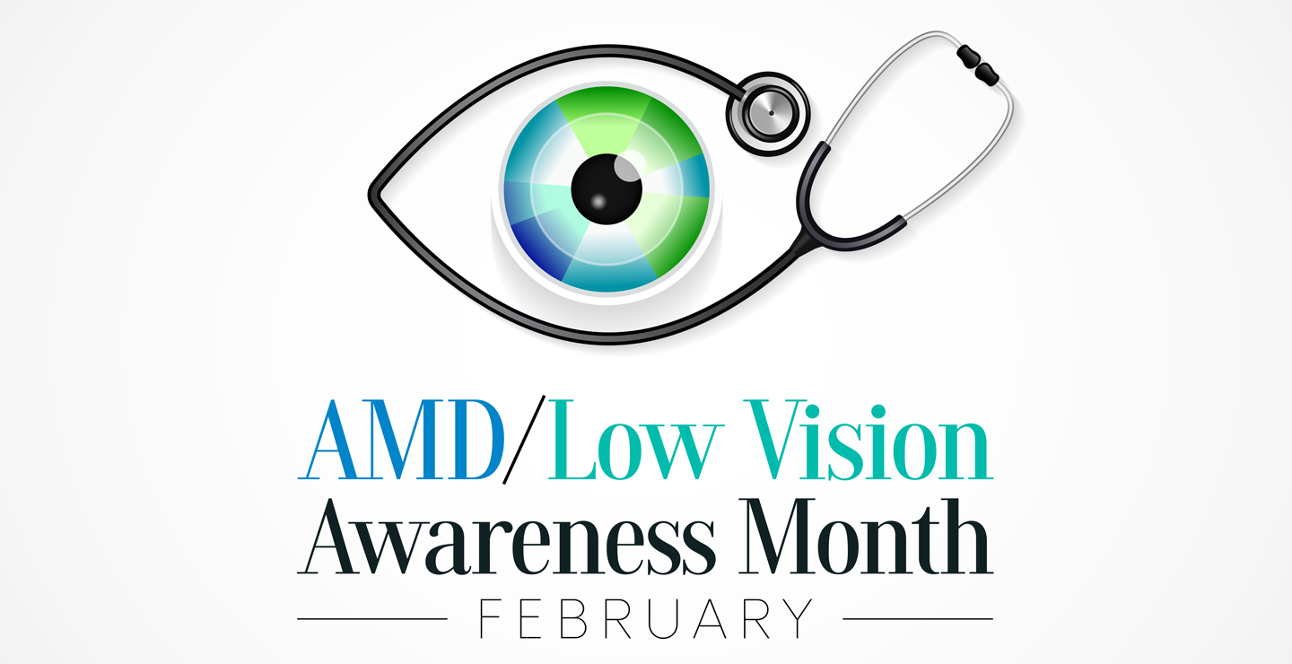
Low vision affects millions of Americans — including many older adults. People with low vision aren’t blind, but because of their vision loss, they may not be able to do everyday tasks like driving or reading even with glasses.

What is Low Vision?

When your eyesight is impaired to the degree that you struggle with daily tasks like reading or cooking – or that you have difficulty recognizing faces, you may have a medical condition referred to as low vision. Someone with low vision can’t simply put on a pair of glasses or contacts and see well; this condition is beyond the typical loss of vision that occurs with aging.

Individuals experiencing low vision can struggle with maintaining independence; hobbies, reading and even socializing may become challenging as vision loss progresses. Because of the impact low vision can have on your life, it is important to have your eyes checked by a low vision specialist if you have any concerns. Since it is unusual to be able to restore vision once it is lost, screenings can help preserve the vision you have and help you access adaptive lenses and devices if you need them. While there is not a cure for low vision,  a low vision specialist can help you adapt and if needed, create a vision rehabilitation program designed to meet your specific needs.

If you are experiencing trouble seeing even with prescription eye glasses, ask your optometrist or ophthalmologist for a low vision evaluation. If your practitioner does not perform this evaluation, request a referral to a low vision specialist in a private practice, at a Braille Institute or a University Eye Care Institute. 

Low vision is often caused by one of these conditions:

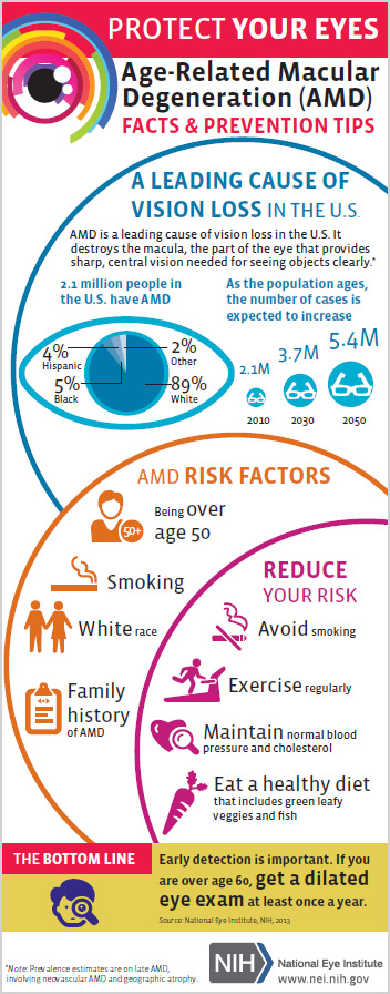
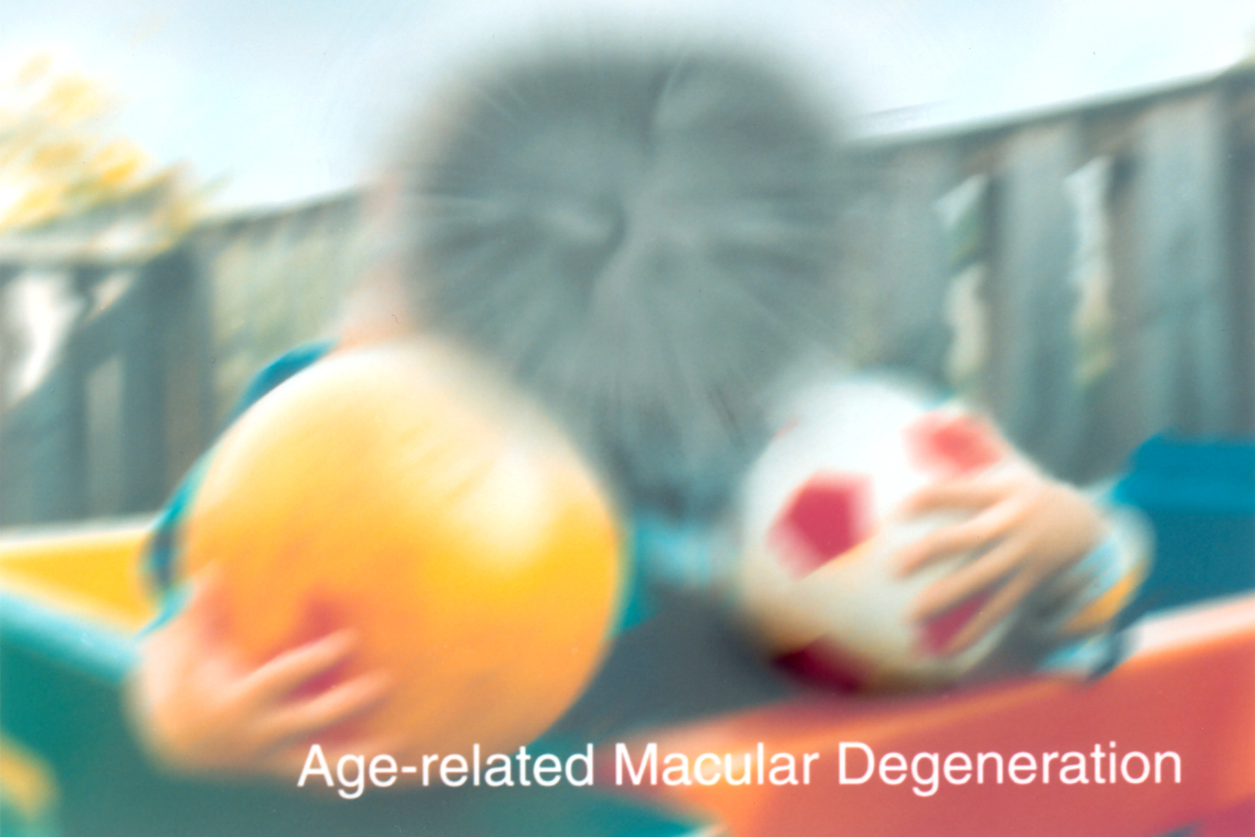
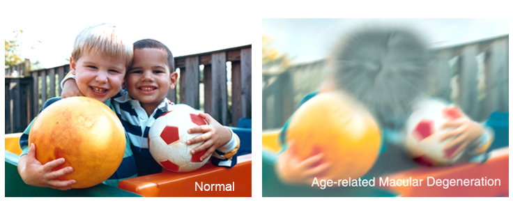
  • Age-related macular degeneration (AMD) –AMD is a progressive eye condition affecting as many as 15 million Americans. The disease attacks the macula of the eye, where our sharpest central vision occurs, affecting reading, driving, identifying faces, watching television, safely navigating stairs and performing other daily tasks. Although it rarely results in complete blindness, it robs the individual of all but the outermost, peripheral vision, leaving only dim images or black holes at the center of vision. Read more
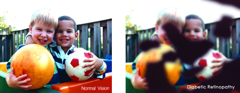
  • Diabetes Diabetic retinopathy is the most common diabetic eye disease and a leading cause of blindness in American adults caused by changes in the blood vessels of the retina.

In some people with diabetic retinopathy, blood vessels may swell and leak fluid or blood inside the eye. In other people, abnormal new blood vessels grow on the surface of the retina. The retina is the light-sensitive tissue at the back of the eye. A healthy retina is necessary for good vision.  Read More 
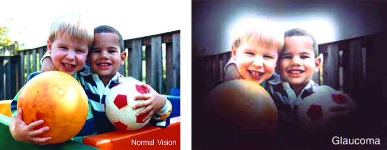
  • Glaucoma Glaucoma is a disease that causes damage to the major nerve of the eye called the optic nerve, a part of the central nervous system that carries visual information from the eye to the brain

The eye experiences a gradual increase of intraocular pressure (IOP) due to an imbalance of the fluid produced in the eye and the amount of fluid drained. Over time, elevated IOP can cause vision loss. The most common form of glaucoma is primary open angle glaucoma which affects about 3 million Americans. However, there are other types including narrow angle, congenital, normal tension, and secondary glaucoma. Read more

Reducing your risk for Low Vision

Practicing healthy daily lifestyle habits can reduce your risk for low vision. A healthy diet, not smoking and maintaining healthy blood pressure are important first steps. Other healthy habits that can lower the risk include:

  • Eat a diet with plenty of green, leafy vegetables such as kale, spinach, and collard greens, and fresh fruit.
  • Fish that is high in omega-3 fatty acids is good for eye health. Try to include it in your diet at least once or twice a week. Types of fish that are high in omega-3 fatty acids include salmon, sardines, mackerel, herring, and albacore tuna.
  • It’s important to keep your body healthy. Maintain a healthy weight, exercise regularly, maintain healthy blood pressure and cholesterol levels. The healthier your body, the more it can foster good eye health. Visit our website for eye healthy recipes Eye Cook.
  • Protect your eyes. Wear sunglasses and a hat with a visor in bright sunlight to protect your eyes from potentially harmful ultra-violet (UV) light and blue light.

Low Vision Reading Apps on Your Smartphone

Reading often is one of the most difficult challenges for visually impaired people. Many people with low vision give up reading altogether, because what used to be an enjoyable, effortless activity now requires thought, preparation and a lot of adjustment. In short, reading is just no fun anymore. 

The American Academy of Ophthalmology (AAO) has listed some of the top apps, devices and resources for people with low vision, below are a few listed for reading app, for more apps visit American Academy of Ophthalmology.

Reading

  • KNFB Reader (Android and iOS, $99.99) – This voice-to-text tool lets you take a picture of any text—books, recipes, product labels—and have it read back to you or converted to braille.
  • Audible (Android and iOS, $14.95 per month plus downloads) – Provides downloadable audiobooks, periodicals, newspapers and more. Their collection currently includes 200,000+ books.
  • Kindle app (Android, iOS, PC and Mac, free) – An e-reader app that allows you to download books. Books can be purchased and directly downloaded from Amazon, or downloaded from your library app into the Kindle app. The free Kindle app can be downloaded onto many iOS and Android devices.
  • Bookshare (Android and iOS, $50 per year) – An online library for people with low vision. Currently, the Bookshare collection contains nearly 850,000 titles, downloadable in a choice of formats including ebooks, audio, braille, and large font. Membership requires verification of your print disability, and includes free downloads.
  • BARD Mobile (Android and iOS, free) – A talking book library that offers access to tens of thousands of titles. You must first enroll in the National Library Service (NLS) for the Blind and Print Disabled at the Library of Congress. Note that this program may only be available in the United States.


Regular eye exams are essential for all adults; it is recommended that adults over the age of 60 have eye exams each year. If your vision can not be improved to the point that you are able to see the things you need to see or read, your eye care professional can refer you to a low vision specialist. This specialist differs from a conventional optometrist and is trained to evaluate your low vision problem and offer solutions to help you retain or regain independence and the ability to do things you enjoy again.


More Low Vision resources

September is Healthy Aging Month

Healthy Aging Month is an annual health observance designed to focus national attention on the positive aspects of growing older.  Aging is a process that brings many changes. Vision loss and blindness, however, do not have to be one of them. There are several simple steps you can take to help keep your eyes healthy for the rest of your life.

Eye diseases often have no early symptoms, but can be detected during a comprehensive dilated eye exam A comprehensive dilated eye exam is different from the basic eye exam or screening you have for glasses or contacts. By dilating the pupils and examining the back of the eyes, your eye care professional can detect eye diseases in their early stages, before vision loss occurs. By performing a comprehensive eye exam, your eye care professional can check for early signs of –

Here are some other tips to help maintain healthy vision and body now and as you age:

  • Eat a healthy, balanced diet. Loading up on fruits and vegetables can help keep your eyes healthy and disease free.
  • Maintain a healthy weight. Being overweight increases your risk for heart disease and diabetes. Complications from diabetes, such as diabetic retinopathy or glaucoma, can eventually lead to vision loss.
  • Don’t smoke. Smoking increases your risk for age-related macular degeneration, cataract, and other systemic diseases, including cancer. Wear protective eyewear when outdoors. Protecting your eyes from the sun’s ultraviolet rays when you are outdoors is important for your eye health. Choose sunglasses that block 99 to 100 percent of both UV-A and UV-B radiation.

Even if you are not experiencing vision problems, visiting an eye care professional regularly for a comprehensive dilated eye exam is the most important thing you can do to reduce your risk of vision loss as you age.


Download “Everyone’s vision can change with age”
A handout with explanation on how vision can change with age.

What You Need to Know About AMD

Age-related macular degeneration (AMD) is the leading cause of vision loss in people over 60. And as we continue to live longer, our chances of being affected, either by being diagnosed, or being a caregiver for someone close who is diagnosed, increases considerably. This infographic from the National Eye Institute clearly defines who is at risk, what the risk factors are and how to reduce your risk – what you need to know about AMD.

need to know about amd

9/3/15

NEI Logo


National Eye Institute
National Institute of Health

Taking Control of Glaucoma

Taking control of glaucoma: The importance of adherence to glaucoma treatment


Glaucoma is known as the “silent thief of sight”: people with glaucoma usually have no symptoms. The only intervention that has been proven to reduce the risk of vision loss in glaucoma is lowering the eye pressure. The most common way to lower eye pressure is with eye drop medications. Glaucoma treatments do not improve vision, but they do to help prevent vision loss from happening.
taking control of glaucoma - eyedrops
What does it mean to be “compliant”?

Adherence (the newer term that is replacing “compliance”) with glaucoma treatment usually means taking your eye drops everyday and at the right times and coming to your glaucoma check ups. Your doctor needs to check your eye pressure regularly, as well as look at your optic nerves and measure your visual fields, to monitor your disease.

Why is it difficult to take eye drops everyday?

Some people with glaucoma only need 1 or 2 eye drops everyday to control their glaucoma, but some may need as many as 4 glaucoma medications, taken multiple times throughout the day. Imagine using 2 eye drops in the morning, 1 eye drop at noon, 1 at dinnertime, and 2 more in the evening, and doing this everyday for years and years, to help protect your sight. It is easy to see how people could miss some drops. Reasons can include forgetting them (or falling asleep before that bedtime drop), the cost of the medications, the side effects from the eye drops, and many others.

Why is adherence important for glaucoma patients?

In one word: blindness. The eye drops lower the eye pressure, which helps protect the eye from loosing vision from glaucoma. If a patient does not put in their eye drops, then the eye pressure will not be as low as it needs to be during that time and eye can be damaged. The damage from glaucoma is not reversible, so prevention is the goal.
Eyedrops2
What can you do?

If you have glaucoma and you take eye drops, use them everyday and as close to the right time as possible. Also, see your doctor for your glaucoma check-ups. Ask about your eye pressure readings, your optic nerve appearance, and your visual fields, so that you know what’s going on with your disease. If you are having any trouble getting your drops in (for example, increased cost due to change in insurance, always forgetting the morning drop, red eyes drawing attention at work), then tell your doctor about it, so you can make changes in your treatment plan.

Friends and family members can help, too. If someone you care about has glaucoma, think about asking them if they need any help with their drops. Some ways you might help: look at videos to see eye drop techniques, put in the drops for them, provide gentle reminders, or go with them to doctor’s visits to be a “second set of ears” on instructions and recommendations.

Taking eye drops for glaucoma is not easy, but it does work. Most people who are treated for glaucoma do not go blind. Take control of your disease, by taking your eye drops and going to your glaucoma check-ups.

For more information about glaucoma and treatment, visit www.nei.nih.gov/health/glaucoma.

1/29/15


Julia Rosdahl - coffee and glaucomaJullia A. Rosdahl, MD, PhD
National Eye Health Education Program Glaucoma Subcommittee
Duke Eye Center, Duke University

Telemedicine For Diagnosing Retinopathy Of Prematurity

8/28/14

Technology now allows patients access to highly-qualified, specialized care, no matter where they live. This potential is demonstrated in the sight-saving treatments that can be used to help children lead normal healthy lives. The following article is posted with the permission of the National Institute of Health (NIH).

Telemedicine is an effective strategy to screen for the potentially blinding disease known as retinopathy of prematurity, according to a study funded by the National Eye Institute (NEI). The investigators say that the approach, if adopted broadly, could help ease the strain on hospitals with limited access to ophthalmologists and lead to better care for infants in underserved areas of the country. NEI is a part of the National Institutes of Health.

retinopathy of prematurity
All babies born before 31 weeks of pregnancy need monitoring for retinopathy of prematurity. c. Photo credit: National Eye Institute

The telemedicine strategy consisted of electronically sending photos of babies’ eyes to a distant image reading center for evaluation. Staff at the image reading center, who were trained to recognize signs of severe ROP, identified whether infants should be referred to an ophthalmologist for evaluation and potential treatment. The study tested how accurately the telemedicine approach reproduced the conclusions of ophthalmologists who examined the babies onsite.

“This study provides validation for a telemedicine approach to ROP screening and could help save thousands of infants from going blind,” said Graham E. Quinn, MD, professor of ophthalmology at the Children’s Hospital of Philadelphia and the lead investigator for the study, which is reported today in JAMA Ophthalmology. The study was conducted by the e-ROP Cooperative Group, a collaboration that includes 12 clinics in the United States and one in Canada.

Some degree of ROP appears in more than half of all infants born at 30 weeks pregnancy or younger — a full-term pregnancy is 40 weeks — but only about 5 to 8 percent of cases become severe enough to require treatment. In ROP, blood vessels in the tissue in the back of the eye called the retina begin to grow abnormally, which can lead to scarring and detachment of the retina. Treatment involves destroying the abnormal blood vessels with lasers or freezing them using a technique called cryoablation. Early diagnosis and prompt treatment is the best prevention for vision loss from ROP, which is why the American Academy of Ophthalmology recommends routine screening for all babies who are born at gestational age 30 weeks or younger or who weigh less than 3.3 pounds at birth.

The study evaluated telemedicine for ROP screening during the usual care of 1,257 premature infants who were born, on average, 13 weeks early. About every nine days, each infant underwent screening by an ophthalmologist, who assessed whether referral for treatment was warranted. Those who were referred were designated as having referral-warranted ROP (RW-ROP). Either immediately before or after the exam, a non-physician staff member in the neonatal intensive care unit (NICU) took images of the infant’s retinas and uploaded them to a secure server at the University of Oklahoma, Oklahoma City. Trained non-physician image readers at the University of Pennsylvania, Philadelphia, then downloaded the photos, independently evaluated them following a standard protocol, and reported the presence or absence of RW-ROP.

Through the telemedicine approach, non-physician image readers correctly identified 90 percent of the infants deemed to have RW-ROP based on examination by an ophthalmologist. And they were correct 87 percent of the time when presented with images from infants who lacked RW-ROP. The examining ophthalmologists documented 244 infants with RW-ROP on exam. After referral, 162 infants were treated. Of these, non-physician image readers identified RW-ROP in all but three infants (98 percent). “This is the first large clinical investigation of telemedicine to test the ability of non-physicians to recognize ROP at high risk of causing vision loss,” said Eleanor Schron, Ph.D., group leader of NEI Clinical Applications. “The results suggest that telemedicine could improve detection and treatment of ROP for millions of at-risk babies worldwide who lack immediate in-person access to an ophthalmologist,” she said.

About 450,000 (12 percent) of the 3.9 million babies born each year in the United States are premature. The number of preterm infants who survive has surged in middle income countries in Latin America, Asia, and Eastern Europe. In these parts of the world, rates of childhood blindness from ROP are estimated at 15 to 30 percent — compared to 13 percent in the United States.

retinopathy of prematurity
NICU care providers take photos of a premature baby’s retinas in the NEI-funded e-ROP study of telemedicine for retinopathy of prematurity. Photo credit: Children’s Hospital of Philadelphia

One advantage of telemedicine ROP screening is that it can be done more frequently than screening by an ophthalmologist. “It’s much easier to examine the retina when not dealing with a wiggling baby,” said Dr. Quinn. “If a baby is too fussy or otherwise unavailable when the ophthalmologist visits the NICU, the exam may be delayed until the ophthalmologist returns — sometimes up to a week later.”

Weekly ROP screening — or even more frequently for high-risk babies — is a realistic goal for telemedicine and could help catch all cases needing treatment, according to the report. In the study, imaging was restricted to occasions when an ophthalmologist examined the baby. In practice, hospital staff could implement an imaging schedule based on the baby’s weight, age at birth, and other risk factors. “With telemedicine, NICU staff can take photos at the convenience of the baby,” said Dr. Quinn.

Telemedicine for evaluating ROP offers several other advantages.

Telemedicine may help detect RW-ROP earlier. In the study, about 43 percent of advanced ROP cases were identified by telemedicine before they were detected by an ophthalmologist — on average, about 15 days earlier.

Telemedicine could save babies and their families the hardship and hazards of being unnecessarily transferred to larger nurseries with greater resources and more on-site ophthalmologists. “Telemedicine potentially gives every hospital access to excellent ROP screening,” Dr. Quinn said.

Telemedicine might also bring down the costs of routine ROP screening by reducing the demands on ophthalmologists, whose time is better allocated to babies who need their attention and expertise. In a separate analysis, the study found that non-physicians and physicians had similar success in assessing photos for RW-ROP. Three physicians evaluated image sets from a random sample of 200 babies (100 with RW-ROP based on the eye exam findings; 100 without) using the standard grading protocol. On average, the physicians correctly identified about 86 percent of RW-ROP cases; the non-physicians were correct 91 percent of the time. The physicians correctly identified about 57 percent of babies without RW-ROP; non-physicians were correct 73 percent of the time.

The cost of establishing a telemedicine ROP screening program includes acquisition of a special camera for taking pictures of the retina, training of NICU personnel to take and transmit quality photos, and establishment and maintenance of an image reading center. “As we move along this road, advances in imaging and grading of images may streamline the process even more,” Dr. Quinn said.

The e-ROP Cooperative Group includes the following clinical sites and resource centers:

  • Children’s Hospital of Philadelphia
  • Johns Hopkins University, Baltimore
  • Boston Children’s Hospital
  • Nationwide Children’s Hospital and Ohio State University Hospital, Columbus
  • Duke University (cost-effectiveness center), Durham, North Carolina
  • University of Louisville, Kentucky
  • University of Minnesota, Minneapolis
  • University of Oklahoma (Inoveon ROP Data Center), Oklahoma City
  • University of Texas Health Science Center at San Antonio
  • University of Utah, Salt Lake City
  • Vanderbilt University, Nashville, Tennessee
  • Hospital of the Foothills Medical Center, Calgary, Alberta
  • University of Pennsylvania (data coordinating center and image reading center), Philadelphia

For more information about ROP.

Click to view a video about e-ROP.

NIH logo without bannerNational Institutes of Health
9000 Rockville Pike
Bethesda, Maryland 20892
301-496-4000,
TTY 301-402-9612

Ways to Reduce the Harmful Effects of Sun Glare

During the height of summer sunshine (and heat!), it’s helpful to discuss the importance of eye protection, including ways to reduce the harmful effects of sun glare.

Fundamentally, we need light to see. Approximately 80% of all information we take in is received through the sense of sight. However, too much light – and the wrong kind of light – can create glare, which can affect our ability to take in information, analyze it, and make sense of our surroundings.

Facts about Sunlight

Every type of light has advantages and disadvantages, and sunlight is no exception:

Advantages:

• Sunlight is the best, most natural light for most daily living needs.
• Sunlight is continuous and full-spectrum: the sun’s energy at all wavelengths is equal and it contains all wavelengths of light (explained below).

Disadvantages:

• It is difficult to control the brightness and intensity of sunlight.
• Sunlight can create glare, which can be problematic for many people who have low vision.
• Sunlight is not always consistent or reliable, such as on cloudy or overcast days.

Visible Light and Light Rays

An important factor to consider is the measurement of visible light and light rays, beginning with the definition of a nanometer:

• A nanometer (nm) is the measurement of a wavelength of light.
• A wavelength is the distance between two successive wave crests or troughs:

Wavelength - glare

• A nanometer = 1/1,000,000,000 of a meter, or one-billionth of a meter. It’s very small!

The human visual system is not uniformly sensitive to all light rays. Visible light rays range from 400 nm (shorter, higher-energy wavelengths) ? 700 nm (longer, lower-energy wavelengths).
Visible Light Spectrum - glare
The visible light spectrum occupies just one portion of the electromagnetic spectrum, however:

• Below blue-violet (400 nm and below), is ultraviolet (UV) light.
• Above red (700 nm and above), is infrared (IR) light.
• Neither UV nor IR light is visible to the human eye.

Ultraviolet Light and Blue Light

Ultraviolet (UV) light has several components:

• Ultraviolet A, or UVA (320 nm to 400 nm): UVA rays age us.
• Ultraviolet B, or UVB (290 nm to 320 nm): UVB rays burn us.
• Ultraviolet C, or UVC (100 nm to 290 nm): UVC rays are filtered by the atmosphere before they reach us.

Blue light rays (400 nm to 470 nm) are adjacent to the invisible band of UV light rays:

• There is increasing evidence that blue light is harmful to the eye and can amplify damage to retinal cells.
• You can read more about the effects of blue light at Artificial Lighting and the Blue Light Hazard at Prevent Blindness.

A new study from the National Eye Institute confirms that sunlight can increase the risk of cataracts and establishes a link between ultraviolet (UV) rays and oxidative stress, the harmful chemical reactions that occur when cells consume oxygen and other fuels to produce energy.

Sunlight and Glare

Glare is light that does not help to create a clear image on the retina; instead, it has an adverse effect on visual comfort and clarity. Glare is sunlight that hinders instead of helps. There are two primary types of glare.

Disability glare

• Disability (or veiling) glare is sunlight that interferes with the clarity of a visual image and reduces contrast.
• Sources of disability glare include reflective surfaces (chrome fixtures, computer monitors, highly polished floors) and windows that are not covered with curtains or shades.

Discomfort glare

• Discomfort glare is sunlight that causes headaches and eye pain. It does not interfere with the clarity of a visual image.
• Sources of disability glare include the morning and evening positions of the sun; snow and ice; and large bodies of water, (including swimming pools).

Controlling Glare

You can protect your eyes from harmful sunlight and minimize the effects of glare by using a brimmed hat or visor in combination with absorptive lenses.

• Absorptive lenses are sunglasses that filter out ultraviolet and infrared light, reduce glare, and increase contrast. They are recommended for people who have low vision and are also helpful for people with regular vision.
• Lens colors include yellow, pink, plum, amber, green, gray, and brown. Ultra-dark lenses are not the only choice for sun protection.
• Lens tints in yellow or amber are recommended for controlling blue light.
NoIR Medical Technologies: NoIR (No Infra-Red) filters absorb UVA/UVB radiation and also offer IR light protection.
Solar Shields: Solar Shields absorb UVA/UVB radiation and are available in prescription lenses.
• You can find absorptive lenses at a specialty products store, an “aids and appliances store” at an agency for the visually impaired, or a low vision practice in your area. Before you purchase, it’s always best to try on several different tints and styles to determine what works best for you.

More Recommendations

• Always wear sunglasses outside, and make sure they conform to current UVA/UVB standards.
• Be aware that UV and blue light are still present even when it is cloudy or overcast.
• Make sure that children and older family members are always protected with UVA/UVB-blocking sunglasses and brimmed hats or visors.

Maureen Duffy-editedMaureen A. Duffy, CVRT
Social Media Specialist, visionaware.org
Associate Editor, Journal of Visual Impairment & Blindness
Adjunct Faculty, Salus University/College of Education and Rehabilitation

Corneal Donor’s Age Not Critical for Transplant Success

6/17/14

In the United States, more than 40,000 corneal transplants are performed each year with a high success rate in comparison to other types of organ transplants. According to the Eye Bank Association of America (EBAA), keratoconus was the leading cause of anterior lamellar keratoplasty (DALK/ALK partial thickness transplant) and was the fourth most common indication for penetrating keratoplasty surgery in 2012 (their last reporting period).
corneal transplant-original size
Advances in technology have led to increasingly successful outcomes for all who need corneal transplants. New long-term research of corneal transplant patients have shown that the age of corneal donors is no longer as important as once thought by eye health providers. According to a study funded by the National Institutes of Health, ten years after a transplant, a cornea from a 71-year-old donor is likely to remain as healthy as a cornea from a donor half that age.

The Cornea Donor Study (see www.ClinicalTrials.gov), funded by NIH’s National Eye Institute (NEI), was designed to compare graft survival rates for corneas from two donor age groups, aged 12-65 and aged 66-75. It was coordinated by the Jaeb Center for Health Research in Tampa, Fla., and involved 80 clinical sites across the United States. The study enrolled 1,090 people eligible for transplants, ages 40-80. Donor corneas were provided by 43 eye banks, and met the quality standards of the Eye Bank Association of America. The study found that 10-year success rates remained steady at 75 percent for corneal transplants from donors 34-71 years old. In the United States, three-fourths of cornea donors are within this age range, and one-third of donors are at the upper end of the range, from 61-70 years old.

Prior to this study, many surgeons would not accept corneas from donors over 65. Since the supply of young donor corneas is limited, these study results are encouraging for those who face a corneal transplant . The high level of success rates using corneas from older donors (over age 60) greatly increases the pool of donated corneas and corneal tissue available for transplant. In 2012, corneal donors under age 31 comprised less than 10 percent of the U.S. donor pool. “Our study supports continued expansion of the corneal donor pool beyond age 65,” said study co-chair Edward J. Holland, M.D., professor of ophthalmology at the University of Cincinnati and director of the Cornea Service at the Cincinnati Eye Institute. “We found that transplant success rates were similar across a broad range of donor ages.”

“Overall, the findings clearly demonstrate that most corneal transplants have remarkable longevity regardless of donor age,” said Mark Mannis, M.D., chair of ophthalmology at the University of California, Davis, and co-chair of the study. “The majority of patients continued to do well after 10 years, even those who received corneas from the oldest donors.”

SOURCE: National Eye Institute Press Releases

For information about Eye Bank Association of America

CathyW headshotCathy Warren, RN
Executive Director
National Keratoconus Foundation

May is Healthy Vision Month

When it comes to our health, we may visit our doctors and nurses regularly to make sure our bodies are healthy. But what about our eyes? They’re not always top of mind, but they’re just as important! HVM Logo-02 May is Healthy Vision Month, an observance coordinated by the National Eye Institute to empower Americans to make their eye health a priority and educate them about steps they can take to protect their vision. What do those steps include?

Get a dilated eye exam. When it comes to common vision problems, many people don’t realize their vision could be improved with glasses or contact lenses. In addition, many common eye diseases have no symptoms. A dilated eye exam is the only way to detect these diseases in their early stages.

Live a healthy lifestyle. Almost everything we do affects our eyes. To keep our vision healthy, it is important to:

    • Eat healthy foods, especially dark leafy greens such as spinach and kale and fish high in omega-3 fatty acids, including salmon, halibut, and tuna.
    • Maintain a healthy weight. Being overweight or obese can increase your risk of developing diabetes and other systemic conditions, which can lead to vision loss.
    • Don’t smoke. It’s as bad for your eyes as it is for the rest of your body.
    • Manage chronic conditions. Many conditions, including diabetes, hypertension, and multiple sclerosis, can greatly impact vision and result in inflammation of the optic nerve, diabetic retinopathy, glaucoma, and even blindness.

Know your family history. Eye health can be hereditary, so it’s important to talk to your family members about their eye health history. Knowing your family history will help you determine whether you are at higher risk for developing an eye disease.

Use protective eye wear. Eye injuries can happen at work, while playing sports, and when doing chores around the house. Prevent those injuries from happening by wearing protective eyewear! Look for safety glasses, goggles, safety shields, or eye guards made of polycarbonate, which is 10 times stronger than other plastics.

Wear sunglasses. The sun’s rays can damage your eyes, so make sure to wear your shades! When purchasing sunglasses, look for ones that block out 99 to 100 percent of both UVA and UVB radiation.

Celebrate Healthy Vision Month by taking these steps today! Then encourage your family and friends to make their vision a priority by helping NEI spread the word. Here are some ideas:

We hope you’ll join in the celebration! And don’t forget to protect your eyes today to see well for a lifetime.

Susan DeRemerSusan DeRemer, CFRE
Vice President of Development
Discovery Eye Foundation

What Are The Differences In The Immune System of An Age-related Macular Degeneration Patient?

A symposium was held in Bethesda, Maryland at the National Institutes of Health on March 6, 2014.  The purpose was to bring together clinicians and researchers from a wide variety of background to discuss the recently discovered differences in the function of the immune system in patients with age-related macular degeneration (AMD) and how it relates to aging.  These differences are important to understand because they may lead to new therapeutic approaches to treat people which are high risk for AMD.

Courtesy of National Eye Institute, National Institutes of Health - immune system
Courtesy of National Eye Institute, National Institutes of Health

  • Three researchers associated with the Discovery Eye Foundation attended the one-day conference.  Cristina Kenney, MD, PhD, has worked in the field of AMD for over twelve years and is a leading expert on the genetics and molecular changes in the mitochondria as it related to the retinal cell death associated with AMD.
  • Lbachir BenMohamed, PhD, is an expert immunologist with a tremendous understanding of how the immune system responds to infections and stress.
  • Anthony Nesburn, MD, has been involved in AMD research at both the clinical and research levels.

By attending this meeting, all three researchers gained insight into the importance of the immune system with respect to maintaining a healthy retina and slowing the progression of AMD.

The highlights of the meeting were the following:

1.  Emily Chew, MD (National Eye Institute) reviewed the clinical aspects of the disease stressing that there are many stages of AMD.

The early stage of AMD is diagnosed based upon the medium-size drusen (about the width of an average human hair) that can be seen underneath the retina.  There may not be any visual changes in these patients.

The intermediate stage of AMD is when subjects have larger drusen and some degree of retinal pigment epithelial cell drop out.  These patients may not have vision loss or other symptoms.

The late stages of AMD which can be categorized into the dry form (geographic atrophy) which has significant loss of the retinal pigment epithelial cells and overlying photoreceptor cells.  Presently there in no treatment for this type of AMD.

These individuals can have changes that cause decreased vision.  The second form of late AMD is the wet form (neovascular), which has growth of abnormal blood vessels beneath the retina that can cause significant loss of vision.  The treatments for this type of AMD are anti-VEGF medications that block the growth of these vessels and help maintain good visual acuity.

2.  Anand Swaroop, PhD (National Eye Institute) reviewed the genetics of AMD and summarized the work of numerous laboratories.  It is now recognized that there are over 20 different genes associated with AMD.  These genes fall into the categories of those involved with Complement Activation, Cholesterol Pathway, Angiogenesis, Extracellular Matrix and Signaling Pathways.  Many of these genes have additive effects, meaning that if a patient has more than one high risk gene, then the likelihood of developing AMD increases.  While we have learned a lot about the genes that are important, we still do not have any gene therapies that can be used to treat AMD.

3.  Six different speakers presented their data related to animal models of AMD and it was agreed that there is not a “perfect” model because most of the animals do not have a macula, the region of the retina that is affected the most by AMD.  However, there is still a lot to be learned by using the models that we do have because if we can better understand the basic pathways involved, then we can block or modify the pathways to prevent the damage.

4.  Jayakrishna Ambati, PhD (University of Kentucky) presented data showing that there is a deficiency of an enzyme called DICER1 in the retinal pigment epithelial cells which leads to increased activation of inflammation via a protein complex called the inflammasome.  He described some of the signaling pathways which are involved in the inflammasome activation.  This is important because these pathways can become targets for treatment of the dry form of AMD.

5.  Jae Jin Chae, PhD (National Human Genome Research Institute) also talked about the role that inflammation plays in the development of AMD.  The data presented reviewed the pathways involved with activation of the inflammasomes which is the first step in a cascade of events that result in inflammatory diseases.  They have identified a calcium-sensing receptor (CASR) which triggers the activation of the NLRP3, a key component of the inflammasome.  Understanding how this series of events works allows researchers to develop medications to block or interfere with the pathway and therefore decrease the levels of inflammation.  

Dr. M. Cristina KenneyM. Cristina Kenney, MD, PhD
Professor and Director of Ophthalmology Research
School of Medicine, Dept. of Ophthalmology
University of California, Irvine