What Is Happening In the Gas-Permeable Contact Lens Industry

The Gas-Permeable Contact Lens

The mainstay of treatment for our patients with keratoconus are gas-permeable lenses.  Corneal gas-permeable (GP) lenses have been the treatment of choice for over 40 years and fit approximately two-thirds the size of the cornea.  Corneal GP lenses translate and pump tears and oxygen under the lens with each blink.  Hybrid lenses have a GP center bonded to a soft skirt that cushions and centers the lens.  Scleral lenses are very large diameter lenses that completely vault the cornea and land on the relatively insensitive scleral tissue.  Whether they are corneal GP, hybrid, or scleral lenses, the commonality is that all of these strategies use the optics and rigidity of GP materials to provide the best vision and comfort for this challenging condition.
gas-permeable contact lens
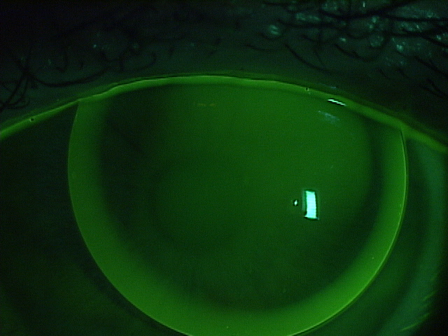
Well-fit corneal GP lens with sodium fluorescein dye

The way that the GP industry works is that GP button manufacturers (there are 6 in the US) sell the raw material, or button, to independent laboratories (there are 39 in the US) who then craft the button using industrial lathes into hundreds of lens designs using their own intellectual property.   GP buttons are used to make corneal GPs, hybrid, and scleral lenses.  Doctors who are skilled in fitting and evaluating the lens designs craft custom made GP lenses for their patients for whatever purpose benefits the patient.

Not many people know that the GP lens industry is on fire right now.  The turmoil began when Valeant Pharmaceuticals purchased Boston Products.  Boston Products manufactures the raw material of GP lenses, GP buttons, and held around 80% of the US market share.

The independent laboratories have a trade association called the Contact Lens Manufacturer’s Association (CLMA) who have an educational wing called the Gas-Permeable Lens Institute (GPLI).  The GPLI is universally beloved by practitioners because its primary function is to educate doctors to become better doctors in a non-branded, good-of-the-industry format.  No lens design is favored over any other.  Education is free to all doctors and expertise in specialty lenses is stressed, so that patients are placed in skilled hands.  Jan Svochak, president of the CLMA, says, “The CLMA represents a longstanding group of Independent Contact Lens Manufacturers working collaboratively where we have shared goals. These include educational resources through the GPLI that work closely with Eye Care Practitioners and Educational Institutions as well as a dedication to protecting and advancing utilization of custom manufactured contact lenses.”

Next Valeant dropped the hammer on the industry.  Overnight and for no apparent reason, they sent a letter to all of the independent laboratories announcing that they were increasing the price on scleral lens-sized Boston buttons by an astounding 60% (and other buttons by multiples).  Simultaneously, they announced that they were dropping out of the CLMA.  This move stunned the CLMA member labs as it blocked them from supplying Paragon CRT lenses to doctors.  Being the market leading GP button manufacturer, dropping out of the CLMA essentially defunded the GPLI and ensured the immanent collapse of the CLMA.

The price increase sent shock waves throughout the industry.  There was a simultaneous but independent reaction from many of the key-opinion leading optometrists who fit GP lenses.  The problem with any increase in price on the GP button level is that these price increases are passed down the line through the laboratories, the doctors and eventually, to the patients.  Valeant saw a huge backlash from optometrists who essentially stopped prescribing their materials.  They admitted making a mistake, and lowered the cost of the buttons, but interestingly, not to the original level.  Instead, there was an average 16% increase in the cost of scleral lens buttons to the laboratories.  Similarly, Valeant did not rejoin the CLMA.  The cost increase has been reported to fund Valeant brand specific education.

The other members of the CLMA came together and saved the association and the GPLI.  Additionally, a key competitor to Valeant, Contamac, rejoined the CLMA.  Contamac is a button manufacturer who formerly held around 8% market share of GP buttons.  At present, key sources within the industry believe that the market share has essentially flip-flopped, so that now, Contamac has rapidly gained market share of the GP button space as doctors have largely abandoned Boston materials in protest of these moves.

In a reactionary panic, Valeant has most recently written to the CLMA, asking to rejoin, but paradoxically with demands.  The CLMA is currently reviewing whether to allow Valeant to rejoin and under what terms.  Long term, it is beneficial for the industry for everyone to work together for the common good. It is unfortunate that a large company has come into the keratoconus treatment area and is raising prices without providing any real value, such as research and development into newer and better tools.  Companies like Valeant ultimately need to realize that they are not in control of an industry.  The patients and doctors are.


Dr. Sonsino is a partner in a high-end specialty contact lens and anterior segment practice in Nashville, Tennessee.  For over 12 years, he was on the faculty at Vanderbilt University Medical Center’s Eye Institute.  Dr. Sonsino is a Diplomate in the Cornea, Contact Lens, and Refractive Therapies Section of the American Academy of Optometry (AAO), chair-elect of the Cornea and Contact Lens Section of the American Optometric Association (AOA), a fellow of the Scleral Lens Education Society, board certified by the American Board of Optometry (ABO), and an advisory board member of the Gas Permeable Lens Institute (GPLI).  He lectures internationally, publishes in peer-review and non-peer-reviewed publications, and operates the website: TheKeratoconusCenter.org.  He consults for Alcon, Art Optical, Allergan, Johnson & Johnson, Optovue, Synergeyes, Visionary Optics, Visioneering, and formerly for Bausch & Lomb.

3/30/16

Sonsino Headshot

Jeffrey Sonsino, OD, FAAO
The Contact Lens Center at Optique Diplomate
Cornea, Contact Lens, and Refractive Therapies,

What You Should Know About Eye Color

One of the most common identifying factors of a person is their eye color. Here is what you should know about eye color.

eye color
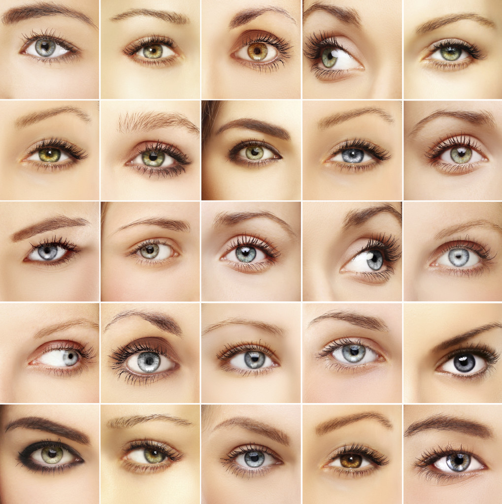
We all know that eye color is genetic, depending on the genes we inherit from our parents. We used to think that brown eyes were “dominant” and blue eyes were “recessive,” but modern science has shown that eye color is not at all that simple. Human eye color originates with three genes, but there may be as many as 50 genes that have influence over eye color. Most people in the world have brown eyes, with the second most common colors being blue and grey. Green is the rarest color.

What gives you your eye color?

The color of your eyes depends on how much of the pigment melanin you have in your iris. The more pigment you have, the darker your eyes will be. Blue, grey, and green eyes are lighter because there is less melanin in the iris. Most Caucasian babies are born with blue eyes because melanin is not present at birth, but develops with age. However, by the age of three their eye color is determined.

A contributing factor is also your stroma, the front layer of the iris that contains fibers that scatter the light that is reflected outward. This helps to account for why you your eyes might appear to change color or intensify depending on what color you are wearing, amplifying the scattered light reflecting back.

Some children are born with eyes that are not the same color. This can be caused by faulty developmental pigment transport, trauma either in the womb or shortly after birth or a benign genetic disorder. However, because there could be a small chance of it indicating eye disease, such as Horner’s syndrome, it is suggested you have an early eye exam to make sure nothing serious is going on.

Can your eyes change color?

Contrary to popular opinion, your eyes do not change color based on your emotions. The iris is a muscle that expands and contracts to control the size of your pupil. It gets bigger in dim lighting and gets smaller in bright lighting. It also shrinks when you are focusing on close tasks, such as reading a book. Certain emotions can also change the pupil size, such as anger, grief or happiness. This can cause the pigments in the iris compress or spread apart, slightly changing the appearance of your eye color. Also because the pupil is black, your eyes appear darker. There are times when your eye color might darken slightly, such as puberty, pregnancy, and aging for 10%-15% of Caucasians with light-colored eyes.

What else does melanin do?

Besides giving our eyes color, melanin helps to protect them from the sun. Because they have less pigment, light eyes are much more sensitive to harmful UV rays from the sun and electronic devices than brown or black eyes. This makes UV protection even more important for babies and people with light-colored eyes.

Finally, because some people believe that the eyes are the windows to the soul, there are some superstitions about eye color. People with blue eyes are thought to have rich imaginations, people with green eyes have sharp minds, hazel eyes indicate a passionate soul and people with brown eyes are calm, but have underlying passion. What does your eye color say about you?

3/17/16


Susan DeRemerSusan DeRemer, CFRE
Vice President of Development
Discovery Eye Foundation

Women’s Eye Health

With this being Women’s History Month, we thought it appropriate to discuss women’s eye heath. Your vision is one of the most important things in your life. Vision loss can make everyday tasks more difficult, impede your work and lead to depression.
women's eye health
According to Prevent Blindness America (PBA), 66% of people who are blind or visually impaired are women. This is primarily due to the fact that women have more risk factors for vision loss than men. Sadly the same study also revealed that only 9% of women realize this. Early intervention can help prevent blindness in many cases. Many blinding eye diseases can be treated to prevent blindness and almost all eye injuries can be prevented.

Here is a closer look at why women are affected more than men, and what condition they need to be aware of for early diagnosis and sight-saving treatments.

Why Women Lose Vision

  • They live longer than men and many eye diseases are age-related. Examples are cataracts, macular degeneration and diabetic retinopathy. The rates of these diseases are increasing as the baby boomer population ages.
  •  

  • Some eye diseases are intrinsically more prevalent among women, like dry eye syndrome which is believed to be linked to hormones. It is 2-3 times more likely in women than men. Hormonal changes can influence vision changes across the life span of a woman, from pregnancy to post-menopause.
  •  

  • Women have higher rates of autoimmune diseases such as lupus, rheumatoid arthritis and multiple sclerosis. The serious side effects of these conditions can affect your eyes, causing vision loss.
  •  

  • Social and economic factors can access to health care for women restricting early detection and treatment which could prevent or limit vision loss.
  •  

  • While behavioral and environmental factors are not restricted to women, poor nutrition and obesity can cause diabetes and subsequent diabetic retinopathy; while smoking is also a proven risk factor for eye diseases such as cataracts and macular degeneration. Women under 23 are the fastest growing segment of new smokers.

women's eye health
Eye Conditions More Prevalent in Women

  • Cataracts are the world’s leading cause of blindness. They are the result of a clouding of your eye’s normally clear lens. They can be treated with cataract surgery, where the cloudy lens is replaced with a clear synthetic lens.
  •  

  • Glaucoma is the second-leading cause of blindness in the Us. It is called the silent thief of sight because there is no warning. It happens when pressure in the eye — intraocular pressure or IOP — is too high, damaging the optic nerve which sends vision signals to the brain. Open-angle glaucoma, the most common type, affects men and women equally. But women are 2-4 times more likely than men to get closed-angle glaucoma. One of the possible reasons for this is that the front chamber between the iris and cornea is shallower in women than men and can block fluids from draining out of the eye, thus increasing pressure. Glaucoma is also genetic, meaning you are at higher risk if someone in your family has had glaucoma. When caught early, there are treatments that can help control your IOP. If it is not controlled early, blindness can result and it is irreversible.
  •  

  • Age-related macular degeneration (AMD) is the number one cause of vision loss in people over 40 in the US. One of the reasons women are at higher risk is because they tend to live 5-7 years longer than men. AMD gradually destroys the central part of your sight that helps you read or drive. There are two types of AMD. Dry AMD, which occurs when drusen accumulate under the retina. There is no treatment for dry, yet accounts for 90% of the cases. Wet AMD occurs when new blood vessels grow and leak between the retina and eye’s outer layer. There is a treatment of anti-VEGF injections for this version of AMD. The earlier the disease is diagnosed and treated, the better the results. Like glaucoma, it is an inherited eye disease.
  •  

  • Diabetic retinopathy is a result of diabetes. When blood sugar levels are too high, the vessels that feed the retina weaken and eventually leak, which cause the macula to swell. In its most dangerous form the retina may detach from the back of the eye leading to blindness. Diabetes is also hereditary, but controllable with early diagnosis and treatment.
  •  

  • Dry eye syndrome occurs when your eyes produce too few tears. Your eyes may burn, sting or feel gritty, making your vision blurry or you may blink more. Contrary to the name, you may appear weepy as the dryness may stimulates more tear production. Thanks to hormones, women are more susceptible. In postmenopausal women, the shift in balance between estrogen and progesterone can be responsible.
  •  

  • Pregnancy-related eye changes, like menopause, are caused by hormone shifts and can cause several temporary eye conditions, such as dry eye and corneal swelling.

3/10/16

Susan DeRemerSusan DeRemer, CFRE
Vice President of Development
Discovery Eye Foundation

The Brain and the Eye – How They Work Together

The Brain and the Eye

The eye works like a camera. The iris and the pupil control how much light to let into the back of the eye, much like the shutter of a camera. When it is very dark, our pupils get bigger, letting in more light; when it is very bright our irises constrict, letting in very little light.

The lens of the eye, like the lens of a camera, helps us to focus. But just as a camera uses mirrors and other mechanical devices to focus, we rely on eyeglasses and contact lenses to help us to see more clearly.

The focus light rays are then directed to the back of the eye, on to the retina, which acts like the film in a camera. The cells in the retina absorb and convert the light to electrochemical impulses which are transferred along the optic nerve to the brain. The brain is instrumental in helping us see as it translates the image into something we can understand.

The Brain and the Eye

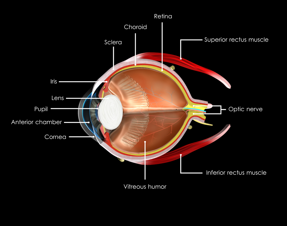
The eye may be small, but it is one of the most amazing parts of your body. To better understand it, it helps to understand the different parts and what they do.

Choroid
A layer with blood vessels that lines the back of the eye and is between the retina (the inner light-sensitive layer that acts like film) and the sclera (the outer white part of the eyeball).

Ciliary Body
The muscle structure behind the iris, which focuses the lens.

Cornea
The very front of the eye that is clear to help focus light into the eye. Corrective laser surgery reshapes the cornea, changing the focus to increase sharpness and/or clarity.

Fovea
The center of the macula which provides the sharp vision.

Iris
The colored part of the eye used to regulate the amount of light entering the eye. Lens focuses light rays onto the retina at the back of the eye. The lens is transparent, and can deteriorate as we age, resulting in the need for reading glasses. Intraocular lenses are used to replace lenses clouded by cataracts.

Macula
The area in the center of retina that contains special light-sensitive cells, allowing us to see fine details clearly in the center of our visual field. The deterioration of the macula can be common as we age, resulting in age related macular degeneration.

Optic Nerve
A bundle of more than a million nerve fibers carrying visual messages from the retina to the brain. Your brain actually controls what you see, since it combines images. Also the images focused on the retina are upside down, so the brain turns images right side up. This reversal of the images Is a lot like what a mirror does in a camera. Glaucoma can result when increase pressure in the eye restricts the flow of impulses to the brain, causing optic nerve damage and makes it difficult to see.

Pupil
The dark center opening in the middle of the iris changes size to adjust for the amount of light available to focus on the retina.

Retina
The nerve layer lining the back of the eye that senses light and creates electrical impulses that are sent through the optic nerve to the brain.

Sclera
The white outer coating of the eyeball.

Vitreous Humor
The clear, gelatinous substance filling the central cavity of the eye.

3/3/16

Susan DeRemerSusan DeRemer, CFRE
Vice President of Development
Discovery Eye Foundation