Our First Three Months Of Eye Care

9/30/14

Discovery Eye Foundation Blog’s First Three Months

It is hard to believe, but this blog has been providing information and insights into eye disease, treatment options, personal experiences of living with vision loss, and other eye-related information for seven months.

All of this would not have been possible without the expertise of remarkable eye care professionals who took time out of their busy schedules to share information to help you cope with vision loss through a better understanding of your eye condition and practical tips. Since so much information was shared in the seven months, here is a look at the first three months, with the additional four months to be reviewed next Tuesday.
Thank You - first three months
I am very thankful to these caring eye professionals and those with vision loss who were willing to share their stories:

Marjan Farid, MDcorneal transplants and new hope for corneal scarring

Bill Takeshita, OD, FAAO, FCOVDproper lighting to get the most out of your vision and reduce eyestrain

Maureen A. Duffy, CVRTlow vision resources

M. Cristina Kenney, MD, PhDthe differences in the immune system of a person with age-related macular degeneration

Bezalel Schendowich, ODblinking and dealing with eyestrain

Jason Marsack, PhDusing wavefront technology with custom contact lenses

S. Barry Eiden, OD, FAAOcontact lens fitting for keratoconus

Arthur B. Epstein, OD, FAAOdry eye and tear dysfunction

Jeffrey Sonsino, OD, FAAOusing OCT to evaluate contact lenses

Lylas G. Mogk, MDCharles Bonnet Syndrome

Dean Lloyd, Esqliving with the Argus II

Gil Johnsonemployment for seniors with aging eyes

We would like to extend our thanks to these eye care professionals, and to you, the reader, for helping to make this blog a success. Please subscribe to the blog and share it with your family, friends and doctors.

Susan DeRemerSusan DeRemer, CFRE
Vice President of Development
Discovery Eye Foundation

Genetics and Glaucoma: Why don’t we have a genetic test for glaucoma?

9/25/14

We have known for a long time that there is a genetic component to glaucoma, since having a family history of glaucoma is one of the most important risk factors for developing the disease. Glaucoma is actually a group of diseases, including some starting at birth or in childhood, as well as the more common types that happen in adults, such as primary open angle glaucoma.dna strand - genetics and glaucoma

In some of the glaucomas of childhood, scientists can trace the cause to a single gene. For example, mutations in the gene for myocillin can cause juvenile-onset open angle glaucoma. And there are genetic tests available for some of these genes. These genetic tests, however, are helpful only for a subset of glaucoma patients. For most glaucoma patients, these tests don’t give us the answers we need.

In the types of glaucoma that happen in adults, both environmental factors and genetic factors contribute to whether or not someone develops the disease. This complex interplay of factors makes testing only one gene or a handful of genes not as helpful. For diseases like adult-onset open angle glaucoma, instead of having just one gene that is mutated and causing the disease, there are many genes involved. All these genes contain tiny differences, some potentially helpful and some potentially harmful. It is an individual’s mix of genes combined with the lifetime of complex environmental exposures, that determines whether he or she will get the disease. In the future, genetic tests that incorporate this group of genes will help doctors, patients, and families better understand their susceptibilities, and hopefully lead to prevention of vision loss from glaucoma.

Genetic testing has the potential to offer a lot of benefits, but is not without risk and unintended consequences. Genetic counselors are trained to help patients, families, and doctors navigate these areas.

For more information about glaucoma, visit www.nei.nih.gov/glaucoma.

References:
Genetics of primary glaucoma. AO Khan. Current Opinion in Ophthalmology. 2011. 22(5):347-55

Julia Rosdahl - genetics and glaucomaJullia A. Rosdahl, MD, PhD
National Eye Health Education Program Glaucoma Subcommittee
Duke Eye Center, Duke University

Lens Care If You Wear Contact Lenses and Use Cosmetics

9/23/14

In a continuation from his article on Proper Contact Lens Care, Mr. Ward, Director of the Emory Contact Lens Service, also offers tips if you wear contact lenses and use cosmetics. Several of these pointers apply even if you don’t wear contact lenses, but want to protect your eyes.you wear contact lenses and use cosmetics

The Bullet List of Contact Lens Care For Users Of Eye Area Cosmetics

    • If possible look for eye makeup specifically labeled for use by contact lens wearers; use premium products.
    • Apply eye area cosmetics after inserting contact lenses (this will help prevent cosmetic contamination of lens surfaces from handling of cosmetics).
    • Remove lenses before removing makeup.
    • Remove makeup daily with mild soap and water; do not use oil or petroleum based make up removers; specifically, avoid moisturizing bar soap and an eye makeup remover that contains mineral oil and cocoa butter.
    • Choose water based makeup; avoid any oil based, or ‘waterproof’ eye area products (oils will travel across the skin and contaminate the tear film).
    • Avoid ‘lash-extending’ mascaras with artificial fibers, and apply mascara only to the end of lashes; do not apply mascara to the base of the eyelash or on the eyelid margin.
    • Do not apply oil-based moisturizers on the eyelids (oils can spread on the skin).
    • Do not apply any makeup to the eyelid margin (shelf), between the eyelashes and the eyeball.
    • Apply face powders sparingly; use pressed powder instead of loose powder; try to stay away from the eye area as much as possible; avoid frosted.
    • Choose liquid or gel eye shadows rather than powders.
    • Use caution with hair styling sprays. If possible, spray aerosols with eyes closed and step back out of the mist before opening the eyes. These gel/wax/lacquer type sprays can significantly coat your contact lenses.
    • Replace eye makeup at least every three months; do not share cosmetics.
    • And, please note that an automobile’s rear view mirror is not intended for makeup application while driving.

    Michael Ward - proper contact lens careMichael A. Ward, MMSc, FAAO
    Director, Emory Contact Lens Service
    Emory University School of Medicine

Proper Contact Lens Care Provides Best Vision, Comfort and Ocular Health

9/18/14

Proper contact lens care is essential for the best contact lens wearing experience. Mr. Ward, Director of the Emory Contact Lens Service has shared some valuable information about taking care of your contact lens in the article below. On Tuesday join us for additional tips for people who wear contact lenses and wear cosmetics.contact lens case - proper contact lens care

Contact lenses provide alternatives to spectacles, and contact lens wearers report better peripheral vision, depth perception and overall vision quality. Contact lenses can correct near-sightedness, far-sightedness, astigmatism and even correct the need for reading glasses. They are also used to manage some ocular surface diseases.

Contact lenses fall into two basic material types: soft contact lenses (SCL) and rigid gas-permeable (GP) lenses. Soft lenses account for the great majority of the contact lens market. GP lenses require more precise fitting and are often used as specialty devices to correct high prescriptions and/or to manage various ocular disorders and may require longer lens-adaptation time. Regardless of lens type, careful attention to lens care instructions can provide good vision and life-long lens wearing comfort.
Proper lens care depends on the lens type, wearing schedule and other factors. Single-use or daily-disposable soft lenses are prescribed to be worn once and discarded. This is theoretically the safest lens wearing modality in that no lens cleaning, lens care or storage case is required for this modality. Other daily wear soft lenses may be replaced every 2 weeks, monthly or by other schedule. Any and all lenses that are removed each day must be cleaned and disinfected prior to their reuse. Your eye care practitioner should provide specific instructions relative to your lens wear and care needs. General lens care instructions and Dos and Don’ts are bullet-listed below.

A word of caution –
Contact lens wear is quite safe as long as proper lens and storage case care are followed. However, improper lens wear and care can put the lens wearer at risk for serious consequences. Sight-threatening microbial keratitis (corneal ulcer) is the most significant adverse event associated with contact lens wear and is largely preventable. The contact lens storage case is the most likely potential reservoir for contact lens related ocular infections. Therefore, lens storage case care should be high on the list of important lens wearing instructions. Contact lens cases are not meant to be family heirlooms; cases should be replaced regularly, at least every 1-3 months.

The Bullet List of Contact Lens Care Recommendations

  • Hand washing: Always wash your hands before handling contact lenses. Use mild, basic soap and avoid antibacterial, deodorant, fragranced or moisturizing liquid soaps (many liquid soaps have moisturizers that can contaminate your contacts from handling).
  • Cleaning, rinsing, and disinfecting: Digital cleaning (rubbing the lens with your finger in your palm) removes dirt and debris and prepares the lens surfaces for disinfection. Rub & rinse thoroughly, even if the product is labeled “No Rub”. Lens storage solutions contain chemicals that inhibit or kill potentially dangerous microorganisms while the lenses are soaked overnight.
    • Contact lenses should be cleaned when removed from the eye.
    • Do not re-use old solution or “top-off” the liquid in the lens storage case. Empty the storage case daily and always use fresh solution.
    • Do not use lens care products beyond their expiration dates. Discard opened bottles after 28 days.
    • Do not allow the tip of the solution bottle to come in contact with any surface, and keep the bottle tightly closed when not in use.
    • Do not transfer contact lens solution into smaller travel-size containers.
  • Keep your contact lens storage case clean (inside and out).
    • All lens storage cases should be emptied, rinsed, wiped, and air-dried between uses.
    • Keep the contact lens case clean and replace it regularly, every one to three months.
    • Do not use cracked or damaged lens storage cases.
    • Take care to remove residual solution from surfaces of lens case and solution bottles.

Other Dos and Don’ts

  • Do not wear your lenses during water activities (swimming, hot tubs, showering, etc).
  • Soft contact lenses should not be rinsed with or stored in water. Soft lenses will change size and shape if exposed to water.
  • Do not put your lenses in your mouth.
  • Do not use saline solution or re-wetting drops in an attempt to disinfect lenses. Neither is capable of disinfecting contact lenses.
  • Wear and replace contact lenses according to the prescribed schedule.
  • Follow the specific contact lens cleaning and storage guidelines from your eye care professional.
  • Do not change lens care products without first checking with your eye care practitioner.
  • Spare rigid (GP) lenses should be stored dry for long term storage { clean, rinse, dry}. New or dry-stored GP lenses should be re-cleaned and disinfected prior to lens wear.
  • Do not store soft lenses in the storage case for an extended period of time. “Spare” soft contact lenses should be new and stored in their original and unopened packaging.
  • Do not sleep in your contact lenses unless specifically approved to do so by your eye care practitioner.

For information from the Centers for Disease Control and Prevention, see:
www.cdc.gov/contactlenses/
www.cdc.gov/contactlenses/cdc-at-work.html

Michael Ward - proper contact lens careMichael A. Ward, MMSc, FAAO
Director, Emory Contact Lens Service
Emory University School of Medicine


The Way Eyes Work

9/16/14

Eyes are an amazing part of your body and not just because of what they do helping you see. The are also fascinating be because of the way eyes work. Here are 20 facts about how your eyes function.
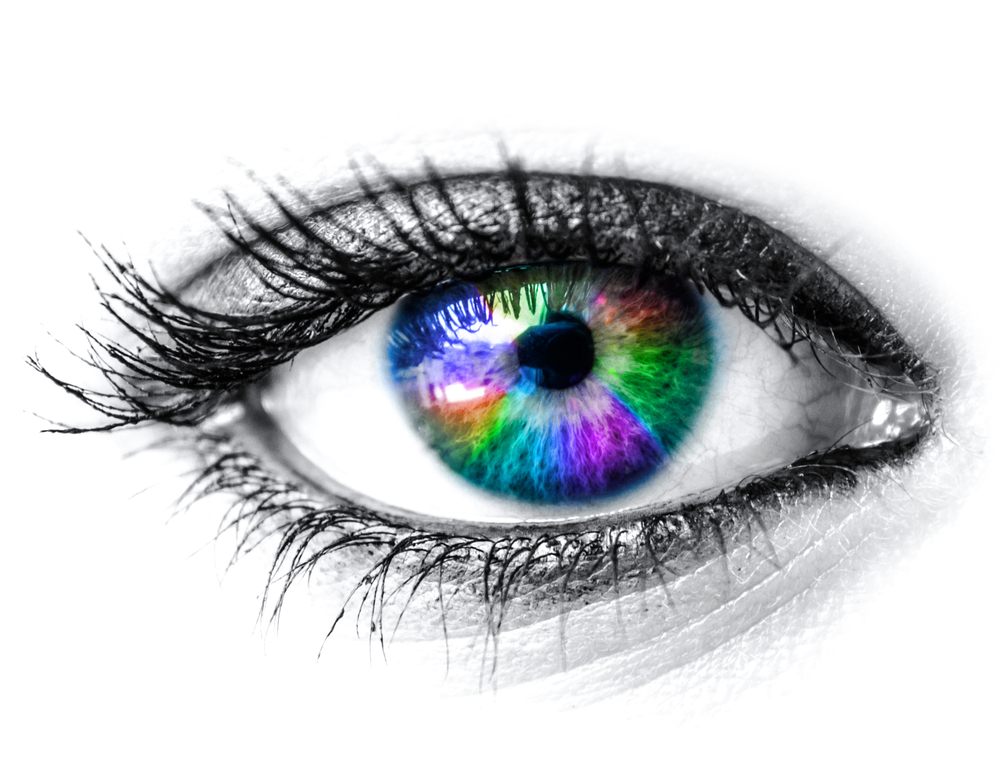
Colorful eye - the way eyes work

      1. The pupil dilates 45% when looking at something pleasant.

2. An eye’s lens is quicker than a camera’s.

3. Each eye contains 107 million cells that are light sensitive.

4. The light sensitivity of rod cells is about 1,000 times that of cone cells.

5. While it takes some time for most parts of your body to warm up their full potential, your eyes are always active.

6. Each of your eyes has a small blind spot in the back of the retina where the optic nerve attaches. You don’t notice the hole in your vision because your eyes work together to fill in each other’s blind spot.

7. The human eye can only make smooth motions if it’s actually tracking a moving object.

8. People generally read 25% slower from a computer screen compared to paper.

9. The eyes can process about 36,000 bits of information each hour.

10. Your eye will focus on about 50 things per second.

11. Eyes use about 65% or your brainpower – more than any other part of your body.

12. Images that are sent to your brain are actually backwards and upside down.

13. Your brain has to interpret the signals your eyes send in order for you to see. Optical illusions occur when your eyes and brain can’t agree.optical illusion - the way eyes work

14. Your pupils can change in diameter from 1 to 8 millimeters, about the size of a chickpea.

15. You see with your brain, not your eyes. Our eyes function like a camera, capturing light and sending data back to the brain.

16. We have two eyeballs in order to give us depth perception – comparing two images allows us to determine how far away an object is from us.

17. It is reported that men can read fine print better than women can.

18. The muscles in the eye are 100 times stronger than they need to be to perform their function.

19. Everyone has one eye that is slightly stronger than the other.

20. In the right conditions and lighting, humans can see the light of a candle from 14 miles away.

Susan DeRemerSusan DeRemer. CFRE
Vice President of Development
Discovery Eye Foundation

Seeing the whole picture: My life after a telescope implant

9/11/14

I’ve lived with my family in Schenectady, NY (outside of Albany) for more than 50 years and I love my home because it’s within walking distance of my grocery store, bank and church. But ten years ago, my vision was severely impacted because of age-related macular degeneration.

Patty and Linda - telescope implant
Patty (on right) and friend Linda have some fun

My children had to help me so much because I just couldn’t see. For example, they had to drive me to the doctor and on errands. I needed help shopping because I couldn’t read labels. After a while, it was very depressing. I started to avoid social situations, like at family parties and church events, because I was embarrassed that I couldn’t recognize faces any longer. I felt terrible about this. I sat and cried every now and then.

But then my doctor told me about a treatment I wasn’t yet aware of called the telescope implant. The device is very small (the size of a pea), and it is implanted in one eye to restore vision. My doctor explained that it works like a real telescope in that it magnifies images, which reduces the blind spot that blocked my straight-ahead vision. The other eye does not get an implant because you need to keep some peripheral vision to help with orientation and balance. This sounded like science fiction! But I wanted to see if it could help me and decided to take a chance on the procedure.

I worked with an entire team of specialists, which were part of a treatment program called CentraSight. My retina doctor, cornea surgeon, low vision optometrist and a low vision occupational therapist all counseled me about what to expect from the outpatient procedure, particularly afterwards. For example, I learned there was a significant amount of occupational therapy required to adjust and become proficient at using my new vision. I also was warned that my sight would not be like it was in my youth, I wouldn’t be able to do everything I used to nor would I be able to see, differently, the minute I opened my eyes.

I had my surgery in February 2013. The cost for the telescope implant and visits associated with the treatment program were covered by Medicare, which was very helpful.

Thinking back, I was nervous on surgery day, but shortly after the procedure, I was back with my family and going out to dinner. The most amazing part is that I was able to see my daughter’s face almost immediately, despite not expecting to see anything right away. It was such a gift! After the surgery, I worked with the low vision specialists for about three months. The exercises varied because you use different techniques to see things when you are standing, sitting or moving around.

It’s been more than a year since my surgery and I am very happy, mostly because I can do so much more now that I can see better. I can read, sew, do canning and work in my garden. I can see the crosswalks, which lets me walk to the grocery store safely. But most importantly, I can see my family and friends. I’m enjoying spending time with the people I care about. I would recommend that people learn more about the telescope implant. There are CentraSight teams all over the country. When you call 877-99-SIGHT or visit www.CentraSight.com a trained CentraSight Information Specialist will point you to the team closest to your home and can even help schedule the appointments for you. The telescope implant isn’t for everyone, but it can make such a difference in your life.

Patty Gadjewski - telescope implantPatty Gadjewski
Schenectady, NY

Rods and Cones Give Us Color, Detail and Night Vision

9/9/14

Function of Rods and Cones

Rods and cones are a vital part of the eye, helping define what we see. Here’s what you should know.
Crayons for rods and cones
1. There are three types of color-sensing cones, red, blue and green. If you are color blind one or more of these cells is missing or not working properly.

2. Men have a higher chance of being color blind than women. 1 out of 12 vs. 1 out of 255.

3. The most common type of color blindness is the disability to tell the difference between red and green.

4. The eye can distinguish between 500 shades of gray.

5. A healthy human eye can detect over 10 million different colors.

6. About 2% of women have a rare genetic mutation that gives them an extra retinal cone allowing them to see more than 100 million colors.

7. During a major depression people see less contrast, making colors appear duller.

8. All babies are color blind when they are born. Color vision begins to develop within a week after birth and by 6 months your baby can see every color you can.

9. Your eyes contain 7 million cones which help you see color and detail.

10. The stars and colors you see when you rub your eyes are call phosphenes.

11. There are 120 million light-sensing cells called rods which help you to see better in the dark.

12. Smoking reduces your night vision.

And these are just fun facts about eyes:

The Mayans believed that cross-eyes were attractive and would make efforts to ensure their children became cross-eyed.

Pirates used to wear a gold earring, believing it improved their sight. They also used eye patches to quickly adjust their eyes from above to below deck. When going below deck where it was dark, they flipped up the eye patch to see with the eye that had not be affected by light.

The phrase “it’s all fun and games until someone loses an eye” comes from Ancient Rome, as the only rule for their bloody wrestling matches was “no eye gouging.”

Susan DeRemerSusan DeRemer, CFRE
Vice President of Development
Discovery Eye Foundation

Evaluating Treatment Claims

9/03/14

The following article on evaluating treatment claims is from the Science-Based Medicine blog is being used with their permission. Since Discovery Eye Foundation provided the inspiration for Dr. Hall, we thought you might enjoy it as well.
Black board evaluating treatment options
I recently wrote about the claim that acupuncture can improve vision in patients with macular degeneration. In response, I received this e-mail:

“At Discovery Eye Foundation we have an education and outreach program for people with age-related macular degeneration, the Macular Degeneration Partnership. We are constantly getting calls from people who have heard of a new “cure” or a way to stop their vision loss. It is always hard to interject reality into the conversation and hear the hope leave their voice, replaced by frustration or despair.”

The e-mail suggested I write an article providing guidelines for consumers to help them evaluate the validity of treatment claims for themselves. On SBM we are constantly stressing the need to apply critical thinking to what you read, and the many pitfalls to be avoided. I’ll try to synthesize some of the principles into a handy list of questions.

What kind of evidence is there?

• If the claim is based on nothing but testimonials, STOP RIGHT THERE. You can forget about it, or at least file it away until there is better evidence. Anecdotes are not evidence; they only serve to suggest promising avenues of research. Science is the only reliable way to determine if a treatment is safe and effective.

• Is the claim based on a gold standard randomized, placebo-controlled, peer-reviewed study or some lesser kind of evidence like case reports? Was it published in a reputable mainstream medical journal? (If you’re not sure how reputable the journal is, you can look it up on tables of journal impact factors.”

• Keep in mind that half of all studies are wrong. There are many factors that can lead to error. Preliminary or pilot studies that are positive are frequently followed by better, larger studies that are negative. We can never rely on one study without confirmation.

• Was it a meaningful clinical study in humans?

In vitrolab studies and animal studies may not be applicable to humans; if promising, they must be confirmed in good human studies. If it was a human study, did it show meaningful outcomes that made a real difference, like a reduction in heart attacks, or did it just show an improvement in lab values or risk factors?

• Were there 10 subjects or 300? Large studies are more trustworthy than small ones. The fewer dropouts, the better.

• Did it use an appropriate placebo control that subjects really couldn’t distinguish from the active treatment?

• Have other studies found similar results? Are there any studies that show the opposite? You can search PubMed and look for them.

Who is making the claim?

  • Is it someone who is likely to be biased?
  • Is it someone who provides the treatment or sells the product?
  • Is it someone with expert medical knowledge or someone like the schoolteacher who claimed she had invented a cure for the common cold?
  • Does the person have a good reputation, or a track record of making questionable statements?
  • Is it someone who quotes or associates with unreliable sources like Mercola.com, the Weston Price Foundation, or the Health Ranger? Quackwatch has a useful list of non-recommended sources of health advice.

Where was the claim reported?

Real medical breakthroughs would be headline news. It’s not likely you would first hear about a cure for diabetes on an afternoon talk show or a Facebook page. Has your doctor heard of it? Are mainstream doctors recommending it? Is it covered on professional medical websites like the American Academy of Pediatrics or disease-focused websites like the American Diabetes Association? Is it sold only through multilevel marketing schemes?

What kind of language is being used?

Is it a sober factual report with caveats, or is it full of hype and buzzwords like “miracle,” “natural,” “known to the ancients,” “quantum,” “amazing,” “revolutionary”? If it really worked, advertising gimmicks wouldn’t be needed to sell it.

Does Gwyneth Paltrow swear it worked for her?

Testimonials can be very passionate but they are notoriously unreliable. Getting better when you use a treatment doesn’t necessarily mean you got better because of the treatment. Symptoms can fluctuate, diseases can resolve without treatment, and placebos can fool people. Perceptions can be wrong (think of optical illusions), the meaning of true perceptions can be misinterpreted, and memories can be inaccurate. Every snake oil salesman has reams of testimonials, and through the centuries there were testimonials galore for bloodletting to balance the humors. People frequently come to believe bogus remedies have worked for them. Barry Beyerstein wrote a classic article about that; it’s essential reading.

Does it make sense?

Is there a plausible mechanism of action? If someone claimed that standing on your head and whistling Dixie would cure diabetes, I think you would be skeptical. If it claims to work by a mechanism incompatible with known scientific principles, the level of evidence would have to be extraordinary for it to outweigh all the evidence those scientific principles are based on. Homeopathy’s claim that water can cure by remembering long-gone molecules, even after the water has been dripped onto a sugar pill and allowed to evaporate, would require extraordinary evidence indeed. If it’s a new antibiotic that is related to an old one, an ordinary level of evidence would suffice.

Is there a double standard?

Are they asking you to accept a “natural” or “alternative” treatment on the basis of the kind of evidence that you wouldn’t want the FDA to accept for allowing marketing of a prescription drug? There is only one science and only one standard of evidence.

Does it sound too good to be true?

Then it probably is too good to be true. Does it promise to cure a hitherto-incurable disease? Does it promise you can eat all you want and still lose weight? Does it promise there are no side effects of any kind? Does it remind you of the spiel of a used car salesman or a TV infomercial? Caveat emptor.

Who disagrees and why?

This is the most important question you can ask. It is rare for 100% of people to agree on anything. If you can find someone who disagrees, you can examine the reasons given for both opinions, and it will usually become obvious which side makes more sense. If you can’t find anyone who disagrees, it might be because it’s too new or because no scientist has taken it seriously enough to bother writing about it. In that case, withhold judgment and keep checking until someone does disagree.

Hope springs eternal, but true hope is better than false hope

If you are a desperate patient, it’s only natural to grasp at any straw of hope; but when the evidence is insufficient, the reasonable approach is to withhold judgment and wait for better evidence. You might think, “If it works, I don’t want to wait” but history teaches us that the great majority of these things don’t pan out. It might not do any harm, but then again it might; there might be adverse effects that haven’t been identified yet, it might raise false hopes only to dash them, and if nothing else it might waste time and money or interfere with getting more appropriate care. When you take an inadequately-tested medicine, you are essentially offering yourself as a guinea pig in a haphazard uncontrolled experiment that doesn’t even keep records. Of course, that’s your privilege; but I hope you would do it with your eyes open, with a realistic understanding of the state of the evidence.

Harriet Hall, MDHarriet A. Hall, MD
Retired US Air Force Physician
Editor of Science-Based Medicine Blog
Author of SkepDoc column in Skeptic Magazine

Charles Bonnet Syndrome – Fast Facts

9/2/14

“The theater of the mind could be generated by the machinery of the brain.”Older woman - Charles Bonnet Syndrome

What is Charles Bonnet Syndrome (CBS)?

  1. It was discovered in 1790 by Charles Bonnet.
  2. 10-40% of low vision individuals experience hallucinations.
  3. Only 1% of them acknowledge it!
  4. Images usually appear suddenly and stop as suddenly. They don’t fade in and out.
  5. Most of the time, the images are not people or things familiar to you.
  6. They may be startling, but they are not frightening or sinister.
  7. It is like watching a movie as the images don’t interact with you, unlike psychological images that interact with you and you with them.
  8. CBS usually stops within 12-18 months.

Why does it happen?

  1. Vision takes place in the brain.
  2. Different parts of the visual brain are triggered by different information. Faces fire up one part of the brain; buildings another and the scientists can see what activity is happening where.
  3. As you lose vision and the visual parts of the brain are not getting any input, they become hyperactive and excitable. This causes them to start to “fire” spontaneously.
  4. Example – If you damage (or lose vision) in a particular area, such as the one for faces, you lose the ability to recognize faces.  That will create abnormal activity in that area and you will hallucinate faces (*see more detailed explanation below).

What Can You Do?

  1. There is no cure or truly effective treatment.
  2. Acknowledge that you are having the visions and talk about them.
  3. Look on them as an experience rather than a problem. It’s fascinating how the brain works, isn’t it?
  4. Having a good sense of humor can help in adjusting well to CBS.
  5. Sometimes, eye exercises — such as looking from left to right without moving one’s head for 15 to 30 seconds — can help stop a hallucination.
  6. Increased room lighting can sometimes prevent an episode of CBS visions if they commonly take place in low light. Changing the lighting in the middle of an episode may stop them.
  7. Stress and fatigue could be contributing factors, so try to get enough rest and reduce stress.
  8. Identify and engage in activities you enjoy; keep up your social life. Reduced social isolation, boredom, lack of stimulation, and low activity seem to increase CBS.

For more information on Charles Bonnet Syndrome, please go to the Macular Degeneration Partnership website.

*Testing for functional brain imagery as individuals hallucinate can find different parts of the brain are activated.

Your brain has a particular area or lobe where vision is interpreted. The light energy that bounces off objects enters the eye and is converted to chemical energy by the retinal cells. That energy is sent through the optic nerve where processing of the vision starts to occur. When it reaches the visual cortex, it is sent to very specific areas of the brain and specific areas of the brain see specific things.

The fusiform gyrus processes faces, but different areas process the parts of faces. While damage in the fusiform gyrus causes you to lose the ability to recognize faces, abnormal activity in that area will  cause you to hallucinate faces. An area in the anterior part of this gyrus is where teeth and eyes are recognized. There are other areas that specifically sees cartoons and another part for buildings and landscapes.

Judi Delgado - age-related macular degenerationJudith Delgado
Executive Director
Macular Degeneration Partnership
A Program of Discovery Eye Foundation