Four Tips For Buying Sunglasses

4/29/14

May will be here this week, and in Southern California we are looking at bright, sun-filled days with temperatures in the upper 80s and low 90s. This means that thousands will be heading to the beaches or their own backyards to enjoy the warm weather.

Now is the perfect time to review one of the biggest contributing factors to vision loss – sun exposure. And it’s not just about sunglasses, but also brimmed hats.

from esty.com
from esty.com

First let’s talk about sunglasses. There are three things to think about when selecting your sunglasses:
1. Lens tint
2. UV protection
3. Glare
4. Frames

Lens Tint
There is a misconception that the darker your sunglass lens, the better protection for your eyes. No true. The color or darkness of your lens is personal preference and often based on the activity you are doing while wearing sunglasses or the sun conditions. At the beach in bright sunlight you are subject to more reflective light and may prefer dark amber, copper or brown lens, if you are on the ski slopes when the skies are overcast you may prefer yellow or orange lens to increase contrast and fight “flat light.” If you are looking to increase contrast on a partially cloudy day, and if you don’t mind distorted color perception, you might prefer amber or rose lenses.

Other considerations include mirrored sun lenses that can block 10-15% more of the sun’s visible rays, or photochromic lenses that darken automatically when you go outside and then quickly become lighter when you come inside.

UV Protection
While darker lenses don’t offer better eye protection, controlling the UV exposure does. Research has found links that extended exposure to UVA and UVB rays can result in eye damage such as cataracts, photokeratitis and macular degeneration. By wearing sunglasses that block these harmful rays your eyes should remain healthier as you age. Also know that some parts of the country receive more UV rays than others – here is a wonderful chart from The Vision Council to let you see how your location rates.

Glare
Another problem when out in the sun, and especially driving, is glare. Making sure your lenses are polarized is a great help. They work by only letting in specific amounts of light at certain angles and reducing the brightness of that light.

Because I am light sensitive I find I use polarized lenses when I am reading outside is helpful. The reflected light from the page of a book can cause me to squint or fatigue my eyes if I read for a long period of time. The only other option is using a paper-ink e-reader which also helps cut down on glare.

Another way to deal with glare is the use of an anti-reflective (AR) coating on your lenses. It reduces eye stain by preventing light from reflecting off lens surfaces. When applied to the back of your lenses it can help with problems when the sun is behind you or to your side.
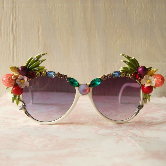
Frames
Not all light hits your eyes from directly in front. It can come through the top, sides and bottom of your frames. The smaller the frames, the more unfiltered light makes its way to your eyes. This is where a brimmed hat can help keep the sun coming in from the top while also providing protection for your face.

Fitovers - Auroa in Claret
Fitovers – Auroa in Claret

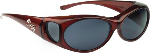
To provide you with the maximum protection, “fit-over” sunglasses, that you can wear over your regular prescription glasses, are a great idea and more economical. Cocoons Eyewear and Fitovers Eyewear are two of several companies that make them. They filter the light from the top, sides and even below to give you the maximum protection and come in a wide variety of lens colors. It is also nice not to have to get new sunglasses when your eyeglass prescription changes.

Whatever frames you choose make sure they fit properly and will not keep sliding down your nose or fall of when being active. You may even want to purchase a band-style foamed neoprene retainer that attaches at both temples, sometimes known as a gator.

Also remember, it is not just the direct sunlight you need to worry about. Water reflects up to 100% of the harmful UV rays, dry sand and concrete up to 25% and even grass reflects up to 3%.

Susan DeRemerSusan DeRemer, CFRE
Vice President of Development
Discovery Eye Foundation

Dry Eye and Tear Dysfunction

4/24/14

With an aging population and increasing awareness of the condition, dry eye has become one of the hottest topics in eye care today. There have been numerous advances in our understanding of the condition as well as several breakthroughs in treatment.
eye drops in eye
Lets start with the most fundamental question. Why do we have tears? Tears serve many functions, but perhaps the most important is that they create a perfectly smooth optical surface at the front of the eye to allow sharp focusing. The sharper your vision the more quickly you can spot and run from a predator or find a supply of food. Hence, a stable tear film is critical for survival. As a result, the body has evolved many ways to help keep the tears healthy and intact.

When I describe the tears to a patient I describe it as a structure. Tears have a foundation that anchors them to the eye’s surface, an elastic protective support section not that different than the walls in a building and even a roof, just like a house has. Like a house, problems with any structural part can adversely impact the entire building.

Over the past few years the most important breakthroughs in understanding dry eye have focused on the outer portion of the tear structure – what you might think of as the roof. When a roof develops holes and it rains, water gets in which can destroy the entire structure of the house. With the tears, holes in the “roof” can let moisture evaporate causing the structure to collapse and become unstable.

The outer layer of the tears consist of lipid or oil produced by the meibomian glands – glands that run radially through the lids and express small amounts of complex oil with each blink. When these glands work properly, the oils they produce coat the tears, preventing evaporation and stabilizing and lubricating. When the glands become blocked – which can occur for a number of reasons including decreased blinking due to excessive computer use, the lack of oil can lead to a down spiral of damage to the eye’s surface with symptoms that can range from annoying to life altering.

Lets talk for a moment about symptoms. We know that dry eye is progressive and, if left untreated will worsen over time, so the sooner we know about it, the sooner treatment can begin. I am often surprised that even with increased awareness, most patients don’t recognize that they suffer from dry eye until the condition is fairly advanced. One reason is that many people think that the symptoms of dry eye are a normal and expected part of aging. For the most part they are not. If you experience, burning, irritation, unstable vision, grittiness or any change in your vision or eye comfort, you should bring this to the attention of your eye care practitioner.

One of the most common signs of dry eye is excessive tearing. Patients often complain that their eyes are too wet and I have to explain that the excessive tearing is the eye’s way of trying to correct for dryness and prevent further damage. Unfortunately excessive tearing further destabilizes the tears and makes matters worse.

Lipiview System
Lipiview System

Perhaps the greatest advance today is the recognition that meibomian gland dysfunction causes or contributes to nearly 90% of all dry eye. Many eye care providers are adding high tech tools like meibography which can show the actual state of the meibomian glands like an X-Ray and LipiView which measure the actual thickness of the lipid layer of the tears. As a result diagnostic ability has improved.

Lipiflow Device
Lipiflow Device

Recently introduced eyedrops like Alcon’s Systane Balance and Allergan’s Refresh Optive Advanced Formula drops can help restore absent or deficient oils in the tears. And new treatments including moist heat goggles like Tranquileyes by EyeEco can help improve comfort. LipiFlow, a technology pioneered by TearScience, offers a breakthrough treatment that can clear blocked and reset meibomian glands. It can produce amazing improvement.

There have also been significant advances for contact lens wearers suffering from dry eye. Scleral lenses are especially helpful for patients with advanced dry eye who also must wear contact lenses to see, such as those who have keratoconus. The large scleral lens serves as a barrier that protects the eye and provides sharp stable vision. In fact, scleral lens designs are used as a treatment for severe dry eye and ocular surface disease.

Finally, if you have been diagnosed with dry eye previously and are still suffering despite treatment, this may be a good time to return to your eye care provider to discuss your condition and explore the possibility of new treatment options.

AArthur B. Epstein, OD, FAAO
co-founder of Phoenix Eye Care
and the Dry Eye Center of Arizona
Fellow of the American Academy of Optometry
American Board of Certification in Medical Optometry
Chief Medical Editor of Optometric Physician™

The Evolving Contact Lens

4/22/14

Contact lenses give a person the ability to see without glasses. If you have keratoconus, they are essential for seeing as regular glasses don’t work with an irregularly shaped cornea. But lately these relatively simple lenses have created a whole new world where they can dispense eye medication, measure blood glucose levels and even help the blind see.

Courtesy Google
Courtesy Google

Monitoring Blood Sugar
You have heard about Google Glasses, but Google is looking beyond the smartphones of eye wear to monitoring health. They are currently working on a lens with tiny wireless chips and glucose sensors that are sandwiched between two lenses. They would monitor glucose levels once a second and use tiny LED lights, also inside the lenses, to flash when the levels are too high or low. And how big are these electronics? They are no larger than a speck of glitter, with a wireless antenna that is thinner than a human hair. While they are still in development – Google has run clinical research studies and is in discussions with the FDA – it could make blood sugar monitor far less invasive than pricking your finger several times a day.

Drug Delivery for Glaucoma
Getting glaucoma patients to regularly use their eye drops to regulate the pressure in their eyes has always been a problem. They forget, don’t want to be bothered, or have a hard time getting the drops into their eyes. This could change with two research projects exploring the use of contact lenses to deliver medication over a prolonged period of time.

Researchers at Massachusetts Eye and Ear/Harvard Medical School Department of Ophthalmology, Boston Children’s Hospital, and the Massachusetts Institute of Technology who are working on a lens designed with a clear central area and a drug-polymer film made with the glaucoma drug latanoprost, around the edge to control the drug release. These lenses can be made with no refractive power or the ability to correct the refractive error in nearsighted or farsighted eyes.

Another team from University of California, Los Angeles have combined glaucoma medication timolol maleate with nanodiamonds and embedded them into contact lenses. When the drugs interact with the patient’s tears, the drugs are released into the eye. While the nanodiamonds strengthen the lens, there is no difference in water content so they would be comfortable to wear and allow oxygen levels to reach the eye.

Seeing in the Dark
Researchers out of the University of Michigan have developed an infrared sensor that could eventually be used in the production of night vision contact lenses. Thanks to graphene, a tightly-packed layer of carbon atoms, scientists were able to create a super-thin sensor that can be stacked on a contact lens or integrated with a cell phone.

Stem Cells for Cornea Damage
Researchers in Australia are working on a way to treat corneal damage with stem cell infused contact lenses. Stem cells were taken from the subject’s good eye and then plated them onto contact lenses (if there is a defect in both eyes, stem cells are taken from a different part of the eye). After wearing for about two weeks the subjects reported a significant increase in sight.
Braille-Tracile-Contacts
Helping the Blind See
And what good are contact lenses if you are blind? At Bar Ilan University in Israel researchers are creating special lenses that translate images into sensations felt on the eye. It works by taking an image with a smartphone or camera, it is then processed and sent to the contact lens. The custom-made lens is fitted with a series of electrodes that use small electric impulses to relay shapes onto the cornea, similar to braille. After some practice, test subjects were able to identify specific objects.

In expanding the uses of contact lenses, these projects seem to be just the beginning, all reported in the first four months of this year. Researchers and developers are working together to find more and better ways help with vision and medical issues, using contact lenses.

Susan DeRemerSusan DeRemer, CFRE
Vice President of Development
Discovery Eye Foundation

The “A B C’s” of Contact Lens Fitting for Keratoconus

4/18/14


Keratoconus is a disease of the cornea in which the cornea loses its natural round shape and becomes distorted with cone-like bulging, progressive thinning, and associated reduction in vision quality. It is the front surface of the cornea that is the most significant focusing element of the eye. The irregularity of the front surface of the cornea in keratoconus is primarily responsible for the poor vision obtained with glasses since light that is focused through glasses lenses once again passes through air prior to reaching the corneal surface where again it is distorted. Contact lenses utilized for vision correction in keratoconus serve to “mask” the irregularity of the corneal surface thus creating a new smooth and regular front surface to the optical system.

Today contact lens correction for keratoconus is NOT synonymous with rigid corneal lenses. There are numerous options available to the contact lens specialist that can provide significant vision improvement for patients with keratoconus. It is the responsibility of the contact lens specialist to determine which design of contact lens is most appropriate for the individual patient. Let’s review the various options available today for the contact lens management of keratoconus.

Corneal rigid gas permeable contact lenses have been the mainstay for contact lens correction of keratoconus for many decades.

Large diameter corneal gas permeable lens fit with clearance of the cone apex. - contact lens
Large diameter corneal gas permeable lens fit with clearance of the cone apex.

These are made of oxygen permeable rigid materials and are typically of a size that is smaller than the overall corneal diameter and they are fit to center as well as possible over the optical center of the cornea. Due to their rigid nature they do a wonderful job in masking the corneal irregularity and provide excellent vision. However, certain limitations have required us to develop other contact lens options for keratoconus. The limitations include; difficult initial adaptation and ongoing comfort issues for some patients, difficulty in centering over the optics of the eye in cases of more advanced and decentered cones, and occasional cases where lenses are not stable on the cornea and will dislocate. Most importantly, corneal rigid lenses that are fit “flat” and bear on the apex of the cone may induce progressive corneal scaring over time. Current fitting philosophy for these lenses attempts to fit with clearance over the cone apex.

Soft contact lenses have a significant role in the treatment of keratoconus. Mild and early cases can be managed with traditional non-specialty soft contact lenses as long as the degree of corneal distortion is not too severe.

Custom specialty soft lens for keratoconus with thick central optical zone and thin periphery.  Courtesy of Visionary Optics. - contact lens
Custom specialty soft lens for keratoconus with thick central optical zone and thin periphery. Courtesy of Visionary Optics.

Typically soft astigmatism correcting contact lenses are required. More advanced cases can be treated with specialty keratoconic designed soft lenses. These lenses typically have the central optic zone thickened in order to mask the corneal irregularity. However, the periphery of these lenses is thinned and provides excellent comfort and eye health response, especially when designed with highly oxygen permeable materials. We have been able to manage many cases of moderate and somewhat severe keratoconus with these specialty lenses. The advantage primarily is in lens wearing comfort and positional stability on the corneal surface. The limitation is typically if these lenses can provide adequate vision correction for the individual.

Piggyback or tandem lens systems utilize a combination of rigid corneal gas permeable lenses and soft lenses. Typically we utilize a thin, highly oxygen permeable soft disposable lens with insignificant power and then fit an appropriate gas permeable rigid lens on top of the soft disposable lens. The rigid lens provides the vision correction and the soft lens provides superior comfort compared to the rigid lens being fit directly on the eye surface and also occasionally helps with centration of the rigid lens over the optics.

Recessed Pillow Lens System (RPLS). Courtesy of EyeVis Eye and Vision Technologies and Fusion Technologies.- contact lens
Recessed Pillow Lens System (RPLS). Courtesy of EyeVis Eye and Vision Technologies and Fusion Technologies.

A variant of this system is called a “recessed pillow lens system” (RPLS) where a custom soft lens is created with a partial thickness cut out or recess that can hold a rigid lens in place. This can be used when a typical piggyback/tandem system cannot allow the rigid lens to center properly.

Hybrid contact lenses are available in specialty designs to address keratoconus. A hybrid lens is constructed with a rigid gas permeable lens center and a soft lens “skirt”. Again, the rigid lens provides the vision correction and the soft skirt provides excellent lens centration and improved comfort. Limitations of hybrid lenses include the inability to fit on more advanced and decentered cones and their potential to tighten up on the eye making removal more challenging and potential for eye irritation. However, new designs and fitting methods have addressed these issues in most cases.

Finally, scleral lenses are an excellent option for keratoconus. These are large diameter rigid gas permeable lenses. They vault the entire cornea and do not touch the corneal surface at all.

Fully customized scleral lens design based on an “Eye Print” impression. Courtesy of EyePrint Prosthetics.- contact lens
Fully customized scleral lens design based on an “Eye Print” impression. Courtesy of EyePrint Prosthetics.

A tear layer is created below the scleral lens and the lens comes to rest gently on the white of the eye (the scleral surface). This results in very good lens comfort and it also protects the corneal surface from the negative effects of contact lens bearing. The rigid material provides excellent optics. Limitations of scleral lenses include the complexity of fitting, difficulty in insertion and removal on occasion and entrapment of mucous and debris below the lens requiring removal and rinsing of the lens for some patients. Overall scleral lens fitting is taking a larger role for keratoconus over the past few years. We are now developing totally customized scleral lens systems based on taking scans or impressions of the ocular surface to design a totally customized lens made uniquely for the individual eye that can address even the most distorted ocular surface.

Today we have multiple contact lens options to address the vision needs of our keratoconic patients. It is the responsibility of the contact lens practitioner to prescribe the most appropriate design for the individual patient.

Barry Eiden OD, FAAOS. Barry Eiden, OD, FAAO
North Suburban Vision Consultants, Ltd., President and Medical Director
www.nsvc.com
National Keratoconus Institute, President and Medical Director
www.nkci.org
EyeVis Eye and Vision Research Institute
sbeiden@nsvc.com

Following the Rainbow

4/15/14

Last week we reviewed the rainbow of fruits and vegetables you can eat to help with eye health.  But you don’t need to limit yourself – enjoy a rainbow of nuts, whole grains and beans as well.

Photo by zcool.com.cn - whole grains
Photo by zcool.com.cn


A reason some people avoid nuts is the number of calories found in nuts. However, a report published in the American Journal of Clinical Nutrition found that adults who incorporate nuts into their diets don’t have to limit their consumption. A review of 31 studies about eating nuts found that people who added nuts to their diets and who replaced other foods with nuts lost more weight, an average of almost one and half pounds.  Nuts have also been shown to be beneficial for stress reduction, heart health, various cancers, cholesterol, brain health and eye health.

Eating more whole grains makes your diet healthier because they are filled with nutrients including protein, fiber, B vitamins, antioxidants, and trace minerals (iron, zinc, copper, and magnesium). A diet rich in whole grains has been shown to reduce the risk of heart disease, type 2 diabetes, obesity, and some forms of cancer.

Beans, lentils and other nutritious legumes are the best sources of lean vegetarian protein. They made up of high-quality carbohydrates that are rich in fiber, zinc, vitamin B6, folate, magnesium, iron, and potassium. These protein-packed, low-fat nutrients can help with osteoporosis; improve heart-health, colon and bowel health; reduce the risks of cancer and age-related macular degeneration; control blood sugars, fight free radicals and even help with weight loss.

RED


  • Red adzuki bean
  • Pinto beans
  • Kidney beans
  • Pecans
  • Buckwheat
  • Amaranth
  • Quinoa
  • Barley

ORANGE/YELLOW

  • Almonds
  • Cashews
  • Millet
  • Chickpeas
  • Butter beans

GREEN

  • Lentils
  • Mung beans
  • Pistachios
  • Pumpkin seeds
  • Lima beans
  • Edamame
  • Sunflower seeds

BLUE/PURPLE

  • Flaxseeds
  • Walnuts
  • Chestnuts
  • Black beans
  • Quinoa
    • Black and wild rice
    • Rye

    WHITE

    • Soy beans
    • Garbanzo beans
    • Rice
    • Barley
    • Sesame seeds
    • Navy beans
    • Oats
    • Quinoa

    To learn more about the benefits of specific nuts, whole grains and beans, go to The World’s Healthiest Foods.  The site is filled with nutritional information, history and recipes for a wide variety of foods from around the world.

    Susan DeRemerSusan DeRemer, CFRE
    Vice President of Development
    Discovery Eye Foundation

    Wavefront Sensing Applied to Custom Contact Lens Research in Keratoconus

    4/10/14

    During a trip to the optometrist or ophthalmologist, a patient will encounter the process of subjective refraction.  This technique involves the clinician asking the patient to make a series of judgments (which is better, one or two?) about the clarity of their vision when looking through a series of lenses.  The choices that the patient makes guide the clinician in identifying an optical prescription which is typically made up of sphere, and potentially, cylinder lenses.

    Why is it that glasses don’t always work for patients with keratoconus?

    In many instances, individuals with keratoconus do not achieve excellent visual performance with spectacles or traditional soft contact lenses.  One cause for the failure of these corrections is that the changes in corneal shape that accompany keratoconus induce refractive errors which traditional spectacles simply cannot correct.  So, even when sphere and cylinder in the keratoconic eye are well-corrected, these “other refractive errors” or “other aberrations” remain uncorrected and can lead to a blurred retinal image and blurred vision.  Collectively these other aberrations can be referred to as higher order aberration, while the aberrations that are typically corrected with spectacles and soft contact lenses are referred to as lower order aberration.

    What kinds of higher order aberrations are present in keratoconus:

    Pantanelli et al. have stated that the level of higher order aberration present in an eye with keratoconus is, on average, approximately 5.5 times higher than the level experienced in a control group.  In an effort to visualize higher-order aberration data, they are commonly represented graphically as shown in the figures below.  Examples of higher order aberration measured in one normal eye are shown in figure A, while an example of higher order aberration from one keratoconic eye are shown in figure B.  The circular nature of the map denotes the boundary of the measurement, which is defined by the round pupil of the eye.  A majority of the higher order aberration map in figure A is green (denoting a relative absence of higher order aberration).  However, the map in figure B displays a much larger variation in color, indicating the presence of higher order aberration  in this individual keratoconic eye in a greater quantity than the normal eye shown in figure A.

    Figure A - normal-keratoconus
    Figure A – normal
    Figure B - keratoconus
    Figure B – keratoconus

    A wavefront aberration map of the “other aberrations” or higher order aberrations of two eyes. Figure A is an example of data for a normal eye and figure B is an example of data for an eye with keratoconus.

    If refraction is not capable of quantifying higher order aberrations, how are they measured?

    One method for obtaining the information regarding higher order aberration shown above is with a wavefront sensor.  The wavefront sensor objectively (without patient feedback) collects information on the optical performance of the eye that can be used to calculate the amount of both lower and higher order aberration present.

    Laboratory-based research related to custom contact lenses:

    Several investigators in the laboratory (e.g. Katsoulos et al., Sabesan et al., Chen et al., Marsack et al.) have reported on work that attempts to further reduce higher order aberration by targeting the eye-specific higher order aberration seen in a given keratoconic eye.  The general philosophy behind these customized lenses is that the aberration pattern measured with the wavefront sensor is a more complete optical prescription for implementation of a custom contact lens.  Figure C demonstrates, in principle, the optical properties of a contact lens designed to correct the higher-order aberration in figure B.  Where the map of the eye (figure B) is red, the map of the correction (figure C) is blue, and vice versa.  When the lens is worn, the net effect as light propagates through the lens-eye system is the cancellation of the higher order aberration in a targeted manner.

    Figure C -keratoconus correction
    Figure C -keratoconus correction

    In principle, this figure pictorially represents the higher order optical properties of a contact lens designed to fully correct the higher-order aberration of the eye represented in figure B.

    What is next:

    Investigators continue to push the technology behind custom contact lenses for keratoconus towards clinical relevance.  However, like every novel intervention strategy, we must manage our expectations.  Complexity in measuring keratoconic eyes, a need for specialized equipment and expertise to design and manufacture the lenses, the infrastructure needed to coordinate the clinical exam and manufacture efforts and cost associated with the process are a subset of the barriers that must be removed if this type of correction is to become more mainstream.  For this reason, it is my opinion that if/when these corrections become commonly available in the clinic, they will likely add to, and not replace, existing forms of corrections that patients and clinicians now utilize to correct vision.

    jmarsack-bio-picJason Marsack, PhD
    Research Assistant Professor
    University of Houston, College of Optometry.
    Dr. Marsack’s work focuses on the relationship between visual performance
    and optical aberration in individuals with highly aberrated eyes.

    Eat A Rainbow

    4/8/14

    When planning what to eat, think of a rainbow.  Eating brightly colored fruits and vegetables helps to give your body the nutrients it needs.  These same nutrients are the disease-fighting components that give fruits and vegetables their array of colors.

    by Sarah DeRemer - rainbow
    by Sarah DeRemer

    Eating a variety of colors can help –

    • Strengthen your immune system
    • Lower risk for certain cancers
    • Help ward off type 2 diabetes
    • Maintain heart heath
    • Improve memory
    • Reduce the risk for some eye diseases


    Red

    The pigments that make some foods red are known as anthocyanins and lycopene.  These are the compounds that fight free radicals and prevent oxidative damage to cells, important to preserving eye health, keeping our hearts healthy and helping to fight cancers. Heat concentrates lycopene levels so cooked tomatoes and tomatoe sauces have higher concentrations than raw fruit.

    Add red to your meals by tossing a handful of raspberries, strawberries, goji berries or pomegranate seeds into your cereal, slicing roasted beets or red bell pepper into a salad, or adding cooked red adzuki or kidney beans to a rice dish.

    Here are examples of red fruits and vegetables:

    • Red apples
    • Adzuki beans
    • Beets
    • Red cabbage
    • Cherries
    • Cranberries
    • Goji berries
    • Pink grapefruit
    • Red grapes

    • Red peppers
    • Pomegranates
    • Red potatoes
    • Radishes
    • Raspberries
    • Rhubarb
    • Strawberries
    • Tomatoes
    • Watermelon

    For a recipe filled with lycopene, try a zesty Orange and Tomato Salsa, one of three salsa crudas using brightly colored fruits.

    Orange & Yellow

    Orange fruits and vegetables contain beta-carotene, a powerful antioxidant that promotes eye health, can delay cognitive aging and protect the skin from sun damage.  Beta-carotene also converts to vitamin A, which is important for night vision and the health of your immune system.  Orange foods also contain vitamin C, another antioxidant that boost the immune system, but also protects against cardiovascular disease.  Yellow fruits and vegetables contain lutein, another nutrient important for healthy vision.

    Some of the sources you can enjoy include:

    • Sweet potatoes
    • Carrots
    • Pumpkin
    • Apricots
    • Cantaloupes
    • Summer squash
    • Citrus fruit

    • Papayas
    • Peaches and nectarines
    • Sweet corn
    • Yellow peppers
    • Mangoes
    • Pineapple
    • Yellow tomatoes

    To incorporate more orange/yellow foods into your diet, replace French fries with crisp, baked sweet potato slices, keep dried apricots, pineapple or mangoes handy for a ready-to-eat snack or add sweet potatoes to black beans or chili for a color and texture boost.

    To get your day off to a great start, try an Apricot-Orange Breakfast Smoothie.

    Green

    Green fruits and vegetables are colored by natural plant pigment called “chlorophyll” and are rich in lutein and zeaxanthin,  pigments that may help your eyes filter damaging light rays, thus protecting against macular degeneration, the leading cause of vision loss in people over 65. Green fruits and vegetables are also a good source of vitamins C & K, fiber, folate and magnesium which contain anti-cancer properties as well as helping promote strong bones and teeth.

    Some examples of the green group include:

    • Green apples
    • Artichokes
    • Asparagus
    • Avocados
    • Green beans
    • Broccoli
    • Brussels sprouts
    • Green cabbage
    • Cucumbers
    • Green grapes
    • Celery

    • Green Pepper
    • Honeydew melon
    • Kiwi
    • Kale
    • Lettuce
    • Limes
    • Green onions
    • Peas
    • Green pepper
    • Spinach
    • Zucchini

    A delicious way to eat your greens is a Summer-time Asparagus, Strawberry and Spinach Salad.

    Blue & Purple

    Blue and purple fruits and vegetables are rich anthocyanins, lutein, zeaxanthin, resveratrol and vitamin C.  These nutrents help protect cells and heal your body. Research suggests they play active roles in promoting eye and heart health, preventing premature aging, reducing inflammation, decreasing cancer cell growth and improving memory.

    Foods such as blueberries, figs, eggplants, plums and grapes get their gorgeous hue from the phytochemical anthocyanin (also found in red foods). Anthocyanins act as powerful antioxidants that protect cells from damage and may help reduce the risk of heart disease, stroke and cancer. Studies have even shown that eating more blueberries is linked with improved memory function and healthy aging.

    Be sure to help yourself to plenty of blue/purple foods, such as

    • Blackberries
    • Blueberries
    • Eggplant
    • Figs
    • Juneberries
    • Plums

    • Prunes
    • Purple grapes
    • Raisins
    • Purple cabbage
    • Bilberries
    • Acai berries

    Here is another easy yet eye-healthy dish from our Eye Cook webpage, Eggplant and Tomato Pasta .

    White

    White fruits and vegetables are colored by pigments called anthoxanthins, which may help lower cholesterol and blood pressure.  Onions also have the flavonoid quercetin, known for its anti-inflammatory properties and cardiovascular health benefits.   As we know, being heart-healthy is also being eye-healthy.

    Some members of the white group, such as bananas and potatoes, are also a good source of potassium, while the hard-shelled coconut is considered a “superfood” because its natural water is loaded with vitamins, minerals, and electrolytes, while the raw coconut meat (flesh), which is found around the inside of a coconut shell contains high levels of lauric acid, for helping reduce cholesterol and promoting brain health.
    Some examples of the white group include:

    • Bananas
    • Cauliflower
    • Coconut
    • Garlic
    • Ginger
    • Jicama
    • Mushrooms

    • Onions
    • Parsnips
    • Pears
    • Potatoes
    • Radishes
    • Shallots
    • Turnips

    This recipe for Dark Chocolate Fondue, not only has cream of coconut, but is wonderful when dipping bananas!

    Susan DeRemerSusan DeRemer, CFRE
    Vice President of Development
    Discovery Eye Foundation

    End of the Day Syndrome

    4/2/14

    “Dr. S., my eyes are red and burning at the end of my work day.”

    “Patient, what sort of work do you do?  Tell me something about your work conditions.”

    “I am a computer graphics artist.  I sit and stare at my twenty-seven inch HD screen for hours on end gently adjusting the composition of each pixel.  My studio is air-conditioned but not humidified, so after some hours of work, I feel dry as a bone.”

    “One more question…can you cry tears?  Say, when you peel and slice an onion?”

    Rule of 20 - blinking

    The need to blink

    Blinking is a complex function of the eyelids that when completed results in a clean, refreshed, re-wetted corneal surface.  The tears that are washed across the outside of the eye with each blink bring oxygen and other nutrients to the outer cell layer aiding in the rebuilding and revitalizing of the surface tissue.

    Blinking is characterized by a full sweep of the upper lid over the eye to meet the lower lid.  The completion of this motion is performed gently without squeezing.  And, to be effective full eye closure needs to be repeated fairly often.  Blink rates vary according to investigators but most sources report an average of between six and ten full blinks per minute under normal viewing circumstances.

    The anti-blink problem of our generation

    In olden times – say the years between 1750-1950 – the most aggravating problem to the ocular surface was a good book or intense study.  The reader would concern himself with the text at hand and slowly his eyes would dry until a “rest break” was necessary.

    Environmental or vocational changes to our lifestyle over the generations have promoted reduced blink rates.  Most recently in this negatively developmental progression is the effect of the television screen, the CRT, the LED screen, the handheld and pocket computer on the blink rate.  It appears that as attention level increases, blinking suffers.  First the eyes close less, then incompletely, and finally rarely only when surface dryness drives the individual to desperate measures.  He must blink or (so he feels) his eyes will pop out of their sockets.

    Adding insult to injury increasingly over the decades is air conditioning – both heating and cooling – when not humidified.  Staring at console screens in dry environs speeds the desiccation of the cornea and results in discomfort.

    The surface of the eye is a biological system.  Living systems require some degree of moisture.  If the cells of the eye – or any biological surface — are permitted to dry out, they will die.  Dead corneal cells fall off the cornea and float in the tears on the surface of the eye until washed away with a blink.  Until the surface is cleaned the dead cells are considered by the eye to be foreign bodies with the consequent irritation and induced reflex to blink.

    When cells die and fall off, the underlying nerve endings send pain signals to the nervous system.  The sensation can be felt as pain, burning, or mere irritation or itching depending upon the severity of cell loss.

    How to handle environmentally induced dry eye

    After the ocular surface is dry most treatments will seem to make matters worse:  to cause burning and stinging, perhaps, even more than the dry eye itself.  Any tear substitute, any amount of blinking will be irritating at first. But, that is really all that can be done at this stage:  wetting and blinking.

    Prevention

    As in many conditions, the best treatment, in fact a cure, for recurrent environmentally induced dry eye is prevention.  For the eye that has a naturally flowing tear supply, the act of blinking is the surest prevention to stinging and burning after a day’s work at the computer.  Additionally, many sources recommend using the ‘rule of 20’:  after each twenty minutes of work, look up from the text or away from the screen; blink and refocus on the page twenty times.  This repetitive exercise simultaneously re-wets the eye and relaxes the focusing mechanism of the eye.

    The result is relaxed and comfortable eyes that can continue to provide important and high quality information for longer hours of work.

    Bezalel-SchendowichBezalel Schendowich, OD
    Chairperson and Education Coordinator, JOS
    Fellow, IACLE
    Member, Medical Advisory Board NKCF
    Sha’are Zedek Medical Center, Jerusalem, ISRAEL