Food for Thought . . .

6/26/14

Can dinner really be delicious medicine for our eyes?

Recent studies have shown compelling evidence that specific nutrients support eye health.* When a vitamin or mineral is given as a supplement, are there the supporting micronutrients and enzymes required for optimal utilization of that supplement by our bodies? Is a nutrient more bioavailable and/or more beneficial to our health as a foodstuff than as a supplement? Are there unintended effects from supplements that are not present when the nutrient is derived from a food source?**

Visionary Kitchen - nutrients
Despite the many thought provoking questions, I personally take supplements as I feel it is difficult to acquire the nutrients strictly through food. Dietary preferences and requirements influence our everyday food choices as well as the quality of food available at our local grocery stores. Thoughtfully designed, well-sourced supplements have been shown to enhance eye health and general well being.
Here are some key nutritional principles which should be kept in mind to maximize the nutrient availability to our eyes and bodies from the foods that we eat:

1. Bioavailability: Vitamins A, D, E and K are fat soluble; the vitamin B-complex and vitamin C are water soluble. Dietary fats aid in the transport of fat soluble vitamins. Of particular importance to eye health are the fat soluble carotenoids in the vitamin A family, lutein and zeaxanthin. Carotenoids are the red, orange and yellow pigments found in fruit and vegetables such as kale, spinach, corn, apricots and orange bell peppers. To maximize their nutritional benefit, combine foods rich in carotenoids with a healthy source of fat such as olive oil, avocados or walnuts. Egg yolk contains the most bioavailable source of lutein and zeaxanthin and is preferentially deposited in the macula.

2. Nutrient Synergy: Nutrient synergy is the interaction of two or more nutrients that work together to achieve a greater effect than a single nutrient alone could. Foods have a vast array of micronutrients. We know that spinach contains a high level of lutein; however, we don’t know precisely how all the nutrients in spinach work together to promote eye health. Epidemiological studies show people who eat spinach have a lower risk for developing Age-related Macular Degeneration (AMD). Levels of lutein and zeaxanthin in the macula can be measured and low levels are a predictor for the risk of developing AMD.

3. Growing and Feeding Practices: The growing and feeding practices of the agriculture industry affect the nutrient profile and nutrient density of our food. Grass-fed versus corn-based animal husbandry, and wild versus farm-raised fish, alters the fatty acid profile. The amount of omega-3 fatty acids found in eggs varies depending upon the chicken’s diet. Ketchup from organically grown tomatoes contains nearly 50% more lycopene than from conventionally grown tomatoes. Choose quality ingredients whenever possible.

4. Cooking Techniques: Steaming, sautéing or pureeing will break down the plant cell walls increasing the body’s access to the lutein found in dark leafy greens. Cooking tomatoes will increase the availability of lycopene. Heat, however, diminishes the amount of vitamin C present. To maximize lutein and vitamin C, consume both fresh and cooked vegetable sources. Excessive heat and lengthy cooking times diminish vitamin content (mineral content will remain intact, however).

5. Whole Foods: Whole foods have benefits such as soluble and insoluble fiber which help to regulate blood sugar. Foods high in fiber have been shown to decrease total cholesterol, triglycerides and VLDL levels. Fiber supports gut health which is integral to nutrient absorption.

6. Select Eye Nutrient Dense Foods: Studies have highlighted lutein+zeaxanthin, the omega-3 fatty acids balanced with omega-6 fatty acids, the vitamin A family, the antioxidant vitamins C and E, as well as the mineral zinc. There a number of other nutrients that play a role in eye health including B vitamins, selenium and other plant based antioxidants. Knowing the food sources of these important nutrients will help you to make better food choices for eye health.

How does this sound for dinner tonight? Grilled wild salmon on a bed of lightly sautéed spinach with caramelized onions!

* AREDS 1, 2; LAST: Lutein Antioxidant Supplement Trial; ZVF: Zeaxanthin and Visual Function
** CARET: Carotene and Retinal Efficacy Trial

author-portraitSandra Young, OD
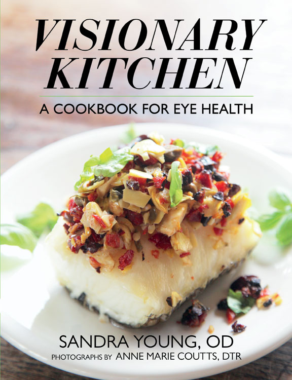
Author of the award winning Visionary Kitchen: A Cookbook for Eye Health
www.visionarykitchen.com

Books About Age-Related Macular Degeneration

6/24/14

Judi Delgado, the executive director of the Macular Degeneration Partnership, is often asked to recommend books about age-related macular degeneration (AMD). People who have been newly diagnosed, along with their family members, are interested to learn about what they can do to save their sight, how the eye disease might progress, and personal experiences from others that have the disease. Here is a list of books about age-related macular degeneration that Judi has put together, including authors, links to Amazon, and the available formats for each title.books about age-related macular degeneration

Books About Age-Related Macular Degeneration

By Doctors:

Macular Degeneration: The Complete Guide to Saving and Maximizing Your Sight
by Lylas G. Mogk and Marja Mogk
Paperback, Nook and Kindle

Macular Degeneration: From Diagnosis to Treatment
by David S. Boyer MD, Homayoun Tabandeh MD
Paperback, Nook and Kindle

By Patients:

The First Year: Age-Related Macular Degeneration:  An Essential Guide for the Newly Diagnosed
by Daniel L. Roberts
Paperback, Large Print and Kindle

Out of Sight, Not Out of Mind: Personal and Professional Perspectives on Age-Related Macular Degeneration
by Lindy Bergman, The Chicago Lighthouse, Jennifer E. Miller
Paperback, Nook and Kindle

Living With Macular Degeneration: What Your Doctors Cannot Tell You
by Edgar C Craddick, Benjamin Joel Michaelis
Paperback, Large Print and Kindle

Twilight: Losing Sight, Gaining Insight
by Henry Grunwald
Hardcover, Paperback, Audio Cassette, Audible, Nook and Kindle

Sunset…A Macular Journey
by F. Leroy Garrabrant
Paperback

Macular Disease: Practical Strategies for Living with Vision Loss
by Peggy R. Wolfe
Paperback, Large Print

Overcoming Macular Degeneration: A Guide to Seeing Beyond the Clouds
By Yale Solomon MD, J.D. Solomon
Paperback and Kindle

Macular Degeneration: Living Positively with Vision Loss
by Betty Wason, James J. McMillan
Hardcover and Paperback

We hope you will find this list useful and share it with people you know dealing with AMD. Also, if you know any books you think others might enjoy, please list them in our comments section.

Judi Delgado - age-related macular degenerationJudith Delgado
Executive Director
Macular Degeneration Partnership
A Program of Discovery Eye Foundation

My New Vision With A Telescope Implant

6/19/14

What it’s like to see with the CentraSight telescope implant

Like many people, I’ve set goals in my life, both professionally and personally and like being an active and engaged member of my community and my country. I love to teach: I taught history, geography and special education for years in Banning, California and now live in Moreno Valley, California, with my wife of 32 years, Kay. I love my country: I’m a proud veteran of both the Army Reserve and the Navy. And I love an open road. My wife and I traveled the country visiting historical monuments in our 32-foot RV. I guess I’ve got what you’d call a real zest for life. But, over the past twenty years, all the things I enjoyed doing in my life, even the simple day-to-day activities, started to decline because I was slowly losing my vision due to age-related macular degeneration. For example, six years ago, my wife took over all the driving because I couldn’t see well enough to drive safely.

Roy Kennedy - telescope implant
Roy Kennedy

That was a real turning point for me. My wife had to help me so much because I just couldn’t see. I needed help shopping because I couldn’t read labels. I started to avoid social situations, like visiting with friends because I was embarrassed that I couldn’t recognize faces any longer. As you can imagine it was heartbreaking for both me and my wife.

But then my doctor told me about a treatment I wasn’t yet aware of called the telescope implant. The device is very small (smaller than a pea!), and it is implanted in one eye to restore vision. My doctor explained that it works like a real telescope in that it magnifies images, which reduces the effect of the blind spot on my straight-ahead vision. The other eye does not get an implant because you need to keep some peripheral vision to help with orientation and balance. This sounded like science fiction! But I wanted to see if it could help me and I decided to give it a try.

I worked with a great team of specialists, who were part of a treatment program called CentraSight. My retina doctor, cornea surgeon, low vision optometrist and a low vision occupational therapist all counseled me about what to expect from the outpatient procedure, particularly afterwards. For example, I learned there was a significant amount of occupational therapy required to adjust and become proficient at using my new vision. I also was warned that my sight would not be like it was in my youth. I wouldn’t be able to do everything I used to nor would I be able to see, differently, the minute I opened my eyes.

I had my surgery in early 2013. The cost for the telescope implant and visits associated with the treatment program were covered by Medicare, which was very helpful. Thinking back, I was nervous on surgery day, but afterwards, I was told by my occupational therapist that I was one of the quickest to recover from surgery. I give lots of credit to my OT folks as well as my wife who helped me with the exercises at home. The most amazing part is being able to see my wife’s face again for the first time in six years! I’ve regained the ability to do many everyday tasks, like reading, working on my computer and watching old Westerns on TV. My wife and I are even back to traveling the open road in our RV (which she drives)!

I would recommend people learn more about the telescope implant to see if it might help them, the way it helped me. There are CentraSight teams across the country. When you call 1-877-99-SIGHT or visit www.CentraSight.com a trained CentraSight information Specialist will point you to the team closest to your home and can even help schedule the appointments for you. The telescope isn’t for anyone, but it can make such a difference in your life. It certainly did in mine.

Roy Kennedy - telescope implantRoy Kennedy

Corneal Donor’s Age Not Critical for Transplant Success

6/17/14

In the United States, more than 40,000 corneal transplants are performed each year with a high success rate in comparison to other types of organ transplants. According to the Eye Bank Association of America (EBAA), keratoconus was the leading cause of anterior lamellar keratoplasty (DALK/ALK partial thickness transplant) and was the fourth most common indication for penetrating keratoplasty surgery in 2012 (their last reporting period).
corneal transplant-original size
Advances in technology have led to increasingly successful outcomes for all who need corneal transplants. New long-term research of corneal transplant patients have shown that the age of corneal donors is no longer as important as once thought by eye health providers. According to a study funded by the National Institutes of Health, ten years after a transplant, a cornea from a 71-year-old donor is likely to remain as healthy as a cornea from a donor half that age.

The Cornea Donor Study (see www.ClinicalTrials.gov), funded by NIH’s National Eye Institute (NEI), was designed to compare graft survival rates for corneas from two donor age groups, aged 12-65 and aged 66-75. It was coordinated by the Jaeb Center for Health Research in Tampa, Fla., and involved 80 clinical sites across the United States. The study enrolled 1,090 people eligible for transplants, ages 40-80. Donor corneas were provided by 43 eye banks, and met the quality standards of the Eye Bank Association of America. The study found that 10-year success rates remained steady at 75 percent for corneal transplants from donors 34-71 years old. In the United States, three-fourths of cornea donors are within this age range, and one-third of donors are at the upper end of the range, from 61-70 years old.

Prior to this study, many surgeons would not accept corneas from donors over 65. Since the supply of young donor corneas is limited, these study results are encouraging for those who face a corneal transplant . The high level of success rates using corneas from older donors (over age 60) greatly increases the pool of donated corneas and corneal tissue available for transplant. In 2012, corneal donors under age 31 comprised less than 10 percent of the U.S. donor pool. “Our study supports continued expansion of the corneal donor pool beyond age 65,” said study co-chair Edward J. Holland, M.D., professor of ophthalmology at the University of Cincinnati and director of the Cornea Service at the Cincinnati Eye Institute. “We found that transplant success rates were similar across a broad range of donor ages.”

“Overall, the findings clearly demonstrate that most corneal transplants have remarkable longevity regardless of donor age,” said Mark Mannis, M.D., chair of ophthalmology at the University of California, Davis, and co-chair of the study. “The majority of patients continued to do well after 10 years, even those who received corneas from the oldest donors.”

SOURCE: National Eye Institute Press Releases

For information about Eye Bank Association of America

CathyW headshotCathy Warren, RN
Executive Director
National Keratoconus Foundation

What Are Floaters? | Causes and Treatment

6/12/14

Floaters can be anything seen in your vision that moves “to and fro” with your eye movement. The movement is not stationary compared to a blind spot which is fixed or stationary in your field of vision.

Example of floaters
Example of floaters
The key is that the position changes with eye movement. Size, shape, color, etc. don’t matter. Anything that moves in your vision is called a floater.

Remember, if you experience new floaters; please see an eye doctor for an examination.

Here are some common causes of floaters:

  • Posterior Vitreous Detachment (PVD)
  • Retinal Tears
  • Blood (Vitreous Hemorrhage) can be caused by;
  • Advanced Diabetic Retinopathy
  • Retinal Tears
  • Retinal Vascular Occlusions
  • Inflammation or Infection
  • Asteroid Hyalosis

Posterior Vitreous Detachment
A posterior vitreous detachment (PVD) is a common cause of floaters. A PVD occurs when the vitreous separates from the retinal surface. This is a normal event and will eventually occur in everyone. Retinal tears are more likely to occur right after a PVD has started.

The vitreous is usually a clear watery gel. As we age, enough of the proteins in the vitreous liquefy and degenerate. Eventually, there is a physical separation of the vitreous from the retina due to this liquefaction.

“Floaters” can develop due to opacities/haziness due to the aging gel which is no longer absolutely clear due to changes in the optical properties of the gel or the interface between the vitreous and water inside the eye.

Tears in the Retina
Retinal tears can cause floaters by either causing a small vitreous hemorrhage, or by dislodging cells into the vitreous which are normally located underneath the retina.

Vitreous Hemorrhage
Blood in the vitreous can also cause floaters. A so-called vitreous hemorrhage may result from either a retinal tear or advanced diabetic retinopathy. Occasionally, retinal vascular occlusions can also lead to bleeding in the vitreous.

Blood in the eye often absorbs, but only your doctor can determine the exact nature of the floaters and if they are associated with a retinal tear. It’s impossible for you to tell the cause of floaters without a proper dilated eye exam.

Inflammation
Certain types of inflammation or infection can cause significant floaters. Actually, these are white cells which migrate to the retina and vitreous and can be seen a floaters.

Asteroid Hyalosis
This is a common entity where lots of fine white opacities are suspended in the vitreous. In most cases, these are not noticed by the patient. Sometimes, however, the so-called asteroid bodies are so dense they prevent good examination of the retina. Other times, especially after a PVD, the asteroid bodies are noticed and patients may complain of floaters.

New Floaters: Evaluation
All new floaters must be examined by your eye doctor within 24-72 hours after occurring. Even if the floaters disappear in that time frame, you should be examined to look for a possible tear in the retina.

Again, you, the patient, can NOT tell the cause of the floaters or if you’ve sustained a retinal tears. Retinal tears can cause a retinal detachment – a potentially blinding problem.

Treatment of Floaters
There are no medicines or eye drops to treat floaters. Most doctors advise simply putting up with the floaters.

For patients who have chronic problems with floaters, I recommend a vitrectomy. A vitrectomy is an eye operation, performed by a retinal specialist. The operation is comparable to a cataract operation in terms of safety and possible complications.

There are a handful of doctors who perform Yag laser to break up floaters.

Randall E. Wong MDRandall V. Wong, MD
Retina Specialist
Fairfax, Virginia

20 Facts About the Amazing Eye

6/10/14

We don’t often give our eyes as much thought as we should, that is until something goes wrong and our vision is affected. But when you learn more about eyes, you realize just how amazing they are. Here are a few facts you may enjoy:

1. Eyes began to develop 550 million years ago. The simplest eyes were patches of photoreceptor protein in single-celled animals.

2. Your eyes start to develop two weeks after you are conceived.
Brown eyes 6.10.14
3. The entire length of all the eyelashes shed by a human in their life is over 98 feet with each eye lash having a life span of about 5 months.

4. To protect our eyes they are positioned in a hollowed eye socket, while eyebrows prevent sweat dripping into your eyes and eyelashes keep dirt out of your eyes.

5. Your eyeballs stay the same size from birth to death, while your nose and ears continue to grow.

6. An eye is composed of more than 2 million working parts.

7. Only 1/6 of the human eyeball is exposed.

8. Corneas are the only tissues that don’t have blood.

9. The human eye weights approximately just under an ounce and is about an inch across.

10. An eye cannot be transplanted. More than 1 million nerve fibers connect each eye to the brain and currently we’re not able to reconstruct those connections.

11. 80% of our memories are determined by what we see.

12. Eyes heal quickly. With proper care, it takes only about 48 hours to repair a minor corneal scratch.

13. There are about 39 million people that are blind around the world.

14. 80% of vision problems worldwide are avoidable or even curable.

15. Humans and dogs are the only species known to seek visual cues from another individual’s eyes, and dogs only do this when interacting with humans.

16. A fingerprint has 40 unique characteristics, but an iris has 256, a reason retina scans are increasingly being used for security purposes.

17. People who are blind can see their dreams if they weren’t born blind.

18. “Red eye” occurs in photos because light from the flash bounces off the back of the eye. The choroid is located behind the retina and is rich in blood vessels, which make it appear red on film.

19. 80% of what we learn is through our eyes.

20. Eyes are the second most complex organ after the brain.


Susan DeRemerSusan DeRemer, CFRE
Vice President of Development
Discovery Eye Foundation

What You Need to Know About Cataracts

6/5/14

Do you feel like your vision is getting worse? Do you feel like colors are not as vibrant as they used to be? Are you having more trouble with glare? If you have any of these symptoms, you may be experiencing the effects of cataracts.

Cataracts are a normal aging process of the crystalline lens in the eye. Well you may ask — what is the crystalline lens? It is easiest to think of the eye as a camera. The eye has a lens (actually two) – the cornea (the front window of the eye) and the crystalline lens (inside the eye). It also has an aperture (the colored iris), and film (the retina). All these structures work together to focus light and allow us to see – just like a camera. When we are young (less than 40), the crystalline lens is flexible. This is why we are able to see distance and then near without the need for reading glasses. The crystalline lens is able to change its shape depending on where one is looking.

Figure 1 – Slit-lamp photo of a visually significant cataract.

As we age, the crystalline lens becomes less flexible, thereby causing one’s near vision to be more blurry. This necessitates the need for reading glasses. As the crystalline lens become less flexible with age, the lens also starts to become more yellow and can also become cloudy. When the yellowing and/or clouding become visually significant, we refer to this as a cataract (figure 1).

Are cataracts dangerous? The simple answer is no. In the vast majority of cases, a cataract can be monitored until it becomes visually significant (drop in vision, glare, decreased contrast, vision related difficulties with day to day activities, etc). However, there are a few instances in which cataract removal is a medical necessity. Routine examinations by your eyecare provider can help you determine if you are at risk for these less common instances.

Figure 2 – Intraoperative photo during cataract surgery (prior to cataract removal).

What can I expect during cataract surgery? Do you have to replace the crystalline lens with anything? Cataract surgery involves removing the clouded crystalline lens (figure 2) and replacing it with an artificial lens known as an intraocular lens (IOL) (figure 3). Surgery generally takes 10-15 minutes under a mild sedative, and you don’t have to stop any of your current medications. Anesthesia is achieved with drops and you will only feel mild pressure during the surgery. IOLs come in different styles – Standard IOLs grossly correct your vision and you can fine tune your vision (distance and near) with glasses post-operatively;

Figure 3 – Intraoperative photo during cataract surgery (after implantation of an IOL).

Toric (astigmatism correcting) IOLs allow for increased spectacle independence, but glasses will still be needed for distance or near; Accommodating IOLs “flex” within the eye to decrease your dependence on distance and near glasses; Multifocal IOLs allow spectacle independence for distance and near. I always counsel patients that there is no perfect IOL and you have to determine which IOL is best for your particular situation. Your doctor can help you decide which IOL is best for you. Generally, cataract surgery is extremely safe. Your doctor will discuss particular risks specific to your eye.

How do I know if cataract surgery is right for me? The best way to know if you have a cataract and if it time to consider surgery is to consult with your local ophthalmologist. If you have experienced a recent drop in vision, that is not correctable with glasses, cataract surgery may be able to restore your vision!

Garg feb 2014 thumbSumit “Sam“ Garg, MD
Medical Director
Vice Chair of Clinical Ophthalmology
Assistant Professor of Ophthalmology
Gavin Herbert Eye Institute – UC, Irvine

Little Bottle, Big Relief

6/3/14

What you need to know about eye drops.

Have you been staring at a computer all day and your eyes are tired? Have allergies been making your eyes watery and itchy? Are your contact lenses irritating your eyes? If you have experienced any of these conditions, you have probably turned to eye drops for relief.

While eye drops are an easy and effective means of treating a number of eye issues, there are many different eye drops available, both over the counter (OTC) and by prescription. It is wise to know what your underlying condition is before trying to get relief.
eye drops 6.3.14
If your eyes are red and you may want to try a decongestant eye drop, which will shrink the tiny blood vessels in the “whites” of your eyes (sclera), but they also cause dryness so may not be a good choice if you wear contact lenses.
For lens wearers you are better off with a re-wetting drop to lubricate the eye and lens making you more comfortable. Another problem with the decongestant eye drops is over use – which can cause irritation and an increased tolerance that could lead to more redness.

If you suffer from allergies and antihistamine eye drop would be the best choice for relief from itchy, watery, red and swollen eyes. They work by reducing histamine in the eye tissue.

Lubricating eye drops, also known as artificial tears, are for short-term relief caused by temporary situations such as eye strain form computer use, being tired or being outdoors in windy and/or sunny conditions. If the condition is chronic, a prescription eye drop will be the best choice.

It is important to remember that if any of the above symptoms worsen or continue for an extended period of time, it is time to see your eye doctor to determine the underlying cause of your issue and to rule out eye disease. Postponing a visit could also lead to an eye infection.

Prescription drops are used to treat a wide variety of eye diseases such as glaucoma, dry eye and the symptoms of ocular herpes. They are also used to help with healing from cataract surgery, corneal transplants, glaucoma surgery and even Lasik. it is extremely important to use them as often as your ophthalmologist recommends to improve healing and prevent infection.

Because of the ease of applying eye drops researchers are working toward using them to treat other eye diseases. Ocular herpes symptoms are sometimes treated with antiviral and steroid drops. But this only is targeted at the symptoms and not the underlying cause, the herpes simplex virus. Lbachir BenMohamed, PhD and Steven Wechsler, PhD at the University of California, Irvine, Gavin Herbert Eye Instittue have been working to determine what reactivate the herpes simplex virus and develop an eye drop that would either stop the reactivation of the virus or kill it.

Using eye drops to treat age-related macular degeneration (AMD) is also being explored. Researchers at the Institute of Ophthalmology at University College London are working with nanoparticles to deliver anti-VEGF drugs such as Lucentis and Avasitn to the back of the eye via drops. “The study shows that Avastin can be transported across the cells of the cornea into the back of the eye, where is stops blood vessels from leaking and forming new blood vessels, the basis for wet AMD.” While researchers in the Department of Ophthalmology, Tufts University School of Medicine in Boston “reported in their “proof of concept” study that topical application of a compound called PPADS inhibits damage to the tissues in the eye that impacts the individual’s ability to see color and fine detail, as well as reduces the growth of extraneous blood vessels in the back of the eye related to AMD.” It would work in both dry and wet AMD reduce the need for direct injections.

Eye drops, when properly applied, can provide temporary relief from symptoms of eye discomfort. But if the symptoms worsen or continue for an extended period of time, consult your eye doctor. To make sure you apply the eye drops correctly check out the article in our February 2013 newsletter for 12 easy steps to get the drops into your eyes and avoid infection.

One final note – keep your eye drops out of reach of children. Eye drops come in small bottles that are the perfect size for small hands and don’t have the same security tops found on other medications. The FDA has warned that ingredients found in some eye drops that relieve redness have caused abnormal heart rate, decreased breathing, sleepiness, vomiting and even comas in children five and younger that have ingested them. If you child has swallowed eye drops, call the Poison Help Line 800-222-1222.

Susan DeRemerSusan DeRemer
Vice President of Development
Discovery Eye Foundation