Silent Thief of Sight – Glaucoma

January is National Glaucoma Awareness Month. The National Eye Institute, through their education program NEHEP, have created this infographic to provide you with information you need to know about this blinding eye disease.

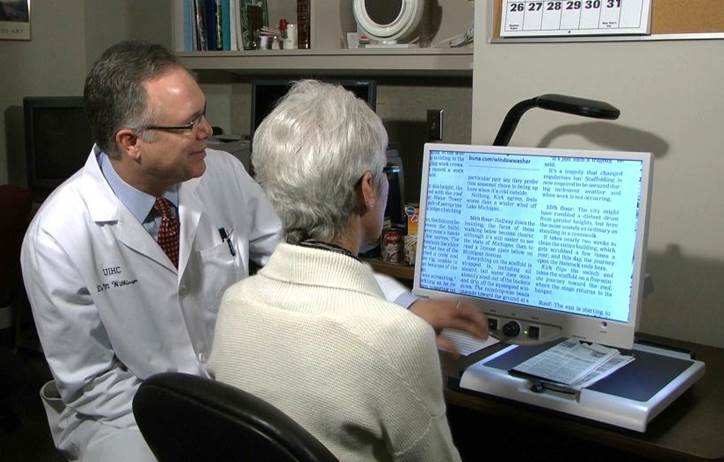
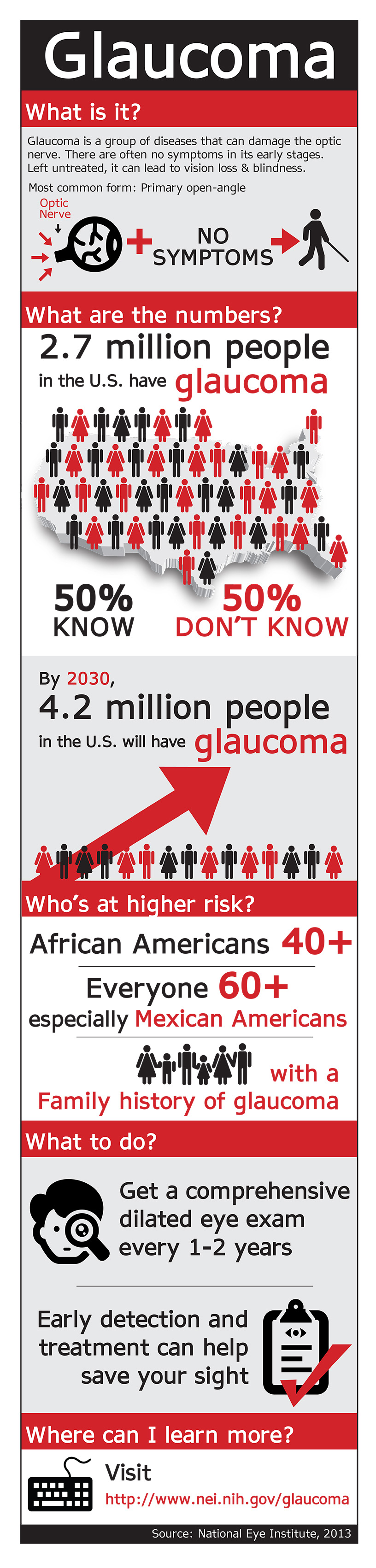
When adults reach their 40s, they often begin to notice small changes in their vision that can affect their daily lives and jobs. It could be difficulty in reading a book or working on a computer. This can be annoying, but it can often be addressed by seeing an eye care professional for comprehensive dilated eye exam. This allows the doctor to detect diseases and conditions that can cause vision loss and blindness and yet have no symptoms in their early stages.

Silent Thief of Sight – Glaucoma

Glaucoma is one of these age-related eye diseases that has no early symptoms, which is why it is called the silent thief of sight. It is actually a group of diseases that can damage the eye’s optic nerve and result in vision loss and blindness. Open-angle glaucoma is the most common form disease.

In addition to an eye exam, you can reduce your chances of losing your vision to glaucoma by also:

  1. Live a healthy lifestyle that includes maintaining a proper weight, eating healthy foods, and not smoking.
  2. Know your family history to determine if you are at a higher risk for some eye diseases.
  3. Protect your eyes against harmful UV rays from the sun or your computer by wearing sunglasses when you are outdoors or computer glasses when using the computer for extended periods of time.
Silent thief of sight glacoma
Courtesy of NEI/NEHEP

1/5/16


Susan DeRemerSusan DeRemer, CFRE
Vice President of Development
Discovery Eye Foundation

It’s All About ME – What to Know About Macular Edema

“You’ve got the macular? I’ve got some but my sister, she’s got all kinds!” Even as the word retina has become commonplace, the macula and its diseases are often feared and misunderstood. The retina is the light-sensitive layer of cells that line the inside of the eye. The many layers of the retina work together to convert what we see into an exquisitely coded signal that travels to the brain. There the message is decoded and directs us to take action – “that’s a fine looking piece of pie!”

The macula is the part of the retina that helps us see fine detail, far away objects, and color. It’s packed with more photoreceptors than any TV or monitor which is why it is prized real estate. It is the small, central area of the retina that’s worth the most – the bullseye of sight. When things happen to the macula, it gets an “r”. Macular edema, macular degeneration, macular hole, pucker, drusen, scar, fibrosis, hemorrhage, and vitreomacular traction are common conditions that involve the macula. When present, distorted vision (metamorphopsia), blank spots (scotoma), and blurred vision are common symptoms.

Four. Ang. - Macular Edema
Figure 1: Fluorescein angiography: Macular edema may be seen as a pinpoint leak (left, large arrow) in mild cases. In serious cases, ME may diffusely involve the macula. Note how the image becomes brighter as more dye leaks from damaged macular capillaries. (Center and Right)

Macular edema refers to an abnormal accumulation of fluid in the layers of the macula. From the side, it looks like the snake that ate the pig. Like a droplet of water on your computer screen, images are distorted by the swollen retina – making it more difficult to see clearly. The more widespread, thicker, and severe the swelling becomes, more likely one will notice visual symptoms. If untreated, chronic macular edema can lead to irreversible damage to the macula and permanent vision loss.

OCT - Macular Edema
Figure 2: Optical Coherance Tomogrphy and Macular Edema: OCT is a useful test to study macular edema (ME)
-The top image has is normal. Note the even layers and gently sloping dip of the macula called the fovea. This eye has excellent vision.
– The middle OCT has ME, black-appearing cysts (arrows) which threaten the normal fovea. This eye also has good vision.
– The bottom OCT shows ME involving the macula. Because ME involves the macular center (the fovea), vision is poor (large red arrow).

Macular edema is not a disease but the result of one. As with other conditions where abnormal fluid accumulates (leg swelling, pulmonary edema, hives, and allergy), macular edema can be caused by many conditions including metabolic (diabetes), aging (macular degeneration), hereditary (retinitis pigmentosa), inflammatory (sarcoidosis, uveitis), toxic, neoplastic (eye tumors), traumatic, surgical, and unknown causes (idiopathic, macular hole, macular pucker, vitreomacular traction). Macular edema occurs when the retina’s ability to keep fluid out of the retina is overwhelmed by the fluid leaking into it. (If more rain falls on the lawn than it can handle, you get puddles of fluid. In the retina, blisters of fluid form and swell the retina – this is macular edema. Fluorescein angiography (Figure 1) and optical coherence tomography (Figure 2) are two common tests to evaluate macular edema.

Macular edema is typically caused by increased leakage or growth of abnormal blood vessels. The most effective treatment strategies address the underlying cause (diabetes, blood vessel occlusion, neovascularization, inflammation, etc) as well as the hyperpermeability of the capillaries in and around the macula. Eye drops, laser, placement of long-acting medication implants, and surgery are effective in many diseases but the mainstay of treatment is now intravitreal injections (IVI). The IVI is an office procedure painlessly performed under topical anesthesia in which medication is placed inside the eye by a very small needle. IVI should be performed by a trained retina specialist with meticulous monitoring of treatment efficacy and of extremely rare but potentially serious complications. IVI is considered one of the most commonly performed procedure in the world.
Lucentis, Eyelea, and Ozurdex are the trade names of the three most common FDA-approved medications for the treatment of the common conditions causing macular edema. Avastin is not FDA approved but has also been extensively studied in large, well-designed, federally-funded clinical trials and is felt to have efficacy and safety no less than any of the other available options. Each option has a considerable track record of success and works by decreasing the amount of fluid leaking from abnormal blood vessels.

Macular edema is a common finding in many diseases of the retina, most which can be treated to improve vision. The physician’s therapeutic armamentarium continues to expand. There has never been a more successful time in the treatment of macular edema and macular disease. While much has been discovered, many promising therapies await.

3/5/15


Dr. Suber HuangSuber S. Huang, MD, MBA
Chair, National Eye Health Education Program
Philip F. and Elizabeth G. Searle – Suber Huang MD Professor
Case Western Reserve University School of Medicine
Past-President American Society of Retina Specialists
CEO, Retina Center of Ohio

Low Vision Resources

What to do when “There’s nothing more that can be done.”

“I’m sorry, but there’s nothing more that can be done. There is no cure for your eye condition.”

In your work as healthcare professionals and health educators, it’s likely you’ve encountered a significant number of adults and older adults who have been on the receiving end of this devastating news.

When an eye care provider says, “There’s nothing more that can be done,” what he or she likely means is, “There’s nothing more I can do for you surgically.” But instead of saying, “There’s nothing more that I can do,” the discouraging message delivered to the patient is, “There’s nothing more that can be done.”

Thus, in many cases, the discussion ends there. Patients will either exhaust their resources searching for an elusive cure or become resigned to a life that is restricted and defined by incurable vision loss.

When receiving a diagnosis of vision loss, many adults who have managed to overcome a host of obstacles in their lives may now believe they are facing an obstacle with no viable solution. A natural, and understandable, initial reaction is to focus instead on the devastating losses that are seen as an inevitable accompaniment to blindness and low vision such as:

  • Loss of independence: “How will I prepare meals, clean my home, or shop? Will I become a burden to my family and friends?” 
  • Loss of confidence and self-worth: “All my life I’ve been physically active and self-reliant. Has my life as an independent person come to an end?” 
  • Loss of privacy: “I won’t be able to handle my finances independently. Will I have to surrender control of my life to someone else?” 
  • Loss of employment: “I’ll have to quit my job. How will I earn a living?” 

It’s important to let your clients and patients know that there is indeed hope—and life—after vision loss. A wide range of vision rehabilitation services enable adults who are blind or have low vision to continue living independently. The term “vision rehabilitation” includes highly trained professionals and comprehensive services that can restore function after vision loss, just as physical therapy restores function after a stroke or other injury.

Patient working with a low vision therapist

Vision rehabilitation professionals include:

Additional vision rehabilitation services can include:

  • Peer support and counseling: talking with peers, sharing common concerns and frustrations, and finding solutions to vision-related problems. 
  • Vocational rehabilitation: vocational evaluation and training, job training, job modification and restructuring, and job placement. 
  • Veterans’ services: vision rehabilitation and related support services for blinded veterans of all ages. 

There are many resources available to help your patients and clients locate vision rehabilitation services. For example, the VisionAware Directory of Services allows you to browse by state and type of service, including counseling resources, support groups, low vision services, independent living skills, and orientation and mobility. The VisionAware “Getting Started” Kit provides tip sheets on specialized services and products that can assist with everyday life after vision loss.

The National Eye Institute’s National Eye Health Education Program (NEHEP) also has low vision education resources. The video, Living with Low Vision: Stories of Hope and Independence, explains how, as a health professional, you can help your patients make the most of their remaining vision and improve their quality of life by referring them for vision rehabilitation services. Share it with your colleagues, too. You can find additional resources and ideas for promoting vision rehabilitation on the NEHEP Low Vision Program page.


Maureen Duffy, CVRTMaureen A. Duffy, CVRT
Social Media Specialist, visionaware.org
Associate Editor, Journal of Visual Impairment & Blindness
Adjunct Faculty, Salus University/College of Education and Rehabilitation