DEF-Funded Retinitis Pigmentosa(RP) and Age-Related Macular Degeneration(AMD) Projects Converge with Promise

Two hallmark DEF-funded projects are converging, providing great hope for those facing vision loss from retinitis pigmentosa (RP) or age-related macular degeneration (AMD).

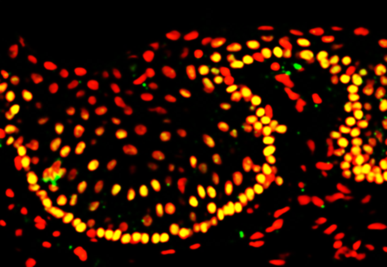
The first project, headed by UC Irvine researchers Drs. Henry Klassen and Jing Yang, concentrates on putting human retinal progenitor cells into the eyes of those with RP in order to rescue damaged retinal cells. That project is currently in Phase II clinical trials, progressing toward FDA approval.

                                                      Tissue-culture model

According to DEF Research Director Dr. Cristina Kenney, if the project is approved by the FDA for use with RP, the next question is: What other diseases might these retinal progenitor cells be used for? That’s where a second DEF-funded project comes in.

Kenney is working on a “personalized” cybrid cell model to screen agents that specifically target the mitochondria in AMD cells. To date, the researchers have different cybrid cell lines representing 60 different individuals with eye diseases. They are looking for novel mechanisms to protect AMD cells from dying.

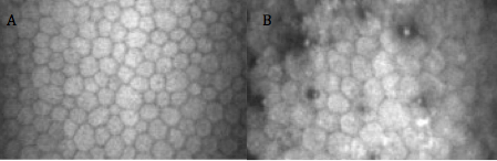
Yang and Kenney are now working together to determine whether the retinal progenitor cells can be the agent that rescues AMD cybrids. “When we take the mitochondria from AMD patients and put them into healthy retinal cells, which makes cybrids, we have shown that these AMD cybrid cells will start to die. So we used that model to ask the question: How do we rescue them?” Kenney says.

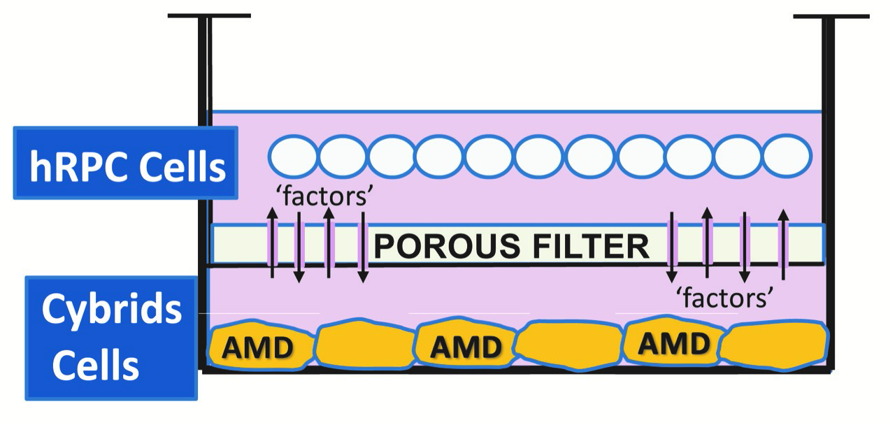
Kenney and Yang developed a tissue culture model, where the retinal progenitor cells are grown in one part of a chamber, and the AMD cybrids are grown in another part of chamber, surrounded by culture medium. There is a porous separator between the two chambers through which the cells can communicate.

 “We are finding that the retinal progenitor cells produce a factor that protects the AMD cybrids,” Kenney says. “This provides promising evidence that these proprietary retinal progenitor cells that are being tested for treating RP also may be helpful in AMD patients.”

“DEF has been supporting both these retina-related projects for quite some time, and it’s very exciting to see them coming together to potentially treat both RP and AMD.”

To help support this sight-saving research, please donate to the Discovery Eye Foundation by clicking button below.

When Is The Best Time For Cataract Surgery?

As you age, cataracts become a concern prompting the question – when is the best time for cataract surgery?

There are decades worth of old wives tales floating around regarding cataracts that often lead to unnecessary fear and apprehension for many patients. These myths involve concepts such as “ripeness”, having to wear eye patches afterwards, danger in “waiting too long, etc. Just as the techniques of cataract extraction have changed over the decades, so have the indications to proceed to surgery.
best time for cataract surgery - people
Firstly, cataracts are a normal part of the aging process. Patients should not be alarmed if they are told that they are developing cataracts, even as early as their fifties. As we age, the natural clear lens inside the eye becomes progressively harder, darker, and cloudier. This dark, cloudy lens is what is referred to as a cataract. Cataracts develop at different rates for different people, and even between the two eyes of the same person. It typically takes many years for the lens to become cloudy enough to impact the clarity of vision. There are many different types of cataracts depending of what area of the lens becomes cloudy, but the typical cataract related to normal aging results in a relatively uniform cloudiness with a denser central core, and is referred to as “Nuclear Sclerosis”. Other varieties of cataracts tend to grow more quickly, are relatively uncommon, and often result from certain conditions other than typical aging.
best time for cataract surgery
Regardless of what type of cataract the patient has, the treatment is the same: cataract extraction with an implant of an intraocular lens. There have been great advances in lens design over the years, and they now result in excellent, stable, predictable vision for the remainder of the patient’s lifetime and do not typically need to be changed once implanted.

Cataracts result in different symptoms that may be more of less relevant to a specific person’s needs, such as:

  • Glare with bright lights
  • Difficulty with fine print
  • Difficulty following the golf or tennis ball
  • Impairment in night driving
  • Difficulty with seeing street signs
  • Seeing the score or small print on the television
  • Fine visual tasks such as threading a needle, etc.

Although cataract surgery is an incredibly successful procedure with only about a 1-2% risk of complications, it still DOES have some risk. Therefore, cataract surgery should only be undertaken when there is something to gain. In other words, the BENEFITS MUST OUTWEIGH THE RISKS. This means that if your symptoms are mild and are not interfering with your activities of daily living, it is not time to accept the risks of surgery. Once your visual impairment progresses to the point that YOU feel your activities of daily living and enjoyment are impaired, this is the time to proceed to surgery. This threshold is very different between people. Some people feel impaired with vision of 20/25, and others still function within their scope of usual activities until they are 20/100! The best first-step in determining if it is time for your surgery is to get an up-to date refraction. This means a detailed check for new glasses. Often, cataract development will change a person’s glasses prescription, and updating this can improve the visual symptoms for months to years. When a new glasses prescription no longer improves the sight adequately, this is when surgery is indicated.

For the most part, putting off cataract surgery does not impact the final outcome. It will not harm you or your eye to leave the cataract alone until you are ready. There are of course certain exceptions to this rule, such as in Fuchs’ dystrophy, pseudoexfolation, untreated narrow-angle glaucoma, and some others. However, these are relatively rare conditions that your doctor will speak to you about if you have any of these diagnoses.

In summary, the time to proceed to cataract surgery is something that you as the patient determine. YOU assess your lifestyle needs and your vision performance within your scope of activities. When you feel you are impaired in these activities, the benefits will outweigh the risks, and it’s time to take them out. You should not feel any pressure to urgency in this process.

Once you have determined you are ready to have cataract surgery, your surgeon will discuss with you your options for intraocular lens implantation including astigmatism neutralizing lenses, standard distance or near-vision lenses, multiple focal distance lenses, accommodating lenses, and others. The current standard approach for cataract surgery is called “phacoemulsification” and uses ultrasound technology to remove the cataract. There are also laser devices that assist in making the incisions and breaking up the lens, which many surgeons now employ in addition to the phacoemulsification. In general cataract surgery only takes a few minutes, is performed with topical anesthesia, is pain-free, and has a very short recovery time. No pirate-patches are used these days! Most patients are very happy with the results, but this requires adequate discussion with the surgeon prior to the procedure to best assess the needs of the individual patient. A well- informed patient who participates in their care results in the best outcomes!

6/18/15

Sameh Mosaed, MD best time for cataract surgerySameh Mosaed, MD
Director of Glaucoma Services, Gavin Herbert Eye Institute, UC Irvine
Associate Professor, Cataract and Glaucoma Surgery, UC Irvine School of Medicine

Fuchs’ Dystrophy: Current Insights

What is Fuchs’ Dystrophy?

Corneal dystrophies are a debilitating group of progressive diseases that can ultimately deprive a person of sight. The cornea, which forms the front of the eye, is a window for vision, and dystrophies due to intrinsic defects in the corneal tissue cause this window to become opaque and hazy. Fuchs’ dystrophy, also known as Fuchs’ corneal endothelial dystrophy (FCED), is amongst the most commonly diagnosed corneal dystrophies requiring corneal transplantation. The ophthalmologist Ernest Fuchs first described the disease in 1910.

Who gets it?

The disease is rare, and it is difficult to predict who will get it. We know that it affects women more than men (3:1 ratio), older adults (older than 50 years of age), and those with a family history. There are forms in which there could be up to a 50% chance of transmission to children of parents with Fuchs’ dystrophy. Most cases, however, occur sporadically.

What causes it and how does it progress?

Although the cause of Fuchs’ dystrophy is still being studied, there are characteristic findings associated with it: small outgrowths on Descemet’s membrane called “guttae” or “guttata”, thickening of Descemet’s membrane, and defects in the endothelial cells (Figure 1).

fuchs dystrophy 1
Figure 1: Fuchs’ dystrophy can affect all layers of the cornea. Layers of the cornea from anterior to posterior, or frontside to backside, include (A) epithelial cells where blisters and bullae may form in late-stage disease, (B) Bowman’s layer where scarring can occur in late-stage disease, (C) stroma where corneal swelling occurs early in disease, (D) Descemet’s membrane where guttae form (arrows) and thickening occurs, and (E) endothelial cells that decrease in number and change shape and size with disease progression.

Descemet’s membrane is a thin corneal layer between the endothelial cell and the stromal layers of the cornea. Endothelial cells make up the backside of the cornea and function as a barrier and pump for keeping fluid out of the cornea and maintaining corneal clarity. As guttae accumulate on Descemet’s membrane, patients experience progressive loss and change in endothelial cells. Dysfunction of endothelial cells causes corneal swelling, which distorts vision. First, the back of the cornea swells, and eventually, swelling can reach the epithelial cells at the front of the cornea. Swelling can range from mild moisture accumulation, to painful “bullae”, or blisters. In very late-stage disease, significant corneal scar tissue can form and dramatically reduce vision. The progression to late stage Fuchs’ varies from person to person, but usually takes a couple of decades.

What are signs and symptoms?

A patient may be asymptomatic for years despite having guttae. Initial symptoms, including blurry, hazy, or cloudy vision, are typically due to corneal swelling from dysfunction of the endothelial cell layer. Patients may also experience glare or halos around light in the early stages just from the density of guttae. New studies suggest that patients can get glare and higher order aberrations from guttae without any corneal swelling. Symptoms tend to be worse on awakening, but usually improve throughout the day. This is because the closure of eyelids during sleep results in the accumulation of fluid in the cornea. For the same reason, humid weather can also worsen symptoms. As the disease progresses, poor vision may last longer into the day. There may be associated pain if blisters develop.

How is it diagnosed?

The presence of any of the above signs and symptoms, especially with a family history of Fuchs’, should prompt a consult with an ophthalmologist who will diagnose the disorder and follow its progression with regular checkups. An ophthalmologist will conduct a microscopic slit-lamp examination of the eyes, looking for guttae and Descemet’s membrane thickening (Figure 2).

fuchs dystrophy 2
Figure 2: Slit-lamp examination showing speckling pattern on the backside of the cornea characteristic of guttae in Fuchs’ dystrophy.

Special tests may be done to measure corneal thickness, a marker of swelling, or count endothelial cells to track disease progression (Figure 3 and 4).

fuchs dystrophy 3
Figure 3: Optical Coherence Tomography (OCT) showing (A) a normal, healthy cornea and (B) corneal swelling typical in Fuchs’ dystrophy.
fuchs dystrophy 4
Figure 4: In-vivo slit-lamp scanning confocal microscopy showing (A) normal endothelial cells and (B) guttae causing endothelial cell loss and change in Fuchs’ dystrophy.

How is it managed?

Management can be medical or surgical depending on symptoms. Patients may have mild or slow progression of disease that can be managed medically including over the counter salt solution drops (5% NaCl) to reduce corneal edema.

When there is late-stage disease, a corneal transplant may be necessary to improve vision. A corneal transplant replaces the patient’s corneal tissue with human donor corneal tissue. Donor corneas are readily available via excellent eye banks throughout the United States. The surgery is outpatient surgery with regular follow-up appointments and suture removal during the subsequent months. The postoperative healing of the cornea and vision stabilization can take up to a year.

Great strides have been made in the last decade in corneal transplantation surgery, giving patients better treatment options. Patients used to be limited to penetrating keratoplasty (PK), a full-thickness replacement of the cornea. We now have newer surgeries known as endothelial keratoplasty (EK), which is a partial-thickness transplant that replaces only the damaged part of the cornea (the endothelial layer). The different types of EK are DSEK (Descemet’s-Stripping Endothelial Keratoplasty) and DMEK (Descemet’s Membrane Endothelial Keratoplasty). The techniques vary by thickness of the transplanted tissue. The type of EK most appropriate is determined by the corneal surgeon and is variable on a case to case basis. Both types of EK surgeries provide comparable long-term visual results. In both surgeries, the patient’s diseased Descemet’s membrane and endothelial cells are stripped from the inner layer of their cornea. The thin lamellar donor graft is then inserted into the eye and positioned onto the back of the patient’s cornea via a gas or air bubble. The patient is then instructed to lie in a face up position for several hours post surgery during which time the bubble supports the graft until the new endothelial cell pumps begin to wake up and naturally adhere to the back side of the recipient cornea. Occasionally, the doctor may replace another air bubble into the eye the next day to allow more time for the graft to adhere. Visual recovery is on the order of 1-2 weeks in DMEK and 2-3 months in DSEK surgery. Rejection risk is still a possibility in EK surgery but has a much lower rate than traditional full thickness PK surgery.

Other surgical considerations depend on the presence of cataracts. Cataract surgery can worsen Fuchs’ dystrophy because of damage to the endothelial cell layer. For this reason, patients with cataracts and Fuchs’ requiring surgical intervention are often recommended to undergo cataract surgery before or at the same time as corneal transplantation to ensure the best outcome for the transplant.

Patients should work with an ophthalmologist to determine the best management plan. Ultimately, vast improvements in treatment options have given many Fuchs’ dystrophy patients the exciting opportunity to regain vision with improved healing times and reduced infection and rejection of the graft.

Citations: Figure 2 and 4 are from Zhang J, Patel DV. The pathophysiology of Fuchs’ endothelial dystrophy—a review of molecular and cellular insights. Exp Eye Res. 2015 Jan

6/4/15

priscilla-thumbnailPriscilla Q. Vu, MS
Medical Student
University of California, Irvine School of Medicine


Farid 3.6.14Marjan Farid, MD
Director of Cornea, Cataract, and Refractive Surgery
Vice-Chair of Ophthalmic Faculty
Director of the Cornea Fellowship Program
Associate Professor of Ophthalmology
Gavin Herbert Eye Institute, University of California, Irvine

Vision and Special Needs Children

In the United States, special needs is a term used in clinical diagnostic and functional development to describe individuals who require assistance for disabilities that may be medical, mental, or psychological. Different types of special needs vary in severity. People with autism, Down syndrome, dyslexia, blindness, ADHD, or cerebral palsy, for example, may be considered to have special needs. Statistics tell us that among children ages 3 to 17, nearly 15 percent have one or more developmental disabilities. For many of these children, the kinds of disabilities they experience may require special approaches to providing care, education and/or other accommodations.
eye exam - vision and special needs children
The way a special needs child behaves or reacts can sometimes be unexpected because he/she processes sensory information differently than other children. Having the right cues in our environment can mean the difference between participation and non-participation in medical care for many of these children. Our work environment should always be arranged in a way to achieve physical and emotional comfort for the child. Patience, attentive listening, caring, and building a trusting relationship with a family and child who may be undervalued in other settings are all critical to providing good medical care.

Vision and Special Needs Children

Children with special needs are referred to pediatric ophthalmologists on a regular basis. Many of these children do not respond appropriately to standard vision screening procedures. Unidentified vision problems in this special population can further impact growth and development. The role of pediatric ophthalmologists is to ensure that the child does not have any eye conditions that could potentially interfere with his/her learning opportunities, personal development and/or overall wellbeing.

Down syndrome:
Down syndrome continues to be the most common chromosomal disorder. Each year, about 6,000 babies are born with Down syndrome, which is about 1 in every 700 babies born. It is estimated that more than 80% of these patients have some clinically significant ocular pathology. Such conditions include refractive error requiring glasses (70%), strabismus (45%), and nystagmus (35%). Other common eye problems include blepharitis, blocked tear duct, eyelid ptosis, cataracts, and keratoconus (irregularly shaped cornea). The improved quality of medical care and educational resources have allowed for a more productive life and a longer life expectancy for people with Down syndrome. Their quality of life can be further enhanced by the proper assessment and correction of eye problems.

Autism:
In 2014, the Centers for Disease Control and Prevention (CDC) released new data on the prevalence of autism in the United States. This surveillance study identified 1 in 68 children (1 in 42 boys and 1 in 189 girls) as having autism spectrum disorder (ASD). Autistic behaviors may include visual components such as lack of eye contact, starring at light or spinning objects, fleeting peripheral glances, side viewing and difficulty attending visually. An eye examination is essential in order to detect an eye condition that could potentially explain these visual behaviors. According to a study published in the June 2013 issue of Strabismus, more than 40 percent of children with autism have strabismus, or crossed eyes. This work is limited in that the researchers did not use a representative sample of children with autism. Still, the prevalence of eye problems in autism appears to be several times higher than that of the general population.
boy eye exam - vision and special needs children
Learning Disabilities:
Dyslexia is the most common neurobehavioral disorder affecting children. Visual abnormalities have not been found to affect the brain’s ability to process visual stimuli and children with learning disabilities have no increased incidence of ophthalmologic disease. However, ophthalmologic consultation should be provided to children who fail vision screening tests. This allows for diagnosis and therapy of treatable ocular conditions such as refractive errors and eye muscle imbalances.

Children with ADHD, cerebral palsy, or any other neurodevelopmental disorders should also have periodic vision screenings. Children who do not pass the vision screening should be referred to an ophthalmologist with experience in the care of children.

Physicians are not the only adults involved in the care of special needs children. At increasing rates, children with special needs are being provided with the same life experiences as their non-disabled peers. Taking part in a sport or joining a group like the Scouts, are popular activities for children. Children with disabilities are encouraged to join in such activities to help improve their health and give them opportunities to make friends. As a result, more and more adults in the community are finding themselves working with these children on a regular basis.

When considering ways to work with special needs children, we have to keep in mind that every child is different. A positive attitude and patience are probably the two most important qualities for anyone who works with these children. Parents of special needs children focus on helping their child to be “the best he/she can be”. The devotion and care these parents provide for their child have been an inspiration, and I always look forward to my visits with them.

4/2/15


Dr. Chantal BoisvertChantal Boisvert, OD, MD
Assistant Clinical Professor
Gavin Herbert Eye Institute, UC Irvine
Pediatric Ophthalmology & Strabismus
Neuro-Ophthalmology

Three Generations of Saving Vision

In Discovery Eye Foundation’s spring e-newlsetter there was an article entitled Surgery for the Surgeon, where a leading ophthalmologist talks about convincing himself to have cataract surgery. That eye doctor was Dr. Nesburn, who was willing to share with us his family’s long tradition of saving vision.

Brainwashed by Medicine

“I was brainwashed from the age of 5,” says Dr. Anthony Nesburn, medical director of The Discovery Eye Foundation (DEF). “My dad would take me on rounds at the hospital and to his office. He introduced me to medicine at a really early age.”

Nesburn saving vision
Dr. Anthony Nesburn in UC Irvine lab
Dr. Henry Nesburn was an ophthalmologist in Los Angeles for more than four decades, and he passed his love of the specialty to his son. “I really looked up to my dad,” the younger Nesburn says. “He loved ophthalmology for the same reasons I do: You get to do medical diagnosis; you get to do wonderful surgery, where you keep people from going blind or restore vision; and you can work with people from newborns to the very elderly — they all need eye care.”

Nesburn received a telegram while he was an undergrad at UCLA, telling him he’d been accepted to Harvard Medical School. His mother started crying: “You’re going to Boston! We’re not going to see you anymore!” While he “was loathe to leave Southern California,” Harvard was too good to pass up.

Drafted by the Army out of his ophthalmology residency at Harvard in 1960, Nesburn joined the Navy instead. He followed in his father’s footsteps again, becoming a Navy flight surgeon. (Henry had volunteered during World War II.)

He went on to a Boston Children’s Hospital fellowship in infectious disease, working with Nobel laureate Dr. John Enders, whose work led to the polio vaccine and changed the face of virology. Nesburn then did his residency at Massachusetts Eye and Ear. “I was part of a special program that allowed us to do research, and I was running a research laboratory while I was a resident,” he says. “It gave me the start I needed.”

“At Mass Eye and Ear, I worked for an up-and-coming ophthalmologist and researcher to prove there was a substance that could treat herpes eye infections. We wrote a paper that included the very first antiviral ever described, and it was against herpes virus. It is the basis for today’s herpes antivirals,” Nesburn says. “I was hooked.”

He went back to Los Angeles and received NIH funding to continue his research on ocular herpes. In 1968, he joined his father’s practice half-time, spending the rest of his time doing research.

Two years later, he received a generous offer, when Rita and Morris Pynoos started DEF to fund his research. The Pynooses were grateful to Nesburn for diagnosing their son, Jon, with keratoconus (KC). “I was a second-year resident at Mass Eye and Ear, and Jon Pynoos was an undergraduate at Harvard. His parents went to see my dad, because Jon couldn’t see well, and no one could figure out what was wrong with his vision. My dad said, ‘Send him over to Tony; he’ll figure out what’s going on!’ I said to myself, ‘Holy mackerel! What happens if he has something really complicated? I’m just a newbie!’” Nesburn remembers. “Jon came in; I looked at him, and the keratoconus was so clear and easy to spot. I couldn’t imagine how his doctors didn’t see it. We got him contact lenses, and he was able to see again. When I came back to LA, the Pynooses wanted to do something to help.”

At first, DEF research focused on KC and the herpes research Nesburn was working on at the time. It soon broadened to include macular degeneration and retinal disease.

“My dad had to retire from the practice of ophthalmology at the age of 70, because of bad age-related macular degeneration (AMD). His mother and older sister had had it, as did several cousins. There was no treatment back then that helped,” Nesburn says.

“AMD is the most common cause of permanent vision loss in the elderly in the developed world. I could see where the need was,” he says. “We moved forward at DEF with two driving mantras: We wanted to do something significant in macular degeneration research and to find the cause of keratoconus.”

As a virologist in research and a corneal surgeon, Nesburn realized he needed a corneal biochemist to help with the KC research. He met Dr. Cristina Kenney at an Association for Research in Vision and Ophthalmology meeting. She joined DEF, and within 15 years, they found the chemical cause of keratoconus; they also got married.

Now nearly 80 years old, Nesburn spends most of his time “wearing three hats”: fundraising for DEF, lab research and clinical practice. His daughter, Kristin, is the third generation to join the family ophthalmology practice.

“While I’m still able, I want to try to make a difference in medicine, particularly in macular degeneration,” Nesburn says. “Macular degeneration affects so many people. This is where I want to put my energy. Luckily, as strong as it is in our family, I don’t have it … yet.

“As a researcher, my interest in putting together a program for macular-degeneration diagnosis and treatment has been because it’s a great public health problem. Yes, if I should ever get it, it might be able to help me or my family, but the first thing, as a scientist, is to try to get something to help humankind. I know it sounds sappy, but it’s true.”

3/26/15


Anthony B. Nesburn, MD, FACSAnthony B. Nesburn, MD, FACS
Medical Director, Discovery Eye Foundation
Professor & Vice Chairman for Research, Ophthalmology
Gavin Herbert Eye Institute, University of CA, Irvine

Cataract Surgery and Keratoconus

1/8/15

The eye works like a camera, specifically a digital camera. There is the front lens of the camera (cornea), the aperture (iris), the film (retina), and a cable to take the image to the brain (optic nerve). This “camera” also has an additional lens – the natural crystalline lens, which lies behind iris. This natural lens is flexible when we are young, allowing us to focus at distance then instantaneously up close. Around age 40-45, this natural lens starts to stiffen, necessitating the need for reading glasses for most people. This stiffening is the beginning of the aging process that eventually leads to formation of a cataract. We refer to the lens as a cataract when it becomes sufficiently cloudy to affect ones quality of vision.cataract surgery and keratoconus-Cataract diagram In general, cataract surgery is one of the safest and most successful of all surgeries performed. The basics of cataract surgery in eyes with keratoconus is very similar to non-keratoconic eyes.

Keratoconus (KC) affects this “camera” by causing the front lens (cornea) to bulge. This causes the optics to be distorted. In many cases, this can be corrected for with hard contact lenses (CL) or spectacles; in other cases a corneal transplant may be necessary. When it comes time for cataract surgery in the setting of KC, there are several factors that need to be considered.

Corneal Stability
The first thing to be considered is the stability of your cornea. In general, KC progresses more in your late teens to early twenties, and then stabilizes with age. A very exciting treatment for KC is collagen crosslinking. This treatment is meant to stiffen the cornea to prevent instability that is inherent to KC. This treatment promises to stop the progression of KC at a young age. Fortunately, with age, the cornea naturally crosslinks and stiffens, therefore when it comes time for cataract surgery, there is little chance of the progression of KC. Your doctor needs to choose the appropriate intraocular lens (IOL) to refocus your eye after surgery. Two of the most important factors in IOL selection are the length of your eye and the shape of your cornea. Long term CL wear can mold your cornea. It is important to assure that you stay out of your CLs long enough for your cornea to reach its natural shape. Depending on how long you have worn your CLs, it may take several months for the cornea to stabilize. This time can be challenging as your vision will be suboptimal (because you can’t wear CLs), and will be changing (as your cornea reaches its natural shape). When your cornea does stabilize, it is important to determine whether the topography (shape) is regular or irregular. This “regularity” is also known as astigmatism. If the astigmatism is regular, light is focused as a line – generally, this distortion can be fixed with glasses. However, if the astigmatism is irregular, light cannot be focused with glasses, and hard CLs are needed to provide optimal focusing. If you have had a corneal transplant, I generally recommend all your sutures to be removed to allow your new cornea to reach its natural shape.

IOL Selection
The second thing to be considered is the type of IOL. IOLs allow your doctor to refocus the optics of your eye after surgery. In many cases, the correct choice of IOL may decrease your dependence on glasses or CLs. There are several factors that are important when considering the correct IOL for a keratoconic patient. The amount and regularity of your astigmatism plays a very significant role in IOL selection. In general, there are four types of IOLs available in the US – monofocal, toric, pseudo-accomodating, and multifocal. In general I do not recommend multifocal IOLs in patients with KC. These IOLs allow for spectacle independence by spitting the light energy for distance and near, however, with an aberrated cornea (which is what happens in KC), these IOLs do not fare well. If there is a low amount of regular astigmatism or irregular astigmatism, your best bet is a monofocal IOL. This is the “standard” IOL that is covered by your health insurance. If you have higher amounts of astigmatism that your doctor determines is mostly regular, you may benefit from a toric (astigmatism-correcting) IOL. These IOLs can significant improve your uncorrected vision and really decrease your dependence on glasses. It is important to realize that monofocal and toric IOLs only correct vision at one distance. With a monofocal IOL you still can wear a CL to fine-tune your vision, however, with a toric IOL, in general you will need glasses for any residual error. There is a pseudo-accomodating toric IOL available, and this may be a good option if you are trying to decrease your dependence on glasses and correct some of your astigmatism. These IOLs are relatively new to the US market.

If You Had A Corneal Transplant
In the setting of a corneal transplant many of the same factors need to be considered – stability of the graft, choice of IOL, etc. In addition, the health of the graft has to be judged. Prior to cataract surgery in my patients with corneal transplants, I make sure to remove all of their sutures and give the cornea time to stabilize (just as if they were a CTL wearer). If you are a CL wearer, the same rule of being out of the TL until the topography is stable applies. The health of a transplant needs to be established prior to undergoing cataract surgery. The cornea has five main layers to it –cataract surgery and keratoconus-corneal structure the back layer (inside) is called the endothelium. This layer is responsible for “pumping” fluid out of the cornea, allowing it to stay clear. In all eyes there is a loss of endothelium cells with cataract surgery. I generally perform a “specular microscopy,” which allows me to visualize and quantify the corneal endothelium prior to surgery. This allows me to risk stratify you before your surgery. It is important to realize that corneal transplants have a lifespan and may have to be repeated in the future.

Keep in mind, there is some uncertainty in biometry (the process of selecting an IOL) in all eyes – this error can be higher in keratoconic eyes. This highlights why assuring stability is important. Equally important is picking the correct IOL for your situation. Also, keep in mind that I have discussed generalities in this article. Your individual case could be different. This is a conversation best left between you and your surgeon. In general, cataract surgery and keratoconus or a corneal transplant can be a very safe and effective way in restoring vision.

Sam Garg, MDSumit (Sam) Garg, MD
Interim Chair of Clinical Ophthalmology and Medical Director
Gavin Herbert Eye Institute at the University of California, Irvine

Nystagmus In Children

8/7/14

Nystagmus is a condition of uncontrolled eye movements. Patients with nystagmus are unable to maintain their eyes in a fixed position of focus. The movements can be pendular, swaying evenly side to side, or, jerk into one direction and drift toward the opposite direction. It can be present early in life or acquired as an adult. It can occur in eyes with poor vision from other anomalous development, or eyes that appear perfectly normal. In almost all patients the vision is compromised to some degree. In some patients, the eye movement is less, and the vision better, in an eccentric position that causes the patient to adopt a face turn, tilt or head posture so they can use this quieter position (“null point”) to navigate during their daily activities. To date there have been no consistently effective treatments for this condition.

Lingua and Grace - nystagmus
Dr. Lingua and Grace Nassar

Treatment efforts have been either medical (drugs to reduce the amplitude of the nytagmus movement) or surgical (to move the “null point” into straight ahead gaze to eliminate a head turn, or, directed at reducing the effective contracture of all the eye muscles to reduce the amount of movement). In general, surgical treatment of nystagmus has been disappointing.

In 2002, Dr. Robert Sinskey, noted cataract surgeon and phacoemulsification pioneer, proposed a revolutionary concept, that nystagmus could only be truly effectively controlled by removing the forward portion of the eye muscle and detach it completely from the eye. Since the twitching eye muscles were controlled by nerves sending that pulsatile information, any operation that allowed the muscles to remain attached to the eye would never quiet the movement. He performed this novel surgery in 2000 and published the results in 2002. It did not receive attention in the nystagmus surgery community, as most experts worried that the surgery would limit normal eye movements excessively. The operation does remove the forward portion of the eye muscle but, surprisingly, the eyes are still able to move to allow reading, computing, and driving.. In 2012, I had the opportunity to view a patient he operated 10 years prior and was impressed with how successful the results were even after 10 years. Coincidentaly, I was caring for a 17 year-old patient with nystagmus who had already undergone the 2 currently accepted eye muscle procedures for nystagmus without success. His movements remained uncontrolled, he could not maintain eye contact with anyone, and is his vision was less than that needed for a drivers license. In 2013, I offered him the Sinskey procedure and the results were remarkable. His nystagmus was quieted, his vision improved (20/25) enough to qualify for a drivers license and to return to school.

Since 2012, we have adapted, augmented and perfected the procedure and performed the surgery on over 12 patients with similar remarkable results. All patients experience a marked reduction in the amplitude of the nystagmus (60-100%), and all patients demonstrate improved vision (1-8 lines of the acuity chart), especially at the reading position.

Visit the YouTube posting “Meet Grace for an example of how this surgery can impact a child’s life and the hopes of their parents. Visit www.eye.uci.edu for further information, contact information and scientific data on the procedure.

Robert Lingua, MDRobert W. Lingua, MD
Director, Pediatric Ophthalmology and Strabismus
Gavin Herbert Eye Institute, UC Irvine

New Hope for Corneal Scarring

5/22/14

There are several etiologies for limbal stem cell deficiency of the front of the eye. These include chemical and thermal burns, Steven-Johnson syndrome (which is an autoimmune severe allergic reaction that causes a burn from within), congenital aniridia, and a few other insults such as contact lens over-wear. All of these cause severe ocular surface scarring and problems with the cornea. Many eyes with these diseases have problems with corneal healing. They do not have the stem cells to support ocular surface health. The scarring can be so severe in many cases that severe corneal blindness can result.

Limbal stem cells from the human cornea, with a protein known as p63 stained yellow. Cell nuclei (which hold the DNA) are stained red.  From eurostemcell.org
Limbal stem cells from the human cornea, with a protein known as p63 stained yellow. Cell nuclei (which hold the DNA) are stained red. From eurostemcell.org

In these cases, a simple corneal transplant will quickly fail and not result in any visual improvement. The reason for this is that the stem cells of the ocular surface have been damaged or burned out.

Visual rehabilitation for these eyes usually requires a limbal-corneal stem cell transplantation. The stem cells can be taken from the other healthy eye of the same patient, a living related donor, and or cadaveric tissue. In most cases systemic immunosuppression medications need to be taken for 1 to 3 years following surgery in order to minimize risk of rejection. Management of these patients is done in conjunction with an immunologist or a transplant specialist who can co-manage and monitor for systemic toxicity while the patient is on the these immunosuppressive medications. As most of these eyes also have concomitant glaucoma and scarring of the eyelids to the globe, co-management with a glaucoma specialist and an oculoplastic specialist is also required.

For patients who cannot be on systemic immunosuppression for other health reasons such as diabetes or cancer, they may require an artificial corneal transplantation. The artificial corneal transplantation is reserved as a last step for visual rehabilitation in these eyes. The only artificial cornea that has shown potential, is the Boston keratoprosthesis. Even this artificial cornea carries a high risk for infection and glaucoma. Very close monitoring of eyes that have an artificial cornea is required to monitor for infection and glaucoma progression. However these eyes do not require systemic immunosuppression.

Eye with Boston keratoprosthesis


The management of eyes with severe ocular surface disease is a difficult one for the cornea specialist. A subspecialist in severe ocular surface disease and limbal stem cell transplantation is required to manage these very sick eyes. At the Gavin Herbert Eye Institute, we have developed a team approach for the management of severe ocular surface disease patients and have successfully treated and are managing many patients who have otherwise no place to go.

Farid 3.6.14Marjan Farid, MD
Director of Cornea, Cataract, and Refractive Surgery
Vice-Chair of Ophthalmic Faculty
Director of the Cornea Fellowship Program
Associate Professor of Ophthalmology
Gavin Herbert Eye Institute, University of California, Irvine

Corneal Transplant Surgery Options

In this day and age of advancing technology, corneal transplants have changed from a long arduous ordeal to a more simple and precise procedure that offers faster visual recovery.  Instead of replacing the entire cornea for any and all corneal diseases, we now perform disease targeted partial corneal transplants.  If the disease involves the back layer of the cornea, we perform endothelial keratoplasty and replace only the diseased inner layer of the cornea.  Conversely, if the problematic portions are the front layers of the cornea, we perform anterior lamellar keratoplasty.  The co-morbidity and risk of rejection from partial corneal transplants are significantly less than the traditional full thickness transplants.

With endothelial keratoplasty, a small incision, about 4-5 mm is made and a sheet of donor endothelial cells are placed into the anterior chamber of the eye.  A large air bubble is then used to float this sheet up so that it opposes the posterior or back portion of the cornea.  The patient is asked to position face up for 24 hours.  Over this period of time, the cells will “stick” on their own and thus no sutures are required to keep the graft in place.,/span>

Figure 1 - corneal transplant
Figure 1

Anterior lamellar keratoplasty is done for superficial scars and opacities of the cornea or for keratoconus, a genetic degeneration of the cornea that is seen in younger individuals.  In this case, the native endothelial cells of the patient are healthy and therefore are left intact while the remainder of the cornea is transplanted.  This significantly lowers the risk of rejection, which is traditionally a much higher risk in young patients.  Multiple sutures are required to maintain this graft in place however, with the advent of femtosecond laser technology, the wound configuration is made in such a way as to promote rapid healing and visual recovery. (Figure 1)  Sutures are removed at an earlier time than with traditional surgery and the eye is able to undergo visual rehabilitation with glasses or contact lenses in 3-6 months’ time.

Corneal transplantation does not require waiting on a list for a donor to become available like it once did.  There are now multiple excellent eye banks across America that harvest, screen, and distribute donor tissue to surgeons.  This way, tissue is readily available and patients only need to schedule a time based on their own and their surgeon’s time schedule.  Post operatively, patients are asked to return to regular activity with the exception of no heavy lifting or bending for a period of 2 months.  Antibiotic and anti-rejection drops are started immediately after surgery and continued for several months after.  No oral medications aside from the patient’s regular medications are required.

Farid 3.6.14Marjan Farid, MD
Director of Cornea, Cataract, and Refractive Surgery
Vice-Chair of Ophthalmic Faculty
Director of the Cornea Fellowship Program
Associate Professor of Ophthalmology
Gavin Herbert Eye Institute, University of California, Irvine