Posterior Vitreous Detachment

10/30/14

Have you ever noticed floaters in your vision? Perhaps they looked like a bunch of small dots or maybe a cobweb swaying back and forth in your visual field. Were the floaters associated with flashing lights that made you think there was a lightning storm coming your way? These are typical symptoms of a posterior vitreous detachment (PVD), and if you have had these symptoms you are far from alone.
Floaters Posterior vitreous detachment
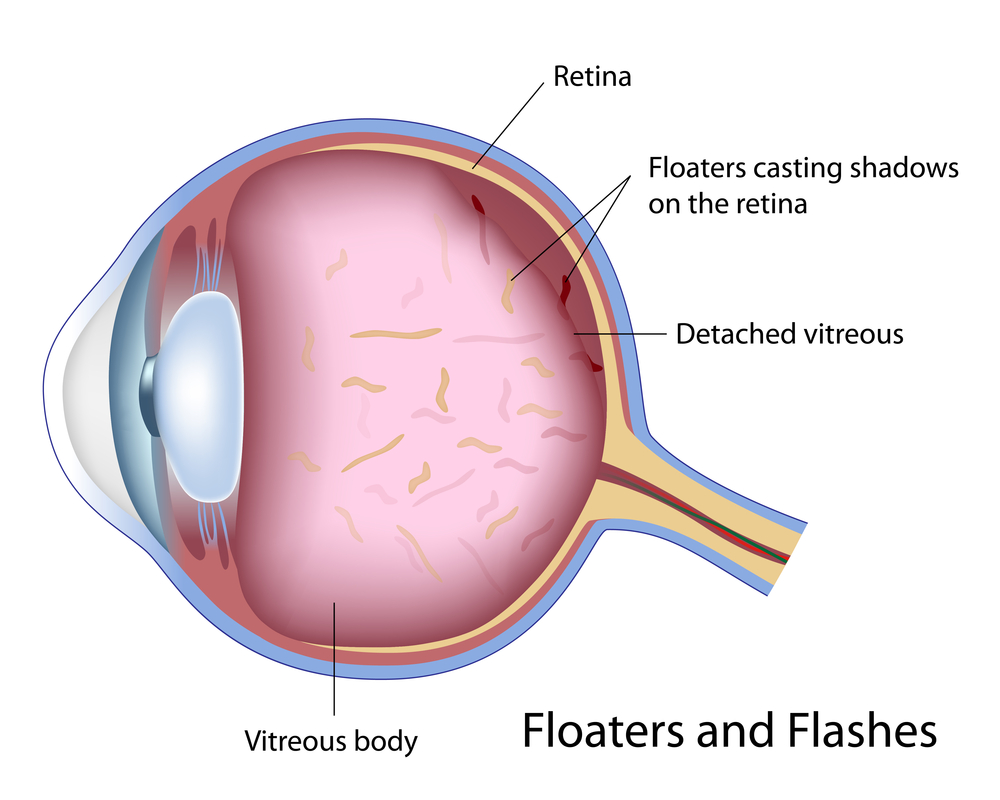
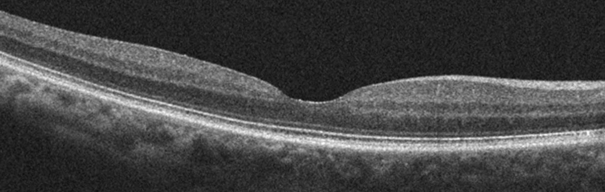
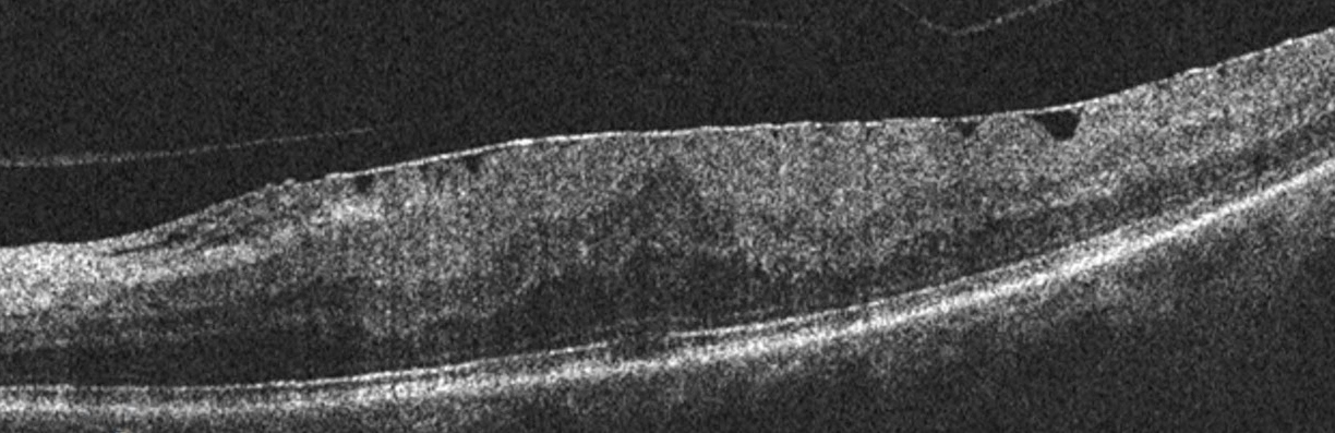
PVD is a natural process that occurs in the majority of people usually over the age of 50. The vitreous is a jelly-like substance that occupies the back portion of the eye. The vitreous is comprised primarily of water, which accounts for 99% of its volume, and the remaining 1% includes proteinaceous substances such as collagen fibers as well as hyaluronic and ascorbic acids. The collagen fibers act as a scaffold to allow the vitreous to maintain a formed shape as well as provide a means for the vitreous to attach to the retina, which is the light-sensitive tissue that lines the inner back wall of the eye and is critical for vision. As we age, changes in these fibers cause the vitreous to lose its shape and eventually pull away from the retina. When the vitreous separates from the retina, this is called a PVD.

As we age, the collagen components of the vitreous can clump together and are free to float in the eye. When the vitreous separates from the retina during the development of a PVD, the floaters may become more noticeable or numerous. It is common for patients to describe floaters of different shapes and sizes, and patients may notice just one or in some cases many. In many people, a PVD develops slowly and there may be no symptoms or just a few annoying floaters. In others, a PVD may occur abruptly and cause more dramatic symptoms that can be very anxiety provoking.

Since the normal process of PVD development involves the vitreous tugging on the retina until it can fully separate, this tugging can result in flashing lights that can commonly appear in the peripheral, or side, vision. These flashing lights are sometimes described as lightning streaks, and patients may notice them more readily in settings with low ambient light. The flashes of light typically resolve once the vitreous has fully separated from the retina and the tugging has ceased.

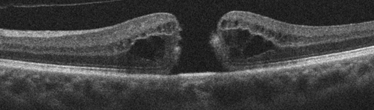
The good news is that PVD is usually harmless in the vast majority of cases, and the annoying floaters will become less bothersome over time. In approximately 5-10% of cases, the vitreous can tug too hard on the retina as it tries to separate and it may pull a hole or tear in the retina. Tears in the retina can predispose to retinal detachment, which is a serious condition that can lead to permanent vision loss.
It is important to recognize that the typical symptoms of a regular PVD are often similar to a PVD with an associated tear. For this reason, it is recommended that all patients with the new onset of floaters or flashes have a dilated eye exam. If a retinal tear or detachment is discovered, early treatment can help prevent loss of vision.

Treatment for PVD usually involves simple observation. With time, the flashes will go away, and the floaters will become less noticeable. More recently, few providers have claimed that floaters can be treated with a laser in order to make them less noticeable. I would caution that this is not mainstream therapy at the current time, and I do not advise my patients to pursue this option. Another treatment possibility is vitrectomy surgery, where the vitreous gel is removed as part of a surgical procedure. Due to safety advances in vitrectomy surgery, this is now a potential option for the rare patient who has floaters that are so numerous and bothersome that they are negatively impacting their activities of daily living. For the vast majority of patients this is not necessary.

When I see a patient with a PVD, I often recommend one follow-up visit in 4-6 weeks to make sure there are no retinal holes or tears that have developed in the interim. If the other eye has not had a PVD yet, I will counsel them that a PVD will most likely develop in that eye within the next few years, and when it does they need to be examined. I will also discuss the retinal detachment warning signs. Patients with retinal detachment will not only have symptoms similar to PVD, including flashes and floaters, but in addition they may also notice what looks like a black shade or curtain that starts in the peripheral vision and extends towards the central vision. My patients are taught that this symptom requires an immediate examination.

In conclusion, PVD is a natural process that the majority of people will experience in their lives. The symptoms can range from having no symptoms at all to many floaters with associated lightning flashes. In the majority of patients, there is no damage to the eye or threat to the vision. A dilated exam is recommended to look for possible holes or tears in the retina, and if these are uncovered, prompt treatment can prevent vision loss.

Dr. Esmaili posterior vitreous detachmentDaniel D. Esmaili, MD
Retina Vitreous Associates Medical Group

Night Blindness

10/28/14

As the number of daylight hours decrease and daylight savings time is about to end, many of us feel that the days are getting much shorter. If you suffer from night blindness, your days are shorter, because getting around or driving at night, are sometimes impossible.

Night blindness is a condition that makes it difficult for a person to see in low-light situations or at night. Some types are treatable, while others are not. You will need to consult your eye doctor to determine the underlying cause of your night blindness to determine what can or cannot be done.

night blindness
Courtesy of wikipedia
There are several things that could cause night blindness:
•Cataracts
•Genetic eye disease
•Vitamin A deficiency
•Diabetes
•Aging eye
•Sunlight exposure

Here is a brief look at each.
Cataracts – This is when the lens of the eye becomes gradually becomes clouded, reducing vision. Besides reducing vision at night you may also experience halos around lights. This is a treatable condition requiring cataract surgery and replacing your clouded lens with a clear artificial lens. Your vision should improve considerably.

Genetic Eye Disease – Both retinitis pigmentosa or Usher syndrome are progressive genetic eye diseases where the rods that regulate light, and cones that control color perception and detail die. Progressive night blindness is one of the first visual symptoms of these two diseases. Currently there is no treatment for them as there is no way to treat or replace the dying rods.

Vitamin A Deficiency – While rare in the US, it can be a result of other diseases or conditions such as Crohn’s disease, celiac disease, cystic fibrosis or problems with the pancreas. Options to help with the deficiency include vitamin supplements suggested by your doctor, or increasing your intake of orange, yellow or green leafy vegetables.

Diabetes – People with diabetes are at higher risk for night vision problems because of the damage to the blood vessels and nerves in the resulting in diabetic retinopathy. Not only can it cause poor night vision, it may also take longer to see normally after coming indoors from bright light outside. There is no cure, but controlling blood sugar levels with medicine and diet can help prevent developing retinopathy or help slow the progression.

Aging Eye – As we age several things happen to our eyes. Our iris, which regulates the amount of light going into the eye, gets weaker and less responsive. This can make adapting from light to dark more difficult and slower. Our pupils shrink slightly allowing less light into the eye. The lens of the eye becomes cloudier, as explained above in cataracts, limiting the amount of light into the eye. We also have fewer rods for light perception. Aside from cataract surgery there is no treatment for age-related night blindness. However, eating a diet rich in fruits and vegetables and low in saturated fat is the best way to slow the progression. Here is more information on how the aging eye is affected.

Sunlight Exposure – If your night vision seems temporarily worse after a trip to the beach or a day on the ski slopes, it probably is. Sustained bright sunlight can impair your vision, especially if you fail to wear sunglasses or goggles.

Night blindness due to genetic conditions or aging cannot be prevented. However if you protect your eyes from extreme sunlight, eat a healthy diet, and monitor blood sugar levels if needed, you can reduce your chances for night blindness.

As we head into the holiday season, you should know that some great sources of vitamin A include sweet potatoes, butternut squash and … pumpkins!

Susan DeRemerSusan DeRemer, CFRE
Vice President of Development

Living With KC Isn’t Easy

10/23/14

The Discovery Eye Foundation Fall 2014 e-newsletter focused on depression and eye disease. At the time we asked for people that were willing to share their stories. Jennifer Villeneuve is one of the many that responded. She is 26 years old and lives in Ontario, Canada. A participant in KC-Link, she talks about the emotional toll keratoconus has taken on her life and living with KC.
Villeneuve with child - living with KC
At age 13, I was diagnosed with keratoconus and given RGP lenses, with which I struggled intensely. ?I became very quiet, and my bubbly personality disappeared. The lenses often got irritated and made my eyes water and turn red, which made me look like I was crying. My doctor didn’t really give me much information on the disease. He just told me my corneas were the shape of footballs instead of circles.

I couldn’t be a normal teenager. I often squinted and had red eyes, which made me very self-conscious. I couldn’t wear makeup or have a free-for-all teenage life. I had to worry about my lenses and what people saw when they looked at me. Some people knew about my KC, but not many. I was just that quiet person who squinted. Because of this, my self-esteem got very low. All in all, my high-school life was hell.

Every time I went to the doctor, he said my pain is normal, the discomfort is typical, and I needed to get used to it. I was also diagnosed with two learning disabilities, in addition to my vision impairment. Throughout high school, I had major anxiety and depression, though I never wanted to admit it. A close teacher even spoke to my mom about the anxiety and low self-esteem. I got through high school, still with the depression being untreated — and still with the same doctor who never even sent me for a topography scan. Each time I went in, it was, “Yup your eyes the same; see ya.”

In college, I was diagnosed with depression and anxiety and was finally getting treated. ?I also went to the University of Ottawa Eye Institute of The Ottawa Hospital in Ontario, Canada. They did topographic scans; my KC had gotten significantly worse. My contacts’ sizing changed twice that year. Again, I couldn’t be normal. College students go out partying, but not me. I stayed in my room by myself. I worried whether I could see enough to go out. What if I drank too much and lost a lens? It wasn’t worth the risk, especially given how expensive they are. In college, I got great help and acceptance. My teachers all knew about my KC, and I was set up with the Centre for Students with Disabilities at Algonquin College in Ottawa. They were my backbone and my support.
Villeneuve with 2 children - living with KC
After my two years in college, I moved on to my career working with children. At each job, I had to explain why I always had a mirror and my contact stuff. At first, I was ashamed and almost embarrassed, explaining why I squinted and that I may not be able to read a kids’ book if the writing is too small. Not only did I get accepted by coworkers, but also by the kids. They knew my eyes were red from my contacts or that my tears meant something was in my eye. I began working in with special-needs children, which was incredible but also challenging — especially in ensuring my eyes were at their best.

At this same time, my vision had gotten worse. I went for corneal crosslinking (CXL) in one eye. I had to take? time off from work, which caused a lot of stress. Unfortunately, there were complications from the CXL. I had a scar in the same eye that caused the crosslinking to be difficult and not as successful. When it came time to do my other eye, I was hesitant, but I needed it. It worked, and the disease slowed down.

A year or so later, the disease had a spike and caused my eye to rub on a lens and make a blister. I had laser surgery to get rid of that and the scar, too. Because KC isn’t covered by insurance in Ontario, I had to pay for every lens, every $11 bottle of solution and countless eye drops. It was expensive, and money was a struggle. I still struggle with the costs of things.

I also have to worry about eye infections. Since I work with kids, they are easy to get, but if I get an eye infection, I can’t wear my contacts, which means no driving, which means no working, which means no money. Things can snowball so quickly.

My vision is up and down. My lenses don’t last as long in my eyes as they used to, and my nighttime vision can be scary. I have to be extra-vigilant. Again, I can’t be normal or go somewhere unfamiliar at night. I’m always concerned. My lens fitter recently recommended scleral lenses, but I can’t afford them. I barely could afford the $2,000 for the CXL.

Living with KC isn’t easy. I can’t help but wonder how long I’ll have the vision I have now. Am I going to be blind in a few years? If I have children, will they have this, or will I even be able to see them? Will I find a guy who would want someone with the possibility of losing vision? I have to stop myself from thinking ahead, or the anxiety gets the best of me.

Janet Villeneuve - living with KCJennifer Villeneuve
Keratoconus Advocate

20 Tips For Cooking With Low Vision

10/21/14

People cook for different reasons. Some just so they can eat, others enjoy the creativity and find it relaxing and for some it is a activity they have fun doing with others. But cooking can become difficult and dangerous as you lose your vision. Here are some useful tips to help keep you safe and give you confidence to enjoy cooking with low vision.

Cooking with low vision
1. To avoid burns, place a pot or pan on the burner before turning it on and, turn the burner off before removing the pot or pan.

2. Turn pan handles inward from the stove or counter to avoid spills and burns.

3. Mark common or often used settings on appliances with bright nail polish, bright colored tape, or raised dots.

4. Purchase a microwave with a sensor reheat feature to automatically reheat food to the proper temperature.

5. Use long oven mitts to protect your hands and arms from hot surfaces.

6. Turn on the oven light while using it to remind you the oven is on.

7. Use a double spatula to help avoid spills when turning food.

8. Use a special measuring cup that enhances contrast.

9. Use a knife with an adjustable slicing guide.

10. Have light and dark cutting boards to be able to provide the most contrast depending on what you are slicing or chopping – such as black for slicing onions and white for chopping kale.

11. Use a large print, low-vision kitchen timer.

12. Use an automated coffee maker that makes a single cup of coffee or tea to eliminate the need to pour boiling liquids and risk burns.

13. Attach low-glare fluorescent lighting to the underside of cabinets to make it easier to see.

14. Outline the end of countertops with colored duct tape, or paint in a color that contrasts with the work surface. Also choose kitchenware that contrast with the countertop.

15. Organize shelves in a logical way, possibly alphabetically, with most use items easily accessible. Be sure to return items to where they were originally.

16. Use a color for the interior of cupboards that contrasts with your dishes to make them easier to see. Do not use clear glasses or dishes as they appear invisible.

17. If you have glaucoma and experience tunnel vision, remove cabinet doors or replace with sliding doors.

18. Place rubber bands around the milk carton to distinguish it from the orange juice carton. This also works well with distinguishing a can of chicken noodle soup from tomato, etc.

19. Avoid overflows by hooking a “liquid Level” indicator to your glass or cup that will play a tune when you are ¾” from the top.

20. For cold drinks you can also use your finger by placing the tip of it over the edge of the glass and stop pouring when you feel the liquid.

Once you have prepared your food, eating can also be a challenge, so here are five bonus tips.

1. Use dinner plates with a raised lip to avoid spills or pushing the food off while eating.

2. Avoid patterns on dinnerware.

3. Make sure your dishes contrast with the table or table linens.

4. Have a system for putting food on your plate such as meat at 6:00, vegetables at 3:00 and starches at 9:00.

5. Use a gooseneck lamp directly over you plate.

Susan DeRemerSusan DeRemer, CFRE
Vice President of Development

15 Things Doctors Might Like Us To Know

10/16/14

The Doctor Patient Relationship

Doctors are human. Professional decorum may not let them speak their minds and for that reason, I have gathered comments heard from eye doctors with whom I have been privileged to associate as a so-called “expert patient”. These may not speak for all, but I do think they represent the majority. Here are 15 of the most important, which we might benefit from hearing.15 - doctor patient relationship

1. Your eyes are growing older, and I can’t turn back the years in an aging retina. I can, however, help you try to maintain your current vision for as long as possible.

2. Your appreciation means a lot. Don’t hesitate to compliment me if I have earned it.

3. I have quite a few patients who need my help and don’t want to be kept waiting. So I don’t have to keep repeating myself, please take notes, or bring someone with you who can refresh your memory later.

4. It is important that you comply with my instructions and show up for appointments if you want the best results.

5. My specialty is eye care. I will, however, try to provide you with resources for other ailments you may have.

6. Please be concise with your questions and stay on topic, so I’ll have time to treat other deserving patients.

7. Don’t believe everything you see in the media unless the source is reliable. If the information isn’t based on good evidence, my response to you may be disappointingly negative.

8. My services may seem expensive, but I have so many expenses and debts resulting from my profession that my bottom line may not be much different than yours.

9. An ethical physician will not choose a particular drug or treatment by how much of a profit he can make prescribing it.

10. I might not agree with everything you say, but rest assured that I will never let it affect the level of care I will provide.

11. Don’t expect me to keep all eye research in my head. I do know where to look it up, so allow me some time to get back to you on some questions.

12. Conferences I attend are sometimes in exotic places, but most of the time is spent going from one event to another, so it is really not a vacation.

13. Even if there is no current effective treatment for your eye disease, I still need to see you regularly to monitor your condition. Regular check-ups are important to your health and preventative care and treatment options are changing all the time.

14. I may not remember you personally, especially in the beginning or if I see you only see you once or twice a year, but your records contain everything I need to know to care for your vision.

15. I may not agree with your decision to try an alternative treatment, but I respect your right to do so. All I ask is that you seriously consider my opinion, and if you decide to go ahead with the treatment on your own, please keep me in the loop so I can monitor your condition.

We should try to understand that doctors are human beings who truly care about our welfare. Doing so could turn a top-down doctor-patient relationship into one of mutual respect and understanding. We might even get a few extra minutes of precious time in the chair, just because we’re a pleasure to have around.

dan robertsDan Roberts
“Expert Patient”

Being A Blind Artist

10/14/14

One thing that was always nurtured in me as a child was art. My father was an architect, and my mother is an art-lover. I remember they would collect all my artwork from preschool and elementary school. I used to have trouble sleeping as a kid, so I started drawing on the wall next to my bed; it would comfort me until I fell asleep. Most parents would probably scold a kid for drawing on the wall, but my parents loved it! In fact, my father would bring his architect friends into the room to “see my daughter’s mural.” Art has always been one area where I don’t feel inadequate. Art is so subjective, and anything can be art, so there is no judgment, and it’s very liberating. I’ve done abstract painting, mosaic tile work, ceramics and, currently, sculpture.

Becoming a Blind Artist

When I became blind, maybe five or six years in, I started going to Braille Institute, and I rediscovered art as a blind person. I saw people there creating art, and I thought, “This is amazing.”
Kooshay scuplture - blind artist
I always wanted to learn how to sculpt, but every time I called art schools about classes, when I told them I’m blind, they told me I had to take private lessons. Fear came up, and they did not know where to put me. Finally, two years ago, I started taking private lessons. My first piece was a portrait of a man, and the school was so amazed, they not only offered to put me in the sculpting class, but they gave me a full scholarship.

My private-lesson teacher wasn’t even a sculptor; she was a painter, but she was so tickled by the idea of a blind person wanting to sculpt that she wanted to be involved. She was a true artist and wanted to try something new. It was a wonderful experience for both of us. My second teacher would blindfold herself to figure out how to teach me. She really went out of her way, and I learned some really great techniques. I started my current class, which is a figure-sculpting class, by sculpting shoes — I have a shoe fetish — but I starting getting jealous of everyone else sculpting figures. I can’t touch the naked models, so now I touch other people’s sculptures and use that as a study to make my own.

I love it. I love the feel of clay. I’m a very tactile person, and I love the sensation of it in my hands. It’s so malleable. I use my hands more than sighted people; I don’t really use tools. I need to feel the clay to shape it, and I think there’s more emotion involved for me. It’s me, the piece and nothing else. Maybe it’s because I can’t see it — it’s like a meditation for me. I get lost in my piece. There is so much emotion — that’s where I get my inspiration. It’s a way to fantasize or fulfill an emotion or need. For me, it’s more about the process — I don’t see the finished product. It’s a very fulfilling way of expressing myself that words can’t; it’s more about expressing what I feel.

Kooshay Malek - seeingKooshay Malek
Marriage and Family Therapist
Los Angeles

What Are A Macular Pucker and Macular Hole?

10/9/14

What is the macula?
The eye is very much like a camera, taking light from the outside world and converting it into picture information that our brains perceive as vision. The retina is the light sensitive layer in the back of the eye that is very much like the film in that camera. The central retina, also known as the macula, is essential for crisp, high definition vision. Conditions that damage or distort the macula can therefore result in blurred or distorted vision. Two common conditions that affect the macula are macular puckers and macular holes.

What is a macular pucker or macular hole?
A macular pucker is a thin layer of scar tissue that forms on top of the retina. The amount of scar tissue can range from mild to severe. Mild macular puckers may be barely noticeable during an eye exam and resemble a fine layer of cellophane resting on the macula. More severe macular puckers can cause wrinkling or distortion of the macula. In contrast to a macular pucker, a macular hole is a small gap that extends through the entire thickness of the macula.

What are the symptoms of a macular pucker or a macular hole?
At first, a macular pucker may lead to mild blurring of the central vision. Because the problem involves the back of the eye, glasses will not completely restore vision. More severe macular puckers may result in wavy or distorted vision. For instance, objects that normally appear straight, such as venetian blinds or a printed line of text, might appear to have a dip or bend in the center. Small macular holes can cause similar symptoms of blurring or distortion. Larger macular holes often result in a central blind spot. This can also result in straight lines appearing broken or having a piece missing in the middle. Patients with a macular pucker or hole do not normally experience difficulty with peripheral vision.

What can cause a macular pucker or macular hole?
Recall that a macular pucker is a scar tissue. Anything that causes scar tissue, such as trauma or inflammation in the eye, can result in scar tissue and hence a macular pucker. Certain diseases that affect the retinal blood vessels such as diabetes can also cause a macular pucker to form. However, one of the most common causes of macular pucker is simple aging of structures within the eye. As the eye ages, the clear jelly that fills it, called the vitreous gel, shrinks. When enough shrinkage occurs, the vitreous gel detaches from its normal position adjacent to the retina. This process of vitreous detachment can cause microscopic damage or inflammation leading to macular pucker formation. In some cases, the vitreous gel does not detach cleanly from the retina. Instead it can put traction on the macula, pulling its delicate structures apart in the center, resulting in a macular hole.

How are macular puckers and macular holes diagnosed?
A simple examination from an ophthalmologist or retina specialist is often enough to diagnose a macular pucker or hole. However, additional testing is often useful in diagnosing subtle cases or monitoring eyes for changes. An optical coherence tomography (OCT) scan is a specialized photograph that allows your physician to look for microscopic changes in the contour of the macula. The following figures show an OCT of a normal macula, a macular hole, and a macular pucker. Note that the normal macula has a central dip known as the fovea, shown in Figure 1. In Figure 2, the dip is replaced by a gap which is a macular hole. Finally, Figure 3 shows a macular pucker where the dip is no longer visible. This is because the macular pucker, seen as a thin white line is distorting the normal shape of the macula.

Normal - Macular Pucker and Macular Hole
Figure 1: Normal Macula
Hole - Macular Pucker and Macular Hole
Figure 2: Macular Hole
Pucker - Macular Pucker and Macular Hole
Figure 3: Macular Pucker

What treatments are available for macular puckers and macular holes?
Macular puckers can be quite mild. For mild cases in patients with minimal symptoms, periodic monitoring may be all that is required. When blurred vision due to a macular pucker begins to affect activities such as driving or reading, treatment in the form of surgery can be considered. Surgery for a macular pucker is known as a vitrectomy. Vitrectomy surgery is usually done under local anesthesia and as an outpatient procedure. During the surgery, fine instruments are used to remove the scar tissue from the surface of the macula. After surgery, patients usually experience an improvement in the blurring and distortion as the eye recovers gradually over a period of months. Some residual waviness can be normal. Vitrectomy is generally very safe although there is a chance of increased cataract growth and a small risk of infection or retinal detachment.

For patients with small macular holes, close monitoring can also be an option since some macular holes can close on their own. For larger holes, there are two options. In select cases where the vitreous gel is actively pulling on the macula, an injection of medication into the eye may cause the gel to release cleanly, allowing the hole to close. In other cases, vitrectomy is recommended. During the surgery, any pulling on the macula is relieved and a gas bubble is placed in the eye to help the hole close. After surgery, patients are asked to look down for a several days to allow the bubble to float up against the hole. Once the body absorbs the bubble, vision is usually significantly improved.
In summary, both macular puckers and holes are common causes of blurry or distorted central vision. If treatment or surgery by a retina specialist is needed, the results are generally quite good and lead to significant restoration of vision.

Liao - Macular Pucker and Macular HoleDavid Liao, MD, PhD
Retina-Vitreous Medical Group

Our Thanks to Guest Bloggers Continues

10/7/14

More Amazing Guest Bloggers

Last week I took the opportunity to thank our very first guest bloggers for helping us launch the Discovery Eye Foundation Blog. We are pleased that so many people appreciate the wide range of eye-related information from eye care professionals, as well as the stories from people that live with eye disease on a daily basis.
Thank you part 2
Here is a round-up of guest bloggers since June 2014 that shared their time, experience and/or expertise to provide you with the best eye-related information.

Sumit “Sam“ Garg, MDwhat you should know about cataracts

Randall V. Wong, MDfloaters, causes and treatments

Roy Kennedyhis personal experiences with the miniature telescope implant

Sandra Young, ODthe importance of getting vitamins and minerals from your food and not just supplements

Jeanette Hassemanliving with keratoconus

Greg Shanetheater for the blind

Caitlin Hernandezblind actress and playwright

Jullia A. Rosdahl, MD, PhDlasers for glaucoma and genetics and glaucoma

Maureen A. Duffy, CVRTways to reduce harmful effects of sun glare

Kooshay Malekwhat is it like to lose your vision and being a blind therapist

Jeffrey J. Walline, OD PhDchildren and contact lenses

Robert Mahoneychoosing a home care agency

Robert W. Lingua, MDnystagmus in children

Buddy Russell, FCLSA, COMTcommon pediatric eye diseases, treatment options for children and pediatric contact lenses

NIH (National Institute of Health)telemedicine for ROP diagnosis

Harriet A. Hall, MDevaluating online treatment claims

Patty Gadjewskithe life-changing effects of a telescopic implant

Michael A. Ward, MMSc, FAAOproper contact lens care and wearing contacts and using cosmetics

Susan DeRemerSusan DeRemer, CFRE
Vice President of Development
Discovery Eye Foundation

Coffee and Glaucoma: “1-2 cups of coffee is probably fine, but…”

10/2/14

The Relationship Of Coffee And Glaucoma

Research shows that drinking caffeinated beverages, especially coffee, causes eye pressure to go up, even just one cup of coffee. The effect is highest in glaucoma patients and people at risk for glaucoma. However, eye pressure goes up only a small amount, so it is probably not a significant risk.
Coffee and glacoma
In some people, though, too much coffee may be causing damage. In people at risk for exfoliation glaucoma (a type of open angle glaucoma where some flake-like deposits are seen on the lens of the eye), drinking three or more cups of caffeinated coffee was associated with an increased risk of developing exfoliation glaucoma. The effect was strongest in women with a family history of glaucoma. This study doesn’t show that coffee causes glaucoma, but does suggest that drinking three or more cups of caffeinated coffee might not be good for your eyes.

One group of scientists applied caffeine eye drops directly on to healthy eyes, and they did not see any increase in eye pressure. This suggests that caffeine doesn’t appear to have a direct effect.

How can you best protect your eyes? Consider going decaffeinated or limit your caffeine consumption. If you are at higher risk for glaucoma or have been diagnosed, be sure to have regular comprehensive dilated eye exams, use medications as directed and see your eye care provider as scheduled.
For more information about glaucoma, visit www.nei.nih.gov/glaucoma.

References
The effect of caffeine on intraocular pressure: a systemic review and meta-analysis. Li M, Wang M, Guo W, Wang J, Sun X. Graefes Arch Clin Exp Ophthalmol 2011. 249(3):435-42.

Effect of caffeine on the intraocular pressure in patients with primary open angle glaucoma. Chandra P, Gaur A, Varma S. Clin Ophthalmol 2011. 5:1623-9.

The relationship between caffeine and coffee consumption and exfoliation glaucoma or glaucoma suspect: a prospective study in two cohorts. Pasquale LR, Wiggs JL, Willett WC, Kang JH. Invest Ophthalmol Vis Sci 2012. 53(10):6427-33.

Effects of caffeinated coffee consumption on intraocular pressure, ocular perfusion pressure, and ocular pulse amplitude: a randomized controlled trial. Jiwani AZ, Rhee DJ, Brauner SC, et al. Eye 2012 26(8):1122-30.

Julia Rosdahl - coffee and glaucomaJullia A. Rosdahl, MD, PhD
National Eye Health Education Program Glaucoma Subcommittee
Duke Eye Center, Duke University