Purpose of Eye Exams for Children

Many children who are 6 – 18 years old are now back in school or will be shortly. But have you given them everything they need to succeed in the school year ahead?

      ? New binders
      ? Notebook paper and dividers
      ? Pencil box filled with pens and pencils
      ? Calculators, protractors and rulers
      ? Backpack to carry it all

These are the tools that children and their parents focus on every year, thinking these will help their child have a fun and productive year. But the list is incomplete. For school-aged children, the AOA recommends eye exams for children every two years if no vision correction is required. Children who need eyeglasses or contact lenses should be examined annually or according to their eye doctor’s recommendations.

“But my child gets and eye screening at school every year…” While this may be true it is important to understand the difference between a screening and an eye exam.

Vision screenings are a short examination that can indicate a vision problem or a potential vision problem; however it cannot diagnose exactly what is wrong with your eyes. It can also easily miss vision issues, giving parents a false sense of security.

With an eye exam, the tests are performed by a trained professional, using specialized equipment looking for specific indicators that could affect your child’s vision. They test much more than how well your child can read letters or symbols at a distance.

Good vision is necessary for a child to succeed at school and not become frustrated or depressed. It has been estimated that as much as 80% of the learning a child does occurs through his or her eyes. Children need to read a book and see a whiteboard, write and use computers every day in the classroom and at home. When a child cannot see clearly, it becomes more difficult to learn.

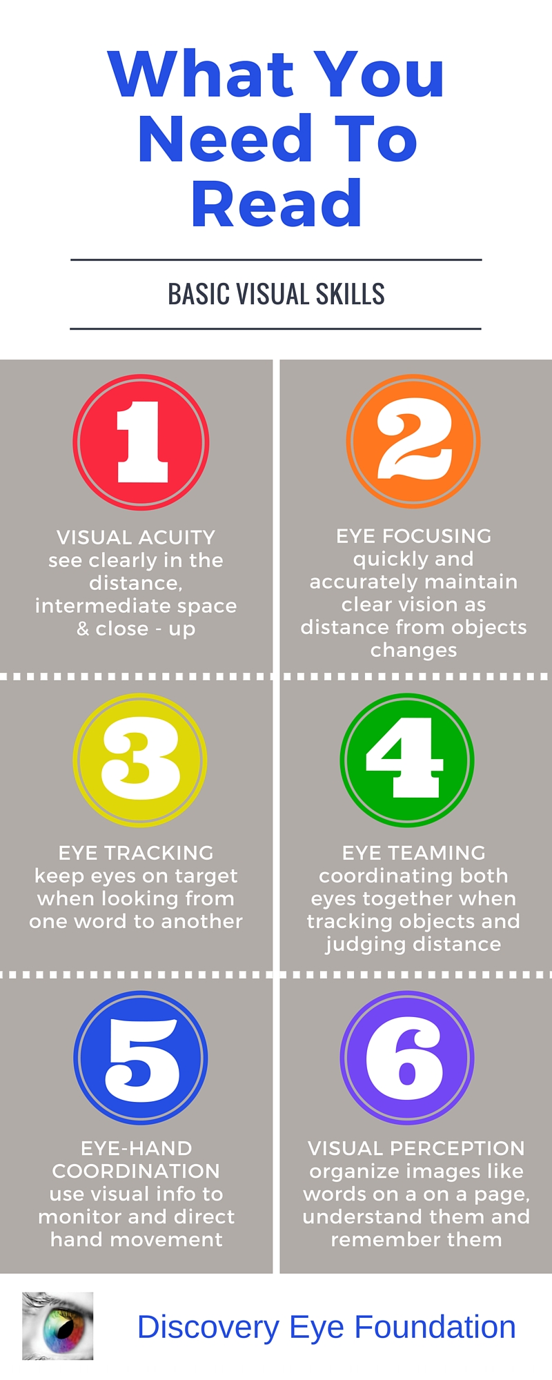
It also goes beyond just seeing clearly. Your child needs their eyesight to understand and respond to what they see. This includes the ability to focus their eyes, use both eyes together, and move them effectively.

Infographic - eye exams for children

Children may not always know they have a vision problem because they think that everyone is seeing the way they do. There are some signs that may indicate a vision issue:

  • Repeated eye rubbing
  • Excessive blinking
  • Short attention span
  • Tilting the head to one side or covering one eye
  • Holding reading materials too close to the face
  • Losing their place when reading
  • Difficulty remembering what they just read
  • Trying to avoid reading or other close activities
  • Numerous headaches

So as you prepare your child to go back to school, give them the best advantage they can have – good vision. Make an appointment with your eye doctor today.

8/27/15


Susan DeRemerSusan DeRemer, CFRE
Vice President of Development
Discovery Eye Foundation

Treatments for Dry Eye Disease

This is the third and final installation of the comprehensive series we have presented on dry eye disease. Dr. Wade first discussed the symptoms you might experience if you have dry eye and the Dr. Garg explained the process of diagnosing they type of dry eye disease you might have. In this article Dr. Farid reviews treatment options based on your diagnosis of dry eye disease.treatments for dry eye

Treatments for Dry Eye Disease

As our understanding of dry eye disease expands, so do treatment options. We now know that dry eye disease is a multifactorial disease. There is no one cause so there is no one magic cure. Treatments aim to improve tear composition, reduce eye surface inflammation, and target eyelid margin disease. Here, we will review many treatment options, but the treatment combination or “cocktail” that is appropriate for you will depend on your specific type of dry eye disease. This is usually determined after some testing by your eye care provider.

Environmental, Dietary, and Medication Adjustments
Before going into specific dry eye treatments, there are modifiable causes and preventative methods to improve dry eyes. Simple changes in the environment, diet, and medications can be easy ways to improve symptoms.

Environmental Changes
As expected, a dry environment will worsen dry eyes. Humidifiers and moisture goggles have been shown to help alleviate these symptoms. Furthermore, situations that cause decreased blinking, such as prolonged use of computer screens, can worsen dry eyes. Patients should take frequent breaks from computer screens and reading, allowing their eyes to rest and resume normal blinking. When in windy, smoky, or dusty situations, sunglasses can act as a barrier to the eyes, reducing dry eye symptoms. Avoiding wind, fans, or any source of air blowing into the eyes may also help.

Dietary Changes
Drinking adequate water keeps patients hydrated and reduces exacerbations of dry eye symptoms. Omega-3 fatty acids, particularly eicosapentaenoic acid (EPA) and docosahexaenoic acid (DHA), have been shown in many clinical studies to improve dry eyes. It is believed that these fatty acids help inhibit inflammatory mediators. Essential fatty acids cannot be synthesized and must be ingested through diet. Foods rich in omega-3 fatty acids include fish, other seafood, and flaxseed oil. Supplements are also available.

Adjusting Medications
Many medications are associated with dry eyes, and patients may benefit from adjusting doses or finding alternative treatments. You can work with your physician to weigh the costs and benefits about modifying your current medications if they are suspected to worsen dry eyes. Some of these medications include: hypertensive drugs, antihistamines, decongestants, antidepressants, acne medications, birth control, and hormone replacement therapy. Eye drops with preservatives, such as glaucoma medications, can also worsen dry eyes.

Lubricating Treatments
For patients with decreased tear production, supplementation of tears or reduction of tear drainage will improve symptoms.

Artificial tears, gels, and ointments
Artificial tears, gels, and ointments are readily available over the counter. Artificial tears are eye drops that are used throughout the day as needed, up to eight times for dry eyes. You must make sure to get “lubricating” drops. There are multiple available brands that are excellent and equivalent in providing artificial moisture. Avoid “redness relief” brands as they are focused on reducing appearance of the vessels in the eyes as opposed to actual dry eye relief. Gels and ointments are thicker and very effective but can blur vision. Non-commercial comparison testing between brands has not been done. Because the ingredients, preservatives, and consistency vary from brand to brand, it is recommended for patients to choose the best option that works well for them based on trial and error.

Preservatives are used in many eye drops to help them to last longer. Unfortunately, they can be irritating to the eyes, especially when used often (more than 4 drops/day) or with other drops containing preservatives (ie. glaucoma medications). Patients who frequently use eye drops are recommended to use preservative-free (PF) drops. Unfortunately, preservative-free drops are more expensive because they come in “single use” containers. You may be able to extend the life of each single use container by spreading out its contents over multiple uses throughout the day.

Lipid-containing lubricants, such as those with castor oil, attempt to mimic the oils found in the tears, reducing evaporative dry eyes, but more research is necessary to study their efficacy.

Punctal Plugs
Your ophthalmologist can place small plugs at a clinic visit into the punctum, a small hole at the upper and inner lids that drain tears from the eyes. One plug can be added initially, and if more tear retention is required, another plug can be added. There are absorbable and non-absorbable types. Absorbable types are made of collagen and last 1 week to 6 months. Once placed, they are not visible or removable. Non-absorbable plugs are usually silicone, and permanent. They are easily placed by your ophthalmologists and remain visible in follow-up exams. Some patients feel the plugs, and there may be mechanical rubbing, especially when the plug is not the proper size. Some patients may also experience excessive tearing with plugs. If problematic, patients can wait for the plugs to dissolve if absorbable or they can be removed with forceps if non-absorbable.

Autologous Serum
Serum is extracted from a patient’s blood and turned into an eye drop. The growth factors, vitamins, and antibodies present in serum are the same as those in natural tears. Evidence is showing significant promise in alleviating symptoms and signs of chronic dry eye disease. A good collaboration between a phlebotomy lab and compounding pharmacy is necessary to make the products. The products must be kept refrigerated or cold between uses. They can be frozen for long-term storage. Each blood draw provides a supply of drops that can last 3-6 months.

Hydroxypropyl Cellulose Ophthalmic Inserts (Lacrisert)
An insert is available for people who find regular artificial tear use to be difficult. A physician can order the inserts through a pharmacy. The patient places the insert in the inferior fornix of the eye, the area between the lower lid and sclera of the eye. It slowly dissolves over 24 hours, giving constant lubrication.

Anti-inflammatory Treatments
Inflammation is now being recognized as a major underlying cause of chronic and worsening dry eye disease. Many patients who have been suffering from chronic dry eyes, particularly those with autoimmune diseases like Sjogren’s Syndrome, will do well on treatments that reduce the amount of inflammation in the tear film and ocular surface. Your physician can order these medications for you if needed.

Cyclosporine A (Restasis)
The FDA approved Restasis for dry eyes in 2002. This is an immunosuppressive and anti-inflammatory eye drop medication. Relief is not instant, and may take 6-8 weeks of sustained use for improvement in dry eye symptoms. Less than 20% of patients may experience a burning sensation with the drops, but the safety and tolerability profile is otherwise excellent.

Antibiotics
Doxycycline and minocycline are used for inflammatory ocular surface and eyelid disease. The antibiotics have a dual effect: they act as anti-inflammatories and anti-microbials. As an anti-microbial, these medications can improve meibomian gland function. They can decrease lid bacterial flora, reducing a cause of meibomian lipid breakdown. A low-dose (doxycycline 20 mg BID) regimen for 1-2 months has been shown to be effective. Side effects are usual mild but include stomach upset, yeast infections, and photosensitivity.

Steroids
Steroids act as anti-inflammatories. Because steroids are associated with complications in long-term use, they are mainly used in short pulses either at the initiation of treatment or as rescue during exacerbations. A short 4-6 week course is generally well tolerated as a “rescue treatment,” or an urgent treatment, to relieve intolerable symptoms quickly before resorting to other treatment options.

Meibomian Gland Treatments
Meibomian glands produce oils that are crucial in preventing our tears from evaporating too quickly. Treatments that target the glands can help patients with meibomian gland dysfunction or lid margin disease.

Warm compresses and lid scrubs
Warm compresses provide heat that warms the oils in the glands, unclogging the glands and improving oil flow. Warm washcloths, small rice bags heated in the microwave, or commercial hydrogel pads are all effective, and no studies have been done to compare the different methods. The heat. The compress should be placed over the eyes for 5-10 minute. Gentle circular or rolling massage of the eyelids can help express the oils from the glands.

Lid scrubs are useful particularly in cases of blepharitis, or mild inflammation of the lids, that cause them to become crusted. There are excellent over the counter commercial lid soap formulations that work well to clean the lid margins. Alternatively, baby shampoo and a washcloth gently applied to the lids can work as well.

Thermal pulsation (Lipiflow)
Lipiflow is an FDA approved in-office treatment for meibomian gland dysfunction and dry eyes. During the 12-minute procedure, a device is placed over the eye and eyelids that provides localized warmth and pressure on the lids (Figure 1).

treatments for dry eye
Image 1 – Lipiflow treatment

The procedure is 100% safe and very effective at clearing out the trapped oil glands and allowing smooth flow to be re-established. After approximately one month, the consistency of the oils in the tear flow will have improved remarkably with associated improvement in dry eye symptoms. With one procedure, the effects last between 12-24 months.

Intense Pulsed Light (IPL)
Originally approved for acne and rosacea dermatologic disease, IPL uses bursts of light to minimize blood vessel size. It can be used off-label for ocular rosacea and meibomian gland dysfunction, but results have not been reported.

Summary
There are many treatment options for dry eye disease. Generally, conservative over-the-counter treatments should be tried first. Commonly, environmental changes, dietary changes, artificial tears, and warm compresses will improve the majority of dry eye symptoms to tolerable levels. However, when these options are exhausted, there are many additional options for patients. Work closely with a trusted health professional to determine the optimal treatment combination as each patient is different.

8/25/15


treatments for dry eyeMarjan Farid, MD
Director of Cornea, Cataract, and Refractive Surgery
Vice-Chair of Ophthalmic Faculty
Director of the Cornea Fellowship Program
Associate Professor of Ophthalmology
Gavin Herbert Eye Institute, University of California, Irvine


treatments for dry eyePriscilla Q. Vu, MS
Medical Student
University of California, Irvine School of Medicine

Dry Eye Diagnosis

As covered earlier by Dr. Wade, the symptoms of dry eye disease (DED) can be variable. Simply put, dry eyes can be separated into two categories: aqueous tear deficient (ATD) or dysfunctional tear syndrome (DTS). More commonly there is a combination of the two that I like to refer to as ocular surface disease (OSD). Lucky for you, as clinicians, we have several tools that will allow us determine what type of DED you have.

Steps In A Dry Eye Diagnosis

First is a review of your symptomatology. This is crucial to determining if you 1) have DED, and 2) what type you have. This determination can drive our treatment plan that is individual to you. In addition, we utilize various questionnaires that can help us hone in on your OSD.

Second is the ocular examination. We use a microscope (slit-lamp) to carefully examine the surface of the eye. When we look at your tear film we are looking to see the amount and health of your tears, how well they are working, and what effect they are having on the ocular surface (conjunctiva and cornea). Not only do we look at your tears, but we pay special attention to your eyelids. In your eyelids, there are oil producing glands called Meibomian Glands. These glands are responsible for creating a key component to the tear film: lipid. Human tears are very complex, but simply put, tears have 3 main components – water, mucus, and oil. I like to describe tears like salad dressing. In order to have tasty salad dressing, there needs to be a balance of oil, vinegar, and spices. Human tears are very much similar. In order for your tears to work properly, There needs to be the proper balance of the aqueous component (water), lipid component (from the Meibomian glands), and mucus component (image 1).

dry eye diagnosis
Image 1 – A relatively healthy meibiomian gland examination

If there is an imbalance in your tears, this will reflect in their function, and ultimately cause signs and symptoms of ocular surface disease. To highlight the appearance and function of the tears on the ocular surface, clinicians often use special stains that can aid us in determining the amount and function of your tears. Two of the most common stains are fluorescein and lissamine green. Each of these stains has particular characteristics that help determine the severity and extent of your ocular surface disease. For example, if you have significant staining near the bottom part of your cornea, your eyes maybe slightly open when you sleep, and therefore you may benefit from using an ointment at nighttime. Alternatively, if your tears appear to break up very quickly on your ocular surface, there is likely an imbalance in the tear composition that may benefit from institution of warm compresses along with tear replacement in the form of artificial tears.

Third is the use of ancillary testing to help confirm our clinical diagnosis. We are fortunate to have access to several commercially available OSD diagnostics at the Gavin Herbert Eye Institute. A brief description of if you have these diagnostics follows.

    1) Schrimer Testing – This is a very simple and common method of determining whether a patient has hey aqueous tear deficiency. Essentially, the eye is numbed and a sterile piece of special paper is placed in the lower outer corner of the eye. After a specified amount of time, the amount of tears is recorded, and if under a threshold value (generally 10 millimeters at five minutes) there is a high suspicion of aqueous tear deficiency. Treatments for this subtype of OSD will be covered in the next blog.
    2) Tear osmolarity – The most available tear osmolarity system is from TearLab. With this test, we look at the integrity of the tears by determining the osmolarity – essentially the ultrastucture of the tears. If the tear osmolarity is high (hyperosmolar), we know that the tears are not functioning properly. With proper institution of treatment, the osmolarity can normalize indicating a healthier tear film. This test is very noninvasive, requiring only a tear sample of 50 nanoliters – less than the volume of a single tear!
    3) InflammaDry – Inflammation has long been accepted as a hallmark of dry eye disease/ocular surface disease. As such, many of our treatment modalities have focused on treating ocular surface inflammation (discussed in the next installment of this blog). Prior to having access to the InflammaDry test, we would have to assume that there was inflammation involved in an individual’s OSD. Now, however, we can test the ocular surface for inflammatory markers and have an answer within just a few minutes. This test not only allows us to custom tailor treatments to an individual, but also we are able to see if our treatments are working. Again, this test is minimally invasive requiring the small sample of tears for testing.
      4) LipiView II – This test allows us to Image of the structure and function of the meibomian glands in vivo. The images obtained allow for several things. First, we are able to determine the extent of meibomian gland dysfunction. Second we are able to determine the extent of meibomain gland drop out (image 2).
dry eye diagnosis
Image 2 – Significant dropout of meibomian glands
    And third we are able to educate our patients so they can see the importance of treatment of their MGD. Again, this information can help us custom tailor treatment options for the individual patient.

In conclusion, as you can see diagnosis of ocular surface disease can be quite intricate. We are fortunate to be in and age where there has been significant improvements in our tools to help us better diagnose our patients and use this information to individualize treatment options. Stay tuned for the next installment of this blog focusing on treatments for ocular surface disease.

8/20/15


Sam Garg, MDSumit “Sam“ Garg, MD
Medical Director and Vice Chair of Clinical Ophthalmology
Assistant Professor of Ophthalmology
Gavin Herbert Eye Institute – UC Irvine

Symptoms of Dry Eye Disease

Introduction

If you could be a fly on the exam room wall of your local ophthalmologist, you would hear patient after patient report symptoms of dry eye disease.

Some patients come in already knowing they have dry eyes. However, the variability of symptoms that can occur from dry eye disease is so wide many patients don’t even use the word “dry.” We will cover some of these symptoms in this article.

symptoms of dry eye
Redness often associated with dry eye

Underlying Factors

Dry eye disease has many underlying factors including an inadequate production of tears, rapid evaporation of tears, poor eyelid function and an imbalance in the tear composition of water, oil, and mucus. Dry eye disease can be associated with systemic conditions such as Sjogrens disease, Sarcoid disease and sleep apnea among many others. It is more common in females especially after hormonal changes such as menopause.

Exacerbating Influences

Many medications can exacerbate dry eye disease. Over the counter antihistamines are one example.

Environmental factors may also worsen dry eye symptoms. These include dry climates, windy weather conditions, smoky environments and the dry air found in airplanes.

Modern life includes hours and hours of focusing our eyes on everything from cell phones to computer screens to television. Prolonged focusing reduces the blink rate resulting in more tear evaporation and worsening of dry eye symptoms. Increased evaporation can also occur with exposure to heating, air conditioning, fans and rolling down the car windows while driving.

Symptoms Fluctuate

It is very common for dry eye symptoms (especially blurred vision) to wax and wane throughout the day. Symptoms can even change from blink-to-blink. Dry eye disease which is predominately due to insufficient tears tends to worsen throughout the day with symptoms worse at night. Dry eye disease that is more associated with blepharitis can be worse in the morning. Blepharitis is associated with burning and itching of the eyes.

Visual Symptoms

The front surface of the eye is the most powerful focusing surface of the eye. Thus, a dry ocular surface will produce visual symptoms. These symptoms can include:

Blurred vision: A decrease or fluctuation in visual acuity. This is manifested in the inability to see fine detail. Objects at both near and far may appear out of focus.

Sensitivity to light: Sensitivity to light is termed photophobia. It occurs because a dry ocular surface has more irregularities than a health surface. These irregularities scatter light entering the eye. This scattered light can cause significant discomfort. The inability to tolerate light may lead to squinting and headaches.

Difficulty with nighttime driving: During low light conditions, such as at night, the pupil enlarges and allows more light into the eye. When the ocular surface is dry, the incoming light becomes unfocused and scattered. Many of these abnormalities are filtered out by the small size of the pupil during the day. However, at night, the larger pupil size allows more light abnormalities to pass through to the retina. This results in nighttime glare and halos. Glare is a decreased tolerance of bright lights. Halos present as circles or auras around a bright source of light. Glare and halos from the headlights of oncoming traffic are especially troublesome.

Physical Symptoms

The front surface of the eye is richly supplied with nerve endings. As such, a dry ocular surface can result in significant symptoms of discomfort. In addition to feeling dry, these symptoms include:

Foreign body sensation: Patients may feel as if there is something present in the eye.

Redness of the eye: Enlarged blood vessels on the ocular surface cause the eye to look red.

Ocular and periocular pain: Pain from dry eye can be mild or severe. Pain from dry eye can be felt on the ocular surface. Pain can also be felt in structures around the eye such as the eyelids or scalp.

Periocular irritation: Stinging, burning, or itching sensations of the ocular surface and eyelids.

symptoms of dry eye
Watery eyes can be a symptom of dry eye

Watery eyes: Patients typically raise an eyebrow or two when I explain how the tearing they are experiencing is from dryness. “How can my eyes be dry if they are watering all of the time?” Although this may seem counter-intuitive, when the ocular surface is very dry it will overproduce the watery component of the tears as a protective mechanism.

Eye fatigue: A tired sensation of the eyes and heaviness of the eyelids.

Decreased tolerance of sustained visual focusing: As noted earlier, any activity that requires prolonged visual attention will decrease the blink rate and increase tear evaporation.

Discomfort while wearing contact lenses: Individuals may experience pain and irritation in the eyes while inserting or wearing contact lenses.

Inability to cry: Tears associated with emotional discomfort or watching a sad movie may be decreased in some types of dry eye disease.

Stringy discharge from the eye: A dry ocular surface can result in the overproduction of a sticky, mucus discharge.

Conclusion

There are many symptoms of dry eye disease. Some symptoms affect vision and others affect ocular comfort. If symptoms persist, an evaluation by your eye care provider can help clarify the cause and offer information on treatment options.

It is important to remember the symptoms of dry eye disease can overlap with the symptoms of other ocular conditions. One example is cataracts which, like dry eye disease, can also cause blurred vision and nighttime glare. The next post in this series will review how dry eye disease (and its sub-types) are diagnosed.

As a final note, while the name “dry eye disease” may sound innocuous, the symptoms of dry eye disease can be very severe in many patients. If you suffer from dry eye disease, you are not alone. Today there are many treatment options which can be very helpful. Significant research is underway to continue improving our ability to treat dry eye disease.


Matthew Wade, MD - toric intraocular lensesMatthew Wade, MD
Assistant Professor of Ophthalmology
Gavin Herbert Eye Institute


Minal Reddy was also a contributor to the is article.

Which Eye Care Specialist Do You Need?

It’s time to get your eyes checked – do you go to an ophthalmologist, optometrist or optician? Your optometrist sees the beginnings of age-related macular degeneration, but is sending you to see and ophthalmologist, why?

One of the most confusing things about taking care of your eyes can be differentiating between an ophthalmologist, optometrist and optician. Each eye care specialist has a very important part to play in the health of your eyes and here is a quick synopsis of what each does so you can choose the best one for your vision issues and treatment.

Ophthalmologist
These specialists are fully trained medical doctors that have completed the eight years of training beyond a bachelor’s degree. Their training has included a full spectrum of eye care, from prescribing glasses and contact lenses and giving eye injections, to carrying out intricate eye surgeries. Many doctors may also be involved eye research to better understand vision, improve eye disease treatments or potentially find a cure. They are easily identified by the MD following their name.

Optometrist
These medical professionals have completed a four-year program at an accredited school of optometry. They have been trained to prescribe and fit glasses and contact lenses, as well as diagnose and treat various eye diseases. They provide treatments through topical therapeutic agents and oral drugs, and are licensed to perform certain types of laser surgery, such as Lasik. They are easily identified by the OD following their name.

Optician
These eye care professionals are not licensed to perform eye exams, medical tests or treat patients. Their purpose is to take the prescription from the ophthalmologist or optometrist and work with you to determine which glasses or contact lenses work best for you. If you suffer from an eye disease like keratoconus, these specialists can make the difference between a relatively normal life, or one that is dictated short periods of vision because of contact lens pain. These eye care professionals may hold and associate optician degree or have apprenticed fore required number of hours.

While each one of these eye specialists has their own area of expertise, they can form a team whose only concerns are your eye health and the ability to see as clearly as possible.

8/11/15


Susan DeRemerSusan DeRemer, CFRE
Vice President of Development
Discovery Eye Foundation

Corneal Transplant Surgery Terms Defined

The following list of corneal transplant surgery terms is provided by the National Keratoconus Foundation (NKCF) website. They have more information on corneal transplants, as well as being a leader in information on keratoconus.

Corneal Transplant Surgery Terms Defined

Medical terminology is a language of its own. It can be confusing and alarming if you don’t know what the words mean. This will help you sort out the language of transplant surgery.
corneal transplant-original size
Atopic or Atopy – conditions associated with allergy like: asthma, eczema, hayfever

Decentration – off centered position

Donor tissue – the part of the cornea that is transplanted. The eye tissue removed upon a donor’s death for transplantation.

Epikeratophakia – a thin disk of donor cornea is sutured to the patient’s cornea after the patient’s epithelium (top layer) has been removed to add thickness.

Filaments – string or ribbon-like threads of epithelial cells and mucous that attach to the outer surface of the cornea.

Graft-host junction – where the donor cornea and patient’s cornea meet and are sutured.

Host tissue – cornea of the patient, the part of the patient’s cornea that is left in place when the central portion is removed to be replaced with the donor tissue.

Hydrops – a condition in which the back surface of a thin cornea ruptures, allowing fluid to waterlog the cornea, making it opaque instead of clear.

Keratic precipitates (KPs) – inflammatory cells that adhere to the innermost layer of the cornea and signal a rejection episode.

Kerato – (prefix) pertaining to the cornea

Keratometer – a device used to measure corneal curvature.
corneal structure
Keratomileusis – patient’s cornea is removed, frozen, reshaped on a computer assisted lathe- then returned to it’s original position.

Keratophakia – the surgical removal part of the surface of the cornea and its replacement with a donor cornea that is reshaped and transplanted to the patient’s cornea.

Keratoplasty – (corneal transplant) is the excision of all layers of the central portion of the cornea and replacement with a clear cornea from a human donor.

Lamellar Keratoplasty – (Non-Penetrating Keratoplasty) the outer two thirds of the cornea is removed and replaced with donor cornea.

LASIK – “laser assisted in situ keratomileusis.” It is a refractive surgery procedure for the correction vision in which a thin layer of the front of the cornea is lifted surgically and the underlying cornea is reshaped using an excimer laser. It is not a procedure applicable to keratoconus.

Penetrating Keratoplasty – (corneal transplant) full thickness layers of the cornea are replaced with a full thickness layer from a donor. The entire thickness of cornea is removed and replaced by full thickness donor corneal tissue.

Sutures – stitches, usually of a nylon material that are used to sew the new cornea tissue in place.

Trephine – a surgical instrument, a cutting tool, used to make a precise circular cut in tissue.

8/6/15


Cathy Warren, RNCatherine Warren, RN
Executive Director
National Keratoconus Foundation
A program of the Discovery Eye Foundation