Is There Life After Vision Loss?

Vision loss does not have to result in isolation and withdrawing from rewarding and enjoyable activities that life has to offer. Jim Vorndran, one of my long-time low vision patients who suffers from macular degeneration wrote: “The greatest obstacle is fear…fear of the unknown and of apparent helplessness. It is difficult to admit to myself and to others that I can no longer do the things I used to do with almost no effort. But once I have begun to embrace this, I am ready to begin to learn to walk once again. “

Is There Life After Vision Loss?

Jim wrote: “Vision loss can be a source of frustration but I also see it as an opportunity, and in some strange way as a gift. It has slowed me down so that I can pay greater attention to what is going on around me.”

living with vision loss
Susan F. was able to start reading magazines after simple reading glasses (stronger for low vision patients) and low vision therapy.
In my ten years of providing low vision care, many patients receive the devastating news of potentially progressive vision loss without being presented with resources or support, and families often do not know how to cope. Their determination to maintain or improve their quality of life is often a source of inspiration.

Macular degeneration is the primary cause of vision loss in the US, followed by diabetic retinopathy and glaucoma. There are steps to take in order to continue enjoying daily life and reduce the isolation that vision loss can bring.

Following is a list of six steps to follow in order to maintain quality of life and to be pro-active in finding ways to cope.

  1. Be informed about your eye condition
  2. Learn as much as possible about your eye condition. Ask your eye doctor what level of vision you have and where you see best in your field of view. Macular degeneration results in loss of central vision while the peripheral vision is still healthy. Glaucoma has the opposite effect. Ask about the prognosis of your condition, treatment options (both medical and rehabilitative),ways to slow down the progression, and current research and clinical trials in this field. Be proactive about how the progression of your condition is affecting your vision. Most eye doctors will have written materials and you can read these on your own, or ask a spouse or family member to read it to you. This will help them to understand your condition as well.

  3. Schedule a low vision evaluation
  4. Many of my patients and their families had never heard about low vision care. Low vision is defined as “vision which is insufficient to do what you want to do”. Most will mention that their glasses are not

    living with vision loss
    Anibal was a vision therapy patient who was able to start walking without a cane after O & M training. Feels more independent. Yellow contrast glasses help.
    working anymore. Vision loss reduces or eliminates the ability to do normal activities of daily living like reading, watching TV, recognizing faces, or driving. Ask your specialist for a referral to a low vision doctor who can help. A low vision specialist can enhance your remaining vision by recommending special glasses, proper lighting, and also low vision therapy to maximize the remaining vision you have. They can help establish a treatment plan taking into account your insurance and other factors.

  5. Establish a “Wish List”
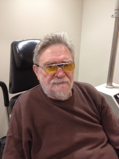
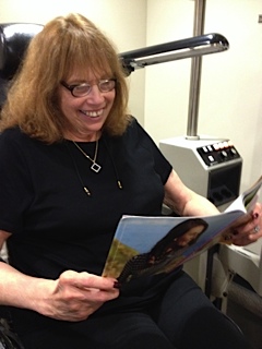
  6. Vision loss does not have to mean discontinuing everything you did before. It might just mean a different way of doing things. It is first important to write a list of things you would like to be able to do. The top three are usually: reading, driving, and watching TV. Other wish list items will include sewing, playing bridge, cooking, etc. Once you write this list, you can then track down the resources that will help you fulfill this.

    For example, while a handful of low vision patients may still be able to continue driving (to be determined by trained specialists), others will need transportation resources such as Access, talking devices, magnification devices or software, books on tape, etc.

  7. Low vision devices and vision rehabilitation
  8. Some of these devices and/or low vision therapy can help you fulfill your wish list. These include simple hand-held lighted magnifiers to more sophisticated assistive-technology devices such as a CCTV, and prescription telescopes. A low vision specialist can help determine if you are a candidate for low vision therapy, or if one of these devices would be appropriate and what your insurance would cover.

  9. Community resources and volunteering
  10. Support groups, such as the Macular Degeneration Partnership, can be a helpful resource to share and gain experiences from others who are coping with the same condition. One of my patients, Bonnie D., started a support group at her church, when she realized that several others also had AMD. It was successful, and they met once a month, learning from each other.

    Some people also find rewarding experiences in volunteering to help others. One patient goes to a nursing home via public transportation twice a month to sit with those who are bedridden and accompany them. She said it helps her to forget her own difficulties and to be more optimistic when she sees there are others who need more help.

  11. Incorporate technology resources
  12. As Jim V. writes “For me, a major challenge has been to overcome resistance to using electronic devices but now that I am freeing myself from that frustrating challenge, I am learning about many opportunities available.” Jim is now an avid iPad user since the font size can be easily increased and the user can switch to voice-over to read what is on the screen. He was able to receive technology training at a local center for the blind and partially sighted. He has discovered magnification software, apps for iPhone an iPad such as ZoomReader.

These are examples of how quality of life can continue even after vision loss. Life can still be enriching, engaging, and enterprising. It all depends on you.

List of Resources:

Braille Institute
Classes, training, on-site demonstrations of assistive-technology
www.brailleinstitute.org

Access Transportation Services
www.accessla.org

Computer Screen Readers
This is a free screen reader for the blind and visually impaired.
www.nvaccess.org

AI Squared
Computer software and other products that magnify and/or read the screen: Zoom Text, Zoom Reader for iPhones
www.aisquared.com

Support Group
Macular Degeneration Partnership
AMD.org

6/25/15

Dr. Limtiaco - living with vision lossLisa Limtiaco, OD
Dr. Richlin, OD & Associates
Beverly Hills, CA

Vision Recap Of Previous Articles of Interest

Besides the comments that we get, one of the best parts of putting together this blog is the wonderful group of guests who share their expertise and personal stories. I want to thank all of the eye care professionals and friends that have contributed to make this blog a success.
Vision Recap
Here is a quick vision recap of some of the articles we had in the past that you may have missed.

Jullia A. Rosdahl, MD, PhDCoffee and Glaucoma and Taking Control of Glaucoma

David Liao, MD, PhDWhat Are A Macular Pucker and Macular Hole?

Kooshay MalekBeing A Blind Artist

Dan Roberts15 Things Doctors Might Like Us To Know

Jennifer VilleneuveLiving With KC Isn’t Easy

Daniel D. Esmaili, MDPosterior Vitreous Detachment

Donna ColeLiving With Dry Age-Related Macular Degeneration

Pouya N. Dayani, MDDiabetes And The Potential For Diabetic Retinopathy

Robin Heinz BratslavskyAdjustments Can Help With Depression

Judith DelgadoDrugs to Treat Dry AMD and Inflammation

Kate StreitHadley’s Online Education for the Blind and Visually Impaired

Catherine Warren, RNCan Keratoconus Progression Be Predicted?

Richard H. Roe, MD, MHSUveitis Explained

Sumit (Sam) Garg, MDCataract Surgery and Keratoconus

Howard J. Kaplan, MDSpotlight Text – A New Way to Read

Gerry TrickleImagination and KC

In addition to the topics above, here are few more articles that cover a variety of vision issues:

If you have any topics that you would like to read about, please let us know in the comments section below.

6/23/15


Susan DeRemerSusan DeRemer, CFRE
Vice President of Development
Discovery Eye Foundation

When Is The Best Time For Cataract Surgery?

As you age, cataracts become a concern prompting the question – when is the best time for cataract surgery?

There are decades worth of old wives tales floating around regarding cataracts that often lead to unnecessary fear and apprehension for many patients. These myths involve concepts such as “ripeness”, having to wear eye patches afterwards, danger in “waiting too long, etc. Just as the techniques of cataract extraction have changed over the decades, so have the indications to proceed to surgery.
best time for cataract surgery - people
Firstly, cataracts are a normal part of the aging process. Patients should not be alarmed if they are told that they are developing cataracts, even as early as their fifties. As we age, the natural clear lens inside the eye becomes progressively harder, darker, and cloudier. This dark, cloudy lens is what is referred to as a cataract. Cataracts develop at different rates for different people, and even between the two eyes of the same person. It typically takes many years for the lens to become cloudy enough to impact the clarity of vision. There are many different types of cataracts depending of what area of the lens becomes cloudy, but the typical cataract related to normal aging results in a relatively uniform cloudiness with a denser central core, and is referred to as “Nuclear Sclerosis”. Other varieties of cataracts tend to grow more quickly, are relatively uncommon, and often result from certain conditions other than typical aging.
best time for cataract surgery
Regardless of what type of cataract the patient has, the treatment is the same: cataract extraction with an implant of an intraocular lens. There have been great advances in lens design over the years, and they now result in excellent, stable, predictable vision for the remainder of the patient’s lifetime and do not typically need to be changed once implanted.

Cataracts result in different symptoms that may be more of less relevant to a specific person’s needs, such as:

  • Glare with bright lights
  • Difficulty with fine print
  • Difficulty following the golf or tennis ball
  • Impairment in night driving
  • Difficulty with seeing street signs
  • Seeing the score or small print on the television
  • Fine visual tasks such as threading a needle, etc.

Although cataract surgery is an incredibly successful procedure with only about a 1-2% risk of complications, it still DOES have some risk. Therefore, cataract surgery should only be undertaken when there is something to gain. In other words, the BENEFITS MUST OUTWEIGH THE RISKS. This means that if your symptoms are mild and are not interfering with your activities of daily living, it is not time to accept the risks of surgery. Once your visual impairment progresses to the point that YOU feel your activities of daily living and enjoyment are impaired, this is the time to proceed to surgery. This threshold is very different between people. Some people feel impaired with vision of 20/25, and others still function within their scope of usual activities until they are 20/100! The best first-step in determining if it is time for your surgery is to get an up-to date refraction. This means a detailed check for new glasses. Often, cataract development will change a person’s glasses prescription, and updating this can improve the visual symptoms for months to years. When a new glasses prescription no longer improves the sight adequately, this is when surgery is indicated.

For the most part, putting off cataract surgery does not impact the final outcome. It will not harm you or your eye to leave the cataract alone until you are ready. There are of course certain exceptions to this rule, such as in Fuchs’ dystrophy, pseudoexfolation, untreated narrow-angle glaucoma, and some others. However, these are relatively rare conditions that your doctor will speak to you about if you have any of these diagnoses.

In summary, the time to proceed to cataract surgery is something that you as the patient determine. YOU assess your lifestyle needs and your vision performance within your scope of activities. When you feel you are impaired in these activities, the benefits will outweigh the risks, and it’s time to take them out. You should not feel any pressure to urgency in this process.

Once you have determined you are ready to have cataract surgery, your surgeon will discuss with you your options for intraocular lens implantation including astigmatism neutralizing lenses, standard distance or near-vision lenses, multiple focal distance lenses, accommodating lenses, and others. The current standard approach for cataract surgery is called “phacoemulsification” and uses ultrasound technology to remove the cataract. There are also laser devices that assist in making the incisions and breaking up the lens, which many surgeons now employ in addition to the phacoemulsification. In general cataract surgery only takes a few minutes, is performed with topical anesthesia, is pain-free, and has a very short recovery time. No pirate-patches are used these days! Most patients are very happy with the results, but this requires adequate discussion with the surgeon prior to the procedure to best assess the needs of the individual patient. A well- informed patient who participates in their care results in the best outcomes!

6/18/15

Sameh Mosaed, MD best time for cataract surgerySameh Mosaed, MD
Director of Glaucoma Services, Gavin Herbert Eye Institute, UC Irvine
Associate Professor, Cataract and Glaucoma Surgery, UC Irvine School of Medicine

Recognising Vision Problems in Children

Children typically have no idea when they have vision problems. Vision problems typically onset gradually, and children tend to believe that everyone sees similar to them, even when they have vision problems. As a result, you will rarely hear a child say any of the following: “My vision doesn’t seem to be as clear as it used to be.” “I started seeing double when reading two weeks ago.” “My eyes are feel uncomfortable when I read.”
vision problems in children
It has been estimated that 80% of what we know is learned through vision. Therefore, children with vision problems may struggle at school simply due to correctable vision issues. Because children rarely complain of vision problems, and vision problems can lead to learning difficulties, it is recommended by the American Optometric Association that children undergo annual comprehensive eye examinations if they are at risk for visual impairment. To determine if your child may be at risk, please go here.

Seeing Vision Problems in Children

Because children don’t often tell you that they have problems with their eyes, there are some signs for which you should watch. The most common vision-related problems differ by age. Beginning with the youngest children, we will provide things to consider that may portend vision issues. Infants’s eyes frequently wander because they have not yet developed the ability to control their eye movements at all times. One eye occasionally pointing in or out is common and should not be considered a problem unless the child continues to do this after eight months of age, unless it is always the same eye, or unless it is constant. A visit to the optometrist is warranted if any of those conditions exist. If the child’s eye crosses most or all of the time, then the child may have congenital esotropia, and surgery in combination with vision therapy may be necessary to correct the problem.

If the child’s eyes begin to cross frequently at around age two years, especially when the child looks at close objects, then the most likely problem is accommodative esotropia. If this is the case, wearing glasses will help to eliminate most or all of the crossed eyes.
vision problems in children
Nearsightedness (myopia) is the most common vision problem in children. It typically onsets between the ages of 8 and 10 years, although that may vary dramatically. Nearsighted children can see clearly at near, but have difficulty seeing distant objects. These children tend to squint when looking at distance objects, and may complain of headaches around their eyes, as a result of squinting. Nearsighted children are frequently detected during school vision screenings or recognized by teachers, and glasses or contact lenses can easily clear up their vision and reduce symptoms.

It is also important to remember that even children who can see details far away may have vision problems. Farsighted children may be able to read distance vision charts, but their eyes must strain to do so. Their eyes must strain even more to see up close. While a child may pass a vision screening at school by reading the bottom line on the chart across the room, he or she may struggle visually. These children often complain of headaches during the week, but less often during the weekend. They may fall asleep after a very short period of reading or struggle to maintain attention because their eyes become strained. Their headaches are typically over the eyes or on the side of the head, and they rarely wake up with a headache. They frequently try to avoid reading because it makes their eyes uncomfortable, although they rarely say that.

Another problem with similar symptoms is convergence insufficiency, which means that children have difficulty bringing their eyes toward the nose when looking at close objects. The symptoms of convergence insufficiency have frequently been misdiagnosed as attention deficit disorder because children find it difficult to attend to any single near task for even a relatively short period of time. Headaches, eye strain, double vision, and tiredness while reading are some of the main symptoms that children will exhibit. Vision therapy frequently reduces or eliminates the symptoms related to convergence insufficiency, and may improve a child’s attention during learning-related tasks.

Teachers are commonly the first people to suspect a vision problem in children because of the demanding visual environment at school. Children rarely complain at home, although homework time is an excellent time to watch your child for potential vision problems. Children with vision problems may rub their eyes frequently, fall asleep after reading for five minutes, perform better when narrating than when writing, cover an eye or close an eye when reading, or simply refuse to read. If your child exhibits these symptoms, then a visit to the eye doctor is warranted and may result in improved attitude and aptitude.

6/11/15

Jeffrey Walline - vision problems in childrenJeffrey J. Walline, OD, PhD
The Ohio State University
Associate Professor
Chair, Research and Graduate Studies

Cataract Prevention

The more you know about cataracts, the easier it is to focus on cataract prevention.

What is a cataract?

At birth, with rare exceptions, most of us arrive in the world with a clear crystalline lens within each eye. The pathway of our visual images start with light passing through the cornea (the clear front window of the eye), through the pupil (the opening in the center of the iris, or colored portion of the eye) and through crystalline lens which functions to focus light onto the center of the retina (the film of the eye). cataract preventionThe retina, via the optic nerve, will then transmit visual images to the brain. When the crystalline lens becomes opacified (cloudy), this system becomes disrupted, and vision becomes impaired. Opacification of the crystalline lens is called “cataract”, and there are many variations in appearance and type and many causes and can present at any age. The word cataract originates from the Greek word “cataracta”, which means waterfall. The ancient Greeks used this term as they noticed a similarity in the appearance of the white opaque rushing water of a waterfall and the appearance of a white mature cataract.

To understand the different types of cataracts and causes, it is important to understand the anatomy of the lens. Using a metaphor, the lens anatomy can be compared to a Peanut M&M candy™. There is an outer candy coating (the lens capsule), a chocolate layer inside (the lens cortex), and a peanut in the center (the lens nucleus).

The most common cause of a cataract is an age related nuclear clouding which is due to long term accumulation of metabolic and oxidative waste products within the lens and possibly UV-B/Sunlight light exposure. Cortical clouding (within the cortex of the lens), due to similar causes, is also a common cause of an age related cataract.

Cataracts can occur earlier in life with poorly controlled diabetes resulting in cortical and nuclear cataract. Patients who are exposed to steroid medications in any form (orally, topically as eye drops, skin creams etc.) are at an increased risk to develop a posterior subcapsular (PSC) cataract which occurs on the posterior lens capsule. PSC cataracts can have a much more abrupt and earlier onset in life than nuclear or cortical cataract. Smoking has also been known to predispose patients to formation of a PSC cataract. Other less common varieties of cataract can occur with any trauma to the eye or even present at birth as a congenital cataract with a large variety of causes.

What can be done to prevent cataracts?

I often joke with patients that a cataract is such a common occurrence that just like birth, death, and taxes, it is an issue we must all face at some juncture in life (hopefully later than earlier). I am often asked if there are any dietary measures or vitamin supplementation to reduce the formation of a cataract, however this is not as well studied as the use of vitamins in the prevention of macular degeneration. Several scientific epidemiological studies following populations over many decades have shown some merit however that using multivitamins regularly (Vitamin B6 and B12, Vitamin C, beta carotene, antioxidants and possibly lutein and zeaxathin) can reduce the degree of lens opacification over time. As with all medications, you should consult with your physician before deciding to use any vitamin supplementation to clarify if you have any contraindication to using them.

There is conflicting evidence regarding the role of UV-B exposure in sunlight as a causative agent for cataracts. There is some support that using sunglasses on a regular basis to block UV-B light may help to reduce cortical cataract formation. Smoking cessation can also help to reduce the formation of cataract. If a patient is diabetic, strict blood sugar control is also an important measure to reduce the formation of a cataract. If possible, reducing or avoiding the use of steroid medication can reduce the formation of a PSC cataract.

What can be done if a cataract is worsenening and glasses cannot help improve vision significantly?

If you are experiencing gradual painless loss of vision, you should consult with your ophthalmologist as cataract can be a common cause. If you are found to have cataract formation, there is generally a shift in the glasses prescription in the early stage. Having your glasses prescription checked to see if your vision can be improved with glasses is the first step in determining how significant your cataract has become. If glasses are not able to sufficiently improve your vision and your daily activities are affected by the decrease in vision your experience, you may be a candidate to have cataract surgery.

Modern cataract surgery has improved a tremendous degree compared to decades earlier. It is the most common and successful surgery in the world, and is typically performed on an outpatient basis with topical anesthetic and often without any sutures or eye patch. Prior to surgery the pupil is dilated, and once in the operating room, a small self-sealing incision is made on the side of the cornea. The surgeon then makes a circular opening in the anterior lens capsule (the candy coating of the peanut M&M), and uses an ultrasound instrument to emulsify and vacuum out the nucleus (the central peanut), and remove the cortex (the chocolate layer). The inside of the lens capsule is polished and an intraocular lens is folded and introduced into the eye through the corneal incision and seated into the remaining lens capsule to conclude the surgery.

Prior to surgery, measurements are taken to determine the power of lens necessary to achieve the best vision after surgery based on the curvature of the cornea and anterior-posterior length of the eye. Intraocular lenses (IOLs) can potentially have several features depending on a patient’s needs. The most common IOL used is a monofocal lens, which does not typically require an additional out of pocket expense. This lens is chosen to have a point of focus either for distance vision (driving, TV) or near vision (reading), but not both. Typically patients who have the monofocal lens will choose to have distance focus and use reading glasses for near vision. There are multifocal/accommodating IOLs available for patients who are appropriate candidates, to allow the patient a larger range of vision at far, near and intermediate (computer) distance and may allow great independence from glasses. There are still other IOLs which can correct astigmatism (a special type of glasses prescription) at the time of cataract surgery. After discussion of the patient’s needs and preferences, the surgeon can best advise their patient regarding which type of IOL may best suit them.

6/11/15

Anand Bhatt, MD - cataract preventionAnand B. Bhatt, MD
Assistant Professor of Glaucoma and Cataract Surgery, Gavin Herbert Eye Institute
UC Irvine School of Medicine

Fuchs’ Dystrophy: Current Insights

What is Fuchs’ Dystrophy?

Corneal dystrophies are a debilitating group of progressive diseases that can ultimately deprive a person of sight. The cornea, which forms the front of the eye, is a window for vision, and dystrophies due to intrinsic defects in the corneal tissue cause this window to become opaque and hazy. Fuchs’ dystrophy, also known as Fuchs’ corneal endothelial dystrophy (FCED), is amongst the most commonly diagnosed corneal dystrophies requiring corneal transplantation. The ophthalmologist Ernest Fuchs first described the disease in 1910.

Who gets it?

The disease is rare, and it is difficult to predict who will get it. We know that it affects women more than men (3:1 ratio), older adults (older than 50 years of age), and those with a family history. There are forms in which there could be up to a 50% chance of transmission to children of parents with Fuchs’ dystrophy. Most cases, however, occur sporadically.

What causes it and how does it progress?

Although the cause of Fuchs’ dystrophy is still being studied, there are characteristic findings associated with it: small outgrowths on Descemet’s membrane called “guttae” or “guttata”, thickening of Descemet’s membrane, and defects in the endothelial cells (Figure 1).

fuchs dystrophy 1
Figure 1: Fuchs’ dystrophy can affect all layers of the cornea. Layers of the cornea from anterior to posterior, or frontside to backside, include (A) epithelial cells where blisters and bullae may form in late-stage disease, (B) Bowman’s layer where scarring can occur in late-stage disease, (C) stroma where corneal swelling occurs early in disease, (D) Descemet’s membrane where guttae form (arrows) and thickening occurs, and (E) endothelial cells that decrease in number and change shape and size with disease progression.

Descemet’s membrane is a thin corneal layer between the endothelial cell and the stromal layers of the cornea. Endothelial cells make up the backside of the cornea and function as a barrier and pump for keeping fluid out of the cornea and maintaining corneal clarity. As guttae accumulate on Descemet’s membrane, patients experience progressive loss and change in endothelial cells. Dysfunction of endothelial cells causes corneal swelling, which distorts vision. First, the back of the cornea swells, and eventually, swelling can reach the epithelial cells at the front of the cornea. Swelling can range from mild moisture accumulation, to painful “bullae”, or blisters. In very late-stage disease, significant corneal scar tissue can form and dramatically reduce vision. The progression to late stage Fuchs’ varies from person to person, but usually takes a couple of decades.

What are signs and symptoms?

A patient may be asymptomatic for years despite having guttae. Initial symptoms, including blurry, hazy, or cloudy vision, are typically due to corneal swelling from dysfunction of the endothelial cell layer. Patients may also experience glare or halos around light in the early stages just from the density of guttae. New studies suggest that patients can get glare and higher order aberrations from guttae without any corneal swelling. Symptoms tend to be worse on awakening, but usually improve throughout the day. This is because the closure of eyelids during sleep results in the accumulation of fluid in the cornea. For the same reason, humid weather can also worsen symptoms. As the disease progresses, poor vision may last longer into the day. There may be associated pain if blisters develop.

How is it diagnosed?

The presence of any of the above signs and symptoms, especially with a family history of Fuchs’, should prompt a consult with an ophthalmologist who will diagnose the disorder and follow its progression with regular checkups. An ophthalmologist will conduct a microscopic slit-lamp examination of the eyes, looking for guttae and Descemet’s membrane thickening (Figure 2).

fuchs dystrophy 2
Figure 2: Slit-lamp examination showing speckling pattern on the backside of the cornea characteristic of guttae in Fuchs’ dystrophy.

Special tests may be done to measure corneal thickness, a marker of swelling, or count endothelial cells to track disease progression (Figure 3 and 4).

fuchs dystrophy 3
Figure 3: Optical Coherence Tomography (OCT) showing (A) a normal, healthy cornea and (B) corneal swelling typical in Fuchs’ dystrophy.
fuchs dystrophy 4
Figure 4: In-vivo slit-lamp scanning confocal microscopy showing (A) normal endothelial cells and (B) guttae causing endothelial cell loss and change in Fuchs’ dystrophy.

How is it managed?

Management can be medical or surgical depending on symptoms. Patients may have mild or slow progression of disease that can be managed medically including over the counter salt solution drops (5% NaCl) to reduce corneal edema.

When there is late-stage disease, a corneal transplant may be necessary to improve vision. A corneal transplant replaces the patient’s corneal tissue with human donor corneal tissue. Donor corneas are readily available via excellent eye banks throughout the United States. The surgery is outpatient surgery with regular follow-up appointments and suture removal during the subsequent months. The postoperative healing of the cornea and vision stabilization can take up to a year.

Great strides have been made in the last decade in corneal transplantation surgery, giving patients better treatment options. Patients used to be limited to penetrating keratoplasty (PK), a full-thickness replacement of the cornea. We now have newer surgeries known as endothelial keratoplasty (EK), which is a partial-thickness transplant that replaces only the damaged part of the cornea (the endothelial layer). The different types of EK are DSEK (Descemet’s-Stripping Endothelial Keratoplasty) and DMEK (Descemet’s Membrane Endothelial Keratoplasty). The techniques vary by thickness of the transplanted tissue. The type of EK most appropriate is determined by the corneal surgeon and is variable on a case to case basis. Both types of EK surgeries provide comparable long-term visual results. In both surgeries, the patient’s diseased Descemet’s membrane and endothelial cells are stripped from the inner layer of their cornea. The thin lamellar donor graft is then inserted into the eye and positioned onto the back of the patient’s cornea via a gas or air bubble. The patient is then instructed to lie in a face up position for several hours post surgery during which time the bubble supports the graft until the new endothelial cell pumps begin to wake up and naturally adhere to the back side of the recipient cornea. Occasionally, the doctor may replace another air bubble into the eye the next day to allow more time for the graft to adhere. Visual recovery is on the order of 1-2 weeks in DMEK and 2-3 months in DSEK surgery. Rejection risk is still a possibility in EK surgery but has a much lower rate than traditional full thickness PK surgery.

Other surgical considerations depend on the presence of cataracts. Cataract surgery can worsen Fuchs’ dystrophy because of damage to the endothelial cell layer. For this reason, patients with cataracts and Fuchs’ requiring surgical intervention are often recommended to undergo cataract surgery before or at the same time as corneal transplantation to ensure the best outcome for the transplant.

Patients should work with an ophthalmologist to determine the best management plan. Ultimately, vast improvements in treatment options have given many Fuchs’ dystrophy patients the exciting opportunity to regain vision with improved healing times and reduced infection and rejection of the graft.

Citations: Figure 2 and 4 are from Zhang J, Patel DV. The pathophysiology of Fuchs’ endothelial dystrophy—a review of molecular and cellular insights. Exp Eye Res. 2015 Jan

6/4/15

priscilla-thumbnailPriscilla Q. Vu, MS
Medical Student
University of California, Irvine School of Medicine


Farid 3.6.14Marjan Farid, MD
Director of Cornea, Cataract, and Refractive Surgery
Vice-Chair of Ophthalmic Faculty
Director of the Cornea Fellowship Program
Associate Professor of Ophthalmology
Gavin Herbert Eye Institute, University of California, Irvine

9 Ways To Relieve Cataract Surgery Stress

It’s an extremely rare person who would not feel nervous before surgery of any kind, even if it’s an outpatient procedure that will only take a few minutes. In the case of cataract surgery, the fear can be even worse than the procedure itself.


9 Ways to Relieve Cataract Surgery Stress
People who are under intense stress can suffer a range of symptoms, including irregular or racing heartbeat, nausea, upset stomach, difficulty breathing, and an inability to sleep. It can even affect your mind, causing you to forget important details about the operation, like advice on how to get ready or what to do after you come home from surgery.

Here are 9 ways to relieve cataract surgery stress:

1.  Just Think About It – Let’s start with the first, and most difficult, suggestion—change your own mind about how you feel. Admittedly, it takes a great deal of discipline that’s hard to muster in the face of great anxiety, but try to remind yourself how your sight will be saved after a relatively short, quick, and easy procedure. What you are about to go through will prevent you from going blind.


2.  Learn Everything You Can – For many, it helps to learn as much as possible about the surgery before it happens. Knowing exactly what’s going to happen and how others have dealt with what you’re going through can be a great relief. Knowledge may be all you need to relieve your anxiety.


3.  Talk to Your Surgeon – It almost always helps to just talk to someone, and who better than your surgeon? Who else knows every detail of the procedure you’re about to undergo? It’s fairly likely that your surgeon has performed many successful surgeries of this kind before, and he or she may have some stories of encouragement for you, as well as important and comforting knowledge of his or her own personal experiences.


4.  Imagine the End Result – It may help if you keep focused on what happens after, as if it’s already done and you can go home, the procedure over. This takes a great deal of imagination, rather than the discipline of thinking rationally about it, but if you have that level of imagination, it’s certainly worth trying.


5.  Alternate Methods – This encompasses a whole range of stress-reducing tactics that are not usually under the medical umbrella. Nevertheless, they have done a great many people a lot of good. Yoga, hypnosis, massage, acupuncture, acupressure, and other treatments have allowed those suffering preoperative stress to sleep better at the least.


6.  Herbal Supplements – A form of alternative treatment involving traditional ingredients to produce a more restful state. The herbs are often just infused into tea and drunk. While these supplements are generally called “all natural,” you should always consult your doctor before taking them, since they can have an effect on other medications you may be taking, including the anesthetic you are given before surgery.


7.  Have Some Fun – Do something fun to take your mind off of what’s about to happen. Whatever that specific thing may be is up to you, since an individual’s idea of fun differs from person to person. Whatever you generally do to take your mind off of things and unwind after a hard day may be just the thing to help you out before undergoing cataract surgery.


8.  Treat Yourself – In the same vein has doing something fun, do something that usually relaxes you. If you like going for long walks, do that the day before the surgery. Or listen to music that fills you with peace (or joy). Take a long bath. If it makes you feel relaxed or calm, it will help you deal with your anxiety.


9.  Distract Yourself – Once you get to the hospital, you’ll probably be waiting around, even if you get there right on time, giving plenty of time for stress to ramp up. You’ll do better if you keep yourself entertained, but the hospital waiting room is probably one of the more boring places on earth. Fix that by bringing something along to entertain yourself, like a book or some magazines, or even stream your favorite movie or podcast. You’ll probably want to bring more than one thing to do, in case the wait is long.


Stress adds complication to the body’s systems, and can therefore cause some complication in the upcoming surgery. Do what you can, whether you kick back with friends or take some herbal supplements, to help yourself get into the best mind space possible. Think about the positive outcomes, and you’ll do well.