Best’s Disease

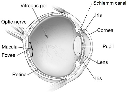
In 1905, Friedrich Best presented a detailed pedigree of an inherited retinal condition referred to as vitelliform dystrophy, or Best’s disease. Best’s disease is an inherited dystrophy of the macula that primarily involves cells known as retinal pigment epithelium (RPE).

best's disease
Friedrich Best

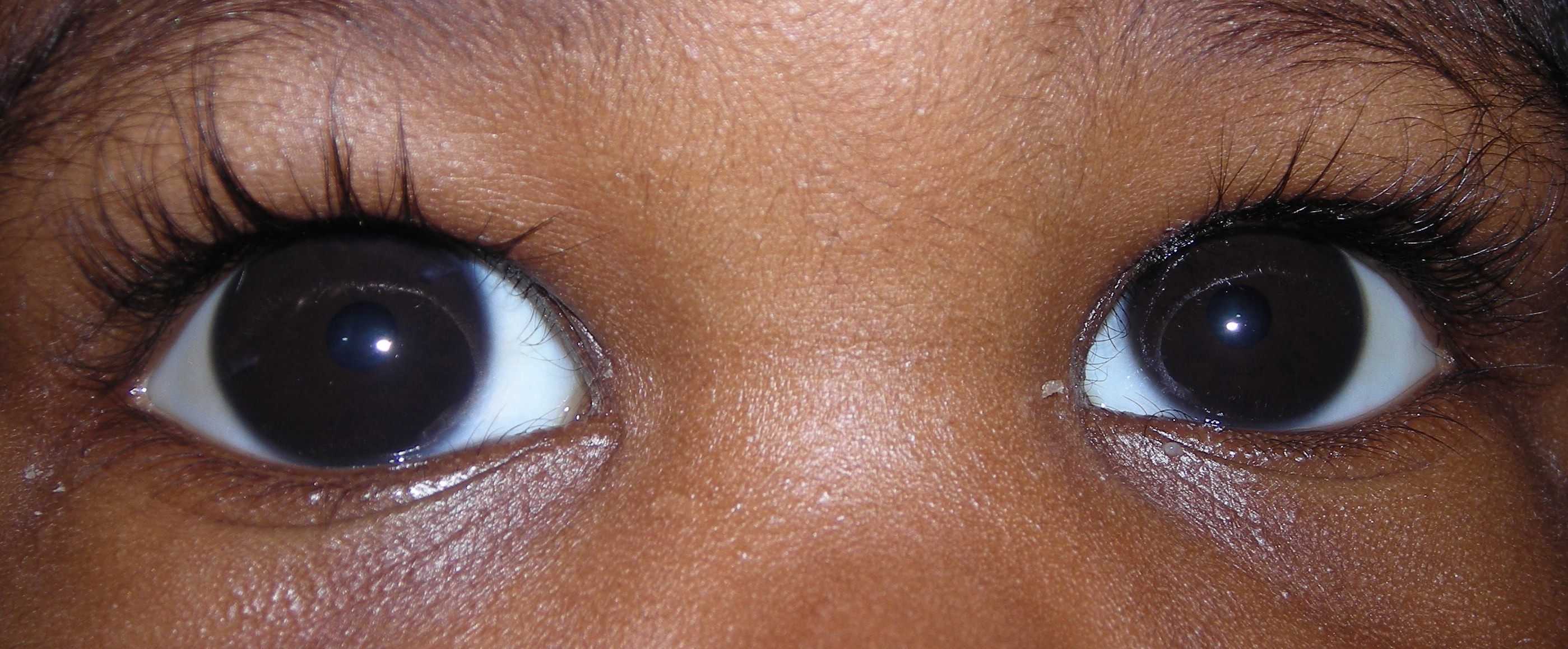
Best’s typically affects both eyes and presents itself either in childhood or early adulthood. Visual acuity is usually minimally affected early on in the course. As the condition progresses, the vision can slowly begin to deteriorate. The rate of progression or the overall amount of progression is difficult to predict. The rate of progression may be also be asymmetric, with one eye progressing at a different rate than the other. Some patients may notice the development of scotoma, or “blind spot”, in their central vision as the condition progresses. Other patients may not progress to later stages or experience vision loss. Loss of peripheral, or side vision, is not expected with Best’s.

Best's disease
Best’s disease
The diagnosis of Best’s disease is primarily based on a careful clinical exam. Taking a careful family history is also important as Best’s is typically inherited in an autosomal dominant pattern. This means that an affected individual has a 50 percent chance of passing the gene to their offspring. It should be noted though that there is highly variable expression, which means there may be some affected individuals in whom the changes are so mild that they never notice any visual disturbance. The causative gene is located on chromosome 11 and has been labeled BEST1 (VMD2). This encodes for a protein known as bestrophin 1, which is located on the membrane of RPE cells. It is believed that this protein is involved in RPE metabolism through its control of chloride channels, although the details are still being elucidated. Thus far, there have been over 200 mutations of the BEST1 gene that have been described.

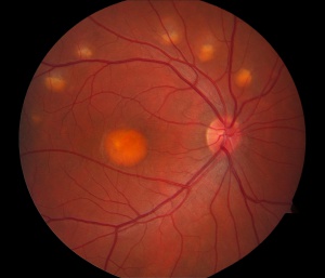
The classic exam finding in Best’s is a circular yellow lesion in the macula. This lesion resembles an “egg-yolk”, and is often referred to as such by ophthalmologists. As the condition progresses, the yellow material begins to break up and the pigmentation of the macula attains a more mottled appearance. This is often referred to as a “scrambled egg” appearance. After many years, there may be evidence of cell loss in the macula, which can negatively impact the visual acuity. In a relatively small proportion of cases, a complication can occur in which abnormal blood vessels grow underneath the macula and begin to leak fluid and/or blood into the macula. This is known as choroidal neovascularization (CNV), and can be vision threatening. Fortunately , CNV can be treated effectively with medications that are injected into the eye as part of a straightforward and low-risk office procedure. Typical signs of CNV would include distortion or blurring of the vision, and it is important to notify your doctor of any sudden changes in vision.

Diagnostic testing is sometimes used to confirm the diagnosis. The electro-oculogram (EOG) is universally abnormal in Best’s, and can be a valuable confirmatory test. Fluorescein angiography and optical coherence tomography can be valuable tests to better evaluate the macula and to also look for the development of CNV. Genetic testing for Best’s is now possible as well.

There is no established medical or surgical management for Best’s disease. In patients who develop CNV as a secondary complication, existing treatment options are effective. Future avenues of therapy hold significant promise, but are in their early stages of development. Stem cell based therapies, for example, have the potential to help restore healthy cells that may have been lost during the disease progression.

10/29/15

Dr. Esmaili posterior vitreous detachmentDaniel D. Esmaili, MD
Retina Vitreous Associates Medical Group

General Differences Between Polarized and Absorptive Lenses

Polarized and Absorptive Lenses

Polarized and Absorptive Lenses
Polarized lenses can be helpful in reducing glare; in fact, they were first developed to help with glare from outdoor sports and activities. Here is a passage from All About Vision that explains the basics of polarized lenses very well.

Light reflected from surfaces such as a flat road or smooth water generally is horizontally polarized. This means that, instead of light being scattered in all directions in more usual ways, reflected light generally travels in a more horizontally oriented direction. This creates an annoying and sometimes dangerous intensity of light that we experience as glare. Polarized lenses contain a special filter that blocks this type of intense reflected light, reducing glare.

Though polarized sunglasses improve comfort and visibility, you will encounter some instances when these lenses may not be advisable. One example is downhill skiing, where you don’t want to block light reflecting off icy patches because this alerts skiers to hazards they are approaching. In addition, polarized lenses may reduce the visibility of images produced by liquid crystal displays (LCDs) or light-emitting diode displays (LEDs) found on the dashboards of some cars or in other places such as the digital screens on automatic teller machines and self-service gas pumps. With polarized lenses, you also may be unable to see your cell phone or GPS device.

Boaters and pilots also have reported similar problems when viewing LCD displays on instrument panels, which can be a crucial issue when it comes to making split-second decisions based strictly on information displayed on a panel. (Some manufacturers of these devices have changed their products to solve the problem, but many have not yet done so.) Many polarized lenses are available in combination with other features that can enhance outdoor experiences.

Absorptive Sunlenses/Sunglasses do a little more than just reduce glare.

These are special wraparound sunglasses that filter out ultraviolet (UV) and infrared (IR) light. I explained those two types of light in my post. In addition to reducing glare, they can also increase contrast, which is important for visibility.

They also come in a variety of tints: dark gray-green, medium amber, medium gray, medium plum, yellow, orange, amber, and light orange. Many of the available tints/colors also have a percentage sign. The percentage sign represents the amount/percent of visible light that is transmitted through the lens. Here are some examples:

  • 32% medium gray
  • 10% medium amber
  • 2% dark gray-green
  • 20% medium plum
  • 65% yellow
  • 49% orange
  • 16% amber
  • 52% light orange

It is the tint – in combination with the amount of light transmission of each tint – that is helpful for people with glare issues. There are a few manufacturer websites that explain the range of absorptive lenses very well.

The first is NoIR Medical Technologies (NoIR stands for “No Infra-red” light.) You’ll see that there are different colors and tints, and many of the colors also have a percentage sign. The percentage sign represents the amount/percent of visible light transmitted through the lens.

Generally, NoIR recommends the following for people with glare problems:

  • 32% Grey
  • 13% Dark Grey
  • 18% Grey
  • 40% Grey-Green
  • 20% Plum
  • 16% Amber
  • 10% Amber
  • 54% Yellow

You can see from the list that the color does not have to be extremely dark for the lenses to reduce glare and light sensitivity.

Also, Eschenback Optik provides a good overview of Solar Shields, another type of absorptive lens product.

Most styles of absorptive lenses also can be fitted over prescription lenses. The bottom line is that it’s probably necessary to visit an office that carries a supply of these lenses and determine which color, tint, and percentage of light transmission is right for your wife. It’s helpful to compare several styles to determine what tint and percentage of light transmission work best.

10/27/15


Maureen Duffy, CVRTMaureen A. Duffy, CVRT, LVT
Social Media Specialist, visionaware.org
Associate Editor, Journal of Visual Impairment & Blindness
Adjunct Faculty, Salus University/College of Education and Rehabilitation

Carrots For Healthy Eyes

Carrots forHealthy Eyes
Lately we have heard quite a bit about carrots and the positive effects they can have on your vision, such as slowing the progression of age-related macular degeneration (AMD). This is because carrots contain pigments called carotenoids. These pigments also give vegetables their colors, in this case orange. But carrots weren’t always orange.

The beginnings of carrots can be tracked back to the dry, hot lands of Iran and Afghanistan in 3000 BC, when the root vegetables were black, white, red and purple. They were bitter and used as a healing remedy for many illnesses, as well as an aphrodisiac.
carrots for healthy eyes
The vegetable grew in popularity because it was still edible even after months of being stored in a variety of conditions. Carrot seeds were soon picked and sold to neighboring Middle Eastern, African and Asian populations. This is when the crossbreeding started and new types of carrots were created.

Across centuries and continents, the carrot evolved, improving the composition, look, flavor and size. After years of selective breeding, in the 17th century a Dutch yellow carrot was engineered to get rid of the bitterness, increase sweetness and minimalize the wooden core. This appears to be the origin of the orange carrot we enjoy today.

Americans didn’t fully use carrots until after World War I when soldiers returning home told about French and other European cuisine which included the carrot. However, it didn’t really become popular until World War II, when England actively encouraged home growing of carrots while the US was engaged in cultivating “Victory Gardens.”

Today the carrot is found around the world in temperate regions. They have a high nutrition value, presence of ?-carotene, dietary fiber, antioxidants, minerals and ability to be prepared in a wide variety of recipes. They have become a staple in many countries.

Currently, the largest producer and exporter of carrots in the world is China. In 2010, 33.5 million tons of carrots and turnips were produced worldwide, with 15.8 million tons from China, 1.3 million tons each from the US and Russia, 1 million tons from Uzbekistan and less than a million from Poland, the United Kingdom and Ukraine.

Because of the popularity and health benefits of carrots, they are now enjoyed in a variety of ways – beyond the simple salad. Here are some recipes you might find interesting to try:

carrots for healthy eyesCrab Toast with Carrot and Scallion – Forget your traditional bruschetta, wow your guests with the appetizer.


carrots for healthy eyesPotato-Carrot Latkes with Lemon-Raisin Topping – Seems perfect with Hanukkah just around the corner.


carrots for healthy eyesRoasted Carrot, Squash and Sweet Potato Soup – This is a more traditional carrot recipe, it is not that hard to find a carrot soup, but this one also has squash and sweet potatoes which are also eye healthy!


carrots for healthy eyesCarrot Farfalle Pasta with Lemon and Herbs – Not only are carrots good for flavor, but they add a nice color to this pasta that could be the base for any number of pasta dishes.


carrots for healthy eyesCarrot Ginger Layer Cake with Orange Cream Cheese Frosting – Most carrot cakes have no frosting or a traditional cream cheese frosting. The idea of an orange frosting makes this cake special.


carrots for healthy eyesCarrot, Ginger, and Lime Juice – Refreshing and healthy.


10/22/15

Susan DeRemerSusan DeRemer, CFRE
Vice President of Development
Discovery Eye Foundation

Tear Film Health is Essential for People with Keratoconus

People afflicted with keratoconus (KC) are often obligated to wear contact lenses in order to obtain functional vision. Unfortunately, wearing contact lenses can have detrimental effects on the ocular surface and tear film layers over the course of decades, ultimately reducing lens tolerance. Therefore, any intervention prolonging the comfortable wear time of contact lenses should be aggressively pursued. The tear film covers the surface of the eye, provides lubrication and is the primary defense against foreign bodies and infection. Without a robust and healthy tear film, safe and comfortable contact lens wear is not possible. This article will describe the structure of the tear film and review simple remedies that can keep it healthy throughout life.

Tear Film Layers

The tear film is a complex, triple layered structure comprised of mucus, water and oil. The surface of the cornea and conjunctiva contain cells specialized to secrete a sticky mucoid substance. These so called goblet cells produce the mucin layer of the tears, which creates a “Velcro” type interface and allows the overlying watery component to stick to the ocular surface without washing away.

The bulk of the tear film is comprised of the watery, or “aqueous” layer which is secreted primarily by the lacrimal gland. This specialized structure is located near the eyebrow. This gland continuously releases small amounts of watery fluid that also contains enzymes and antibodies to help fight infection and wash away contaminants.

The lipid layer is the final, outermost layer of the tears. If the tear film is the first line of defense for the ocular surface, then the lipid layer is the first line of defense for the entire tear film and the ocular surface combined. Because of that role, it is extremely important and helps stabilize the tear film by preventing evaporation. This thin, lipid based layer is released by the meibomian glands, which are modified sebaceous glands that reside in the upper and lower lids. In each lid there are 20-30 glands. These glands open up onto the lid margin and through the action of a complete blink, release the lipid secretion to ocular surface which gets spread with the upward motion of the upper eyelid.

Each one of these layers contributes to the structure of the tear film, and a problem with any one of these structures (goblet cells, lacrimal gland or meibomian glands) will negatively impact the corresponding tear layer.

Tear Film
Image 1 -Layers of the tear film across the ocular surface & Meibomian glands of the eyelids. (Picture courtesy of TearScience™)

Tear Film Issues

Because the tear film is so thin, each individual component is necessary to maintain the integrity of the tears as a whole. When any layer of the tear film is deficient, the tear film becomes unstable and the ocular surface becomes irritated and can progress to developing classic symptoms of dry eye. This includes burning, stinging, redness, tearing, fatigue and contact lens intolerance.

Deficiencies in the mucin layer are uncommon, and are typically the result of chemical or thermal insult, or scarring. An aqueous deficiency, primarily from a lacrimal gland related etiology, is also relatively uncommon, and can arise from autoimmune and inflammatory causes such as Sjögren’s Syndrome. The most common reason for a poor tear film is linked with excessive evaporation of our tears due to a lack of sufficient lipid secretions from non-functioning or obstructed meibomian glands. It is understood that many factors contribute to why these glands stop performing optimally.

One factor has been linked to our habitual working environments. The compressive force exerted by the muscles of our eyelids that control blinking are essential for lipid secretion. However, the use of computers or wearing contact lenses has been shown to negatively impact our blinking habits, both by reducing the number of blinks and making blinks less complete. With an incomplete blink, the upper and lower lids do not make contact. The negative consequences of this are 1) the meibomian glands do not release their lipid contents, 2) the lower part of the eye is chronically exposed to the air, increasing evaporative stress and 3) dead skin cells accumulate on the lid margin which can clog the meibomian gland openings.

When increased evaporation of the tear film occurs chronically, the integrity of the entire ocular system becomes compromised over time and problems to the health of the eye become permanent attributes. This condition is known as Meibomian Gland Dysfunction or MGD and is linked with 86% of all dry eye sufferers.

Image 2 - Histology slide of a Meibomian gland with a terminal duct blockage
Image 2 – Histology slide of a Meibomian gland with a terminal duct blockage
Contact lenses have been shown in multiple studies to have a negative impact on the integrity of the tear film. To begin with, placement of a lens onto the eye divides the tears into two sections, referred to as the “post” (behind) and “pre” (in front) lens tear films.

The characteristics of the post lens tear film can differ depending on the type of lens that is worn. For example, soft lenses and scleral lenses have very little turnover of this post-lens tear film. This can cause issues related to the build up of toxic waste and bacterial elements that ultimately aggravate the corneal surface. Conversely, rigid gas permeable lenses are designed to have substantial tear turnover behind the contact lens with every blink.

The pre-lens tear film is also greatly affected by the type of lens material, as well as the interaction between the lid and the contact lens surfaces. Eye doctors know that without a healthy tear film, chances for contact lens intolerance increases. The rate of contact lens intolerance substantially increases as patients enter their fourth decade of life, primarily because of MGD caused by years of poor blinking habits.

Tear Film Care

Fortunately, simple interventions can prevent and/or limit the severity of MGD altogether or help to manage it once it occurs. Just like brushing and flossing one’s teeth can prevent gum disease, attention to complete blinking and lid margin hygiene can improve the tear film and prevent contact lens intolerance problems.

Because partial blinking is strongly linked with developing MGD, it is vitally important that the two lids touch when blinking. It is best to practice this several times throughout the day as well as when you are reading or using the computer.

Akin to flossing the teeth, it is also important to clean the lid margins with a Q-tip soaked in saline solution or a bit of mineral oil by gently brushing the Q-tip across the lid margin 10-20 times each night. It is easiest to get the lower lid.

Finally, performing warm compresses daily can provide heat to the Meibomian glands to soften the hardened oil that can plug the meibomian gland ducts. Warm compresses need to be done continuously for at least 10 minutes with consistent heat in order to attain a temperature that is sufficient to melt the oil that clogs the glands. We recommend folding 5-6 small towels or facecloths into a rectangular shape and wrapped together into a circular bundle, similar to the appearance of a cinnamon roll. The towels should be damp and moist, placed in a microwaveable safe dish with a lid and heated for approximately 1 minute and 50 seconds. After removal, wait a minute or two and then proceed to use the outermost cloth and cover the rest. Replace the first cloth after two minutes and grab the next outer most towel from the bundle, continuing this until all towels are used. In this way, the temperature can be adequately maintained for the full 10 minutes. The high temperatures applied to the lid are transferred to the cornea and very often cause temporary deformation, a phenomenon characterized by transient visual blur immediately following compress application. Therefore, it is vitally important, especially for patients with keratoconus, that pressure never be exerted onto the globe of the eye with a compress or massage administered to the lids of closed eyes after a compress.

It is becoming apparent that MGD is developing in patients at earlier ages. Because of this, the condition has likely been present for decades by the time the patient becomes symptomatic. It may take significant time and effort to rehabilitate not only the glands themselves, but also to reduce the resulting inflammation of the ocular surface.

Meibography is the technique used to image Meibomian glands. In chronic cases of MGD, we see abnormal changes to gland structure, in the form of atrophy or loss of gland tissue and/or dilation of glands where obstructed material causes glands to become widened. In severe cases, the prognosis for recovery is guarded.

The visual clarity that contact lenses provide for patients with keratoconus is incredibly important. But the ability to comfortably wear contact lenses is reliant on our body’s ability to provide a sufficiently thick protective tear film. Taking a small amount of time daily to attend to the lipid producing Meibomian glands by proper blinking habits, exfoliation of the lid margin with a Q-tip and warm compresses will help to extend the number of hours, and ultimately the number of years, that contact lenses can be safely and comfortably worn.

10/20/15

tear filmAmy Nau, OD
Korb and Associates, Boston, MA
Contact lens fitting for keratoconus, other ocular surface disorders and dry eye


tear filmDavid Murakami, MPH, OD, FAAO
Tear Science, Inc.
Researcher, Dry Eye

Increased Awareness for Saving Vision

The following is a survey done by Essilor (a French company that produces ophthalmic lenses along with ophthalmic optical equipment) and a large marketing research firm in the UK, YouGov. While the focus in on people living in the UK, the results would probably be similar to the US population. Even with increased access to the Internet, many people are still not aware of the risks associated with eye disease and what they can do to help retain their vision. Increased awareness of informational resources are important for saving vision.
saving vision
There are a number of websites with easy to understand information about taking care of your vision that I have listed under Resources to Help Save Vision at the bottom of this article. And while there are eye diseases that are hereditary, you can slow the onset and progression by making good lifestyle choices about smoking, diet and exercise. Your eye care specialist is also an excellent source of information about what you can to do reduce your risk of vision loss, at any age.

Increased Awareness for Saving Vision

A YouGov poll conducted with Essilor reveals that most Britons are unaware of damage to their eyes by surrounding objects, activities, and devices. This widespread lack of awareness means fewer people seeking methods of prevention and avoidance, and for those that are aware of risks, most are not informed of existing preventative measures.

The poll has shown* that many British people remain uninformed about the various ways in which eyes are damaged by common daily factors, despite evidence that eye health is affected by blue light, UV rays (reflected from common surfaces), diet, obesity, and smoking.
Of the 2,096 people polled, the percentage of respondents aware of the link between known factors affecting and eye health were:

  • Poor diet – 59%
  • Obesity – 35%
  • Smoking tobacco – 36%
  • UV light, not just direct from the sun but reflected off shiny surfaces – 54%
  • Blue light from low energy lightbulbs and electronic screens – 29%

More than one in ten people were completely unaware that any of these factors could affect your eyesight at all.
saving vision
72% of respondents own or wear prescription glasses but only 28% knew that there were lenses available (for both prescription and non-prescription glasses) to protect against some of these factors; specifically, blue light from electronic devices and low energy light bulbs, and UV light from direct sunlight and reflective surfaces.

76% admitted they haven’t heard of E-SPF ratings – the grade given to lenses to show the level of protection they offer against UV.

Just 13% have lenses with protection from direct and reflected UV light, and only 2% have protection from blue light (from screens, devices, and low energy bulbs).

Poll results showed that younger people were most aware of the dangers of UV and blue light, yet least aware of how smoking tobacco and obesity can affect your eye health. Within economic sectors, middle to high income people are more aware of the effects of smoking & obesity on eyesight than those with low income –

  • 39% of people with middle to high income compared to 33% of people with low income are aware of the impact of smoking tobacco.
  • 38% of people with middle to high income compared to 31% of people with low income are aware of the impact of obesity.

Awareness of the impacts of smoking and obesity on eye health is significantly higher in Scotland (47% & 49% respectively) than anywhere else in the UK (35% & 33% in England and 40% & 38% in Wales).
Essilor’s Professional Relations Manager, Andy Hepworth, has commented: “The lack of awareness about these common risks to people’s eyes is concerning. Not only would many more glasses wearers be better protected, but also many people who do not wear glasses would likely take precautions too, if made aware of the dangers and the existence of non-prescription protective lenses.”

To see the full results of the poll, please visit the Essilor website.

For more information on the protection offered from blue light and UV through specialist lens coatings, for both prescriptions and non-prescription glasses, please see here for UV & Blue Light Protection options.

*All figures, unless otherwise stated, are from YouGov Plc. Total sample size was 2,096 adults. Fieldwork was undertaken between 21st and 24th August 2015. The survey was carried out online. The figures have been weighted and are representative of all GB adults (aged 18+).

Resources To Help Save Vision
All About Vision
Macular Degeneration Partnership
National Eye Institute (NEI)
Prevent Blindness

10/16/15


Susan DeRemerSusan DeRemer, CFRE
Vice President of Development
Discovery Eye Foundation

Business Opportunity for Blind Adults

Food for Thought – Business Opportunity for Blind Adults

For those not in the know, the acronym sounds like a popular sandwich. However, for Louisville, KY resident, George Bouquet, The Hadley School for the Blind’s and the National Association of Blind Merchants’ joint“BEPLT” program (Business Enterprise Program Licensee Training) is more like a dream come true.

George Bouquet - Business Opportunity for Blind
George Bouquet

Hadley is the largest provider of distance education for people who are blind and visually impaired worldwide and the BEPLT program is part of the school’s Forsythe Center for Employment (FCE) and Entrepreneurship. Under the Randolph-Sheppard Act, legally blind adults are given first right of refusal on operating state and federal government vending facilities including cafeterias, snack bars, convenience stores, micro markets, and vending machines and rest stop vending areas nationwide. In February 2014, Hadley’s FCE partnered with the National Association of Blind Merchants (NABM) and the National Federation of the Blind Entrepreneurs’ Initiative (NFBEI) to bring the academic portion of training to would-be blind vendors. Individual state Business Enterprise Programs provide the hands-on component of the blind vendor training. Bouquet is Hadley’s first graduate from the school’s new BEPLT program.

Born with both Pierre Robin Syndrome, which often results in a smaller-than-normal lower jaw, a cleft palate, a tongue that falls back in the throat, and difficulty breathing, as well as Stickler Syndrome, which causes hearing loss, eye abnormalities and joint problems, Bouquet has struggled with health issues throughout his 54 years. Although he was born without eye lenses, he was not born blind. Rather, his vision worsened over time. Bouquet worked in several food service positions since high school and had wanted to become a Randolph-Sheppard vendor even before he would have qualified as legally blind!

There are only so many blind vendor licensee training slots available and many more people compete for them than such programs can accommodate. The first time Bouquet applied to receive the training was in February 2014. Unfortunately, he was not accepted into a program. However, he was fortunate to gain some blind vendor experience by working under friends who already held the license. In early 2015, Bouquet’s counselor told him of another opportunity to apply for vendor training. This time he was accepted and Bouquet began Hadley’s BEPLT program in April 2015. Bouquet was so motivated to graduate from the program that he completed approximately two modules (one-lesson online courses) per week. Hadley’s BEPLT students complete a 10-module program and then take their state’s physical training component. After passing both elements, graduates are eligible to bid for the opportunity to become a blind vendor in their community.
George Bouquet - Business Opportunity for Blind
“The [Hadley BEPLT] program offers a lot of useful information. It will really help anyone wanting to undertake vending,” he said. Bouquet then acknowledged that the material about food borne illnesses helped him to realize the tremendous responsibility he would be accepting by running a government food service area. “As a manager, you need to decide what you are willing to delegate to other people,” he added.

For Bouquet, becoming a blind vendor allows him to hire and train his 25 year-old unemployed son, who inherited most of his visual and hearing problems. This training is Bouquet’s first step toward creating a legacy of financial independence.

10/13/15

Sheryl BassSheryl Bass, MA, MSW
The Hadley School for the Blind

Accommodative Esotropia

This article is reprinted with the permission of Dr. Kenneth W. Wright, Medical Director of the Wright Foundation for Pediatric Ophthalmology & Strabismus. Check out his website for information on over 20 pediatric eye disease and conditions.
Accommodative Esotropia

Normal Binocular Vision

Normally, both eyes are aligned on the same visual target and the images from each eye are merged in the brain to form a single three-dimensional image, or binocular vision. The brain’s process of merging or “fusing” images from each eye into one image is called binocular fusion. The perception of three-dimensional depth is called stereoscopic vision. Stereoscopic vision is the highest level of binocular vision and requires intricate processing of information from both eyes. Binocular vision develops during early infancy, and proper alignment during this time is necessary for normal binocular development to occur.

Accommodative Esotropia

Esotropia means one eye is turned in towards the nose, or crossed eyes. Patients with esotropia have one eye aligned on the visual target, but the other eye is turned in towards the nose. When the eyes are crossed only one eye is aligned with the target and the child is forced to use only one eye for vision. Accommodative esotropia is a type of esotropia caused by significant farsightedness (hypermetropia). Most think that farsighted people can see well only in the distance. In children, this is not true. Children have the ability to focus great amounts, so most children can see well for distance and near even without glasses. Focusing (medically termed accommodation) is the process of increasing the lens power of the eye to see clearly. Linked to focusing is the natural reflex of convergence (eyes move in). As one focuses on an approaching near object, the eyes move in to stay on target. This process of focusing and convergence used for near vision is termed the near reflex. Accommodative esotropia occurs because the farsighted child has to over focus to see clearly. When eyes over focus, the natural reflex is for the eyes to cross. You can experience this by trying to see the tip of your nose. When you look at the tip of your nose you have to over focus and consequently your eyes cross. Since more focusing is needed to see near objects, the crossing tends to be greater when looking at close objects.

Accommodative Esotropia eye turns
Right eye turns in because patient is farsighted and not wearing glasses.
Accommodative Esotropia corrected
Eyes are in excellent alignment after prescribing glasses.


The onset of accommodative esotropia is most commonly seen between the ages of 2 and 4; however, even infants can have crossed eyes. This situation is usually first noticed when the child is tired, sick, or looking at an object very near to their face. Some children cross when they are tired and this is because they cannot sustain the effort to keep the eyes straight. The crossing is usually intermittent at first, but can quickly become constant. There may be a parent or a close relative with the same problem; however, in many cases there is no family history of crossed eyes.

During the examination, three important determinations are necessary. The first determination is to make sure the vision is normal in each eye. This is done by assessing the visual behavior of the preverbal child, or simply having the verbal child read the eye chart. Secondly, the amount of crossing is evaluated. This is measured using prisms while the child is viewing an object. Thirdly, the need for glasses is measured, and this requires drops to dilate the pupil and relax the child’s focusing. These drops take approximately 20 to 30 minutes to work and will blur vision for 1 to 3 hours, but the pupil may stay large for much longer. After the eyes have been dilated, the eyeglass prescription is calculated using a special light (retinoscope) along with lenses. Determining the proper lens power in young children is difficult and may require repeat exams and changes in the eyeglass lenses.

Effects of Esotropia on Visual Development

Esotropia occurring in young children and infants results in the immature brain turning off the information from the deviated eye. This mechanism of turning off visual areas of the brain connected to the deviated eye is called “suppression.” Thus, patients with esotropia use one eye at a time (monocular vision) and do not have binocular fusion or stereoscopic vision. Suppression disrupts normal binocular visual development and if not treated early, causes permanent loss of binocular vision and stereoscopic vision. Early treatment of esotropia is critical to stimulate binocular development.

How Do Patients with Esotropia See?

If the esotropia is acquired in late childhood (after 7 to 9 years of age) or in adulthood, it will cause double vision. Esotropia occurring in infants and young children, however, does not cause double vision, as the young, immature brain has the ability to suppress the information from the deviated eye. The child uses one eye at a time to see and avoids double vision. The fact that the eyes are crossed disrupts normal binocular visual development and often causes permanent loss of binocular vision and stereoscopic vision. Early treatment of esotropia is critical in order to stimulate binocular development.

Treatment of Accommodative Esotropia

Glasses

The goal of treatment is to align the eyes, stimulating them to work together to establish binocular vision and stereoscopic vision. Children and infants who are significantly farsighted are best treated with glasses. If the glasses align the eyes, then surgery is not necessary, and the treatment is to continue with the glasses. The full, hypermetropic (farsighted) prescription is usually given via eyeglasses, and most parents are surprised at how well these children adjust to the glasses. When properly worn, most children adapt to the glasses like “fish to water.” The glasses not only straighten the eyes, but also relax the child’s vision, as they no longer have to over focus. In patients with accommodative esotropia, glasses must be worn full-time. Older children over 4 to 5 years may have blurred distance vision when they first put on their glasses. This is because they had a strong habit of over-focusing and continue to do so even when wearing the glasses. Over several days, most children will relax their over-focusing and enjoy the comfort the glasses afford. In those children who do not adapt to the glasses, drops can be used to relax focusing, or a reduced prescription power can be given. In most cases, however, the best treatment is to give the full power. The eyes usually straighten within a few days to a few weeks after wearing the glasses. If the eyes are still crossing with the glasses and the child is not using the eyes together after several weeks, then eye muscle surgery is usually required. Occasionally, an initial response to glasses is that the eyes “break down” and cross for distance and near. In this situation, surgery in addition to the glasses may be required.

Bifocal Glasses

In certain children, glasses will align the eyes for distance viewing, but the eyes will still cross for near work. These patients can be helped with bifocal glasses. A bifocal is a small powerful lens placed in the lower part of the eyeglass lens. This more powerful lens will further relax near focusing to straighten the eyes for near work. Chin-up posturing for near work indicates that the child is using the bifocals correctly and is viewing through the bifocal lens for near work.

Amblyopia

Some children with crossed eyes have a strong fixation preference for one eye (dominant eye) and constantly have one eye turned in. Constant use of only one eye can lead to vision loss of the deviated, or non-dominant, eye. Poor vision occurs as visual areas in the brain connect with the dominant eye and are then subsequently suppressed. If left untreated, the deviated eye will progressively lose vision over time. This poor vision caused by brain suppression is called amblyopia. Amblyopia occurs only in young children when the visual areas are immature and still developing. Children who have a difference in the strength of the glasses in one eye as compared to the other have an increased risk of developing amblyopia in the more farsighted eye. Approximately 20 to 40% of patients with esotropia will also have amblyopia of the non-preferred eye.

Patching

If amblyopia is present, patching of the good eye is indicated to promote visual stimulation of the amblyopic eye and improve vision. Patching does not straighten the eyes and is not indicated if vision is equal. Another way to promote stimulation of the amblyopic eye is to blur the vision of the “good eye.” This can be done by placing a blurring lens over the good eye, or by administering drops to blur the good eye. In most cases, patching the good eye with an adhesive patch is the most practical treatment. Patching is continued until vision improves in the weaker eye, usually taking a few weeks to several months. In the vast majority of patients, vision can be improved if the parents and child are compliant with the treatment.

10/8/15

Dr. Kenneth WrightKenneth W. Wright, MD
Medical Director, Wright Foundation for Pediatric Ophthalmology & Strabismus
Clinical Professor of Ophthalmology, USC Keck School of Medicine

Vision Loss and Depression

On Tuesday, September 29, 2015, the National Eye Institute (NEI) hosted a Twitter chat on vision loss and depression. Here are some highlights of that discussion, along with some great resources to learn more about dealing with vision loss and depression.
vision loss and depression

  • Many studies show that people with vision loss or low vision are at risk for depression, although not everyone with vision loss gets depressed.
  •  

  • A person with low vision is defined as someone who finds it difficult to do daily tasks even with regular glasses, contacts, medications or surgery.
  •  

  • The number of Americans with low vision will increase over 70% by 2030. Broken down by ethnicity, African Americans with low vision will increase 93% and Hispanics with low vision will increase 190% during the same period. This is due to the rapidly aging Boomer population. 88% of Americans with low vision are age 65 and older.
  •  

  • Symptoms of depression include persistent feelings of sadness, anxiety, irritability and fatigue. It is a common and serious illness that interferes with daily life. Each year, about 6.7% of American adults experience major depressive disorder. Women and men experience depression differently, with women 70% more likely to experience depression than men.
  •  

  • People 65 and older are at particular risk for developing serious depression related to vision loss and yet it is often underdiagnosed and undertreated. Older adults may have other, less obvious symptoms of depression or they may not be willing to talk about their feelings. Many overlooked because sadness is not their main symptom. It is important to remember that while depression is a common problem among older adults, it is not a normal part of aging.
  •  

  • An estimated 29-58% of those who suffer significant vision loss have major depressive disorder one year later. People with vision loss are 2x more likely to be depressed than someone without vision loss. Depression can be very disabling and may reduce the effectiveness of low vision rehabilitation interventions.
  •  

  • A recent study confirmed age-related macular degeneration (AMD) is a big contributor to depression risk, as it accounts for about 45% of low vision cases.
  •  

  • Older adults w/ vision loss are also 3X more likely to report difficulty in 1) walking, 2) managing medications, and 3) preparing meals. In fact about 39% of people with severe vision loss experience activities of daily living ADL limitations, compared to 7% of those with better vision. ADLs include eating, bathing, dressing, toileting, walking and continence.
  •  

  • A link between depression and vision loss was also found in people as young as 20 according to a recent study. It looked at over 10,000 adults in the US and found they were approximately 2x more likely to be depressed.
  •  

  • A decline in vision can also be associated with lower emotional, physical, and social functioning. To help those with low vision avoid depression it is important that they remain active and engaged in the world around them. And while people may become depressed because of vision loss, other causes of depression may also be present.
  •  

  • An integrated approach to depression management in older adults with impaired vision may be the best course of action. Behavioral activation helps people recognize that loss of the activities they enjoyed that have led to depression and encourages them to find ways to re-engage with these activities. After 4 months, behavioral activation reduced the risk of depression by 50% compared to the control group. Behavioral activation can be used alone, or as part of psychotherapy called cognitive-behavioral therapy (CBT).
  •  

  • Cognitive behavioral therapy (CBT) helps people with depression restructure negative thought patterns and to correct distorted thinking that is often part of depression. But it is important to remember that the best approach to treating depression is to personalize it for each individual.
  •  

  • Often, the combination of pharmacotherapy and psychotherapy is a very effective option for depression treatment. Other time-limited psychotherapies, including interpersonal therapy (IPT) are effective in treating depression in people of all ages.
  •  

  • Collaboration between eye care and mental health professionals can help people with vision loss improve their quality of life.

Resources
Understanding depression

How to live with low vision

Living with Low vision – How you can help webinar

Update on depression and AMD

Association of vison loss and depression in those over 20

Sadness impairs color perception

Rehab helps prevent depression from age-related vision loss

10/6/15

Susan DeRemerSusan DeRemer, CFRE
Vice President of Development
Discovery Eye Foundation

Primary Congenital Glaucoma

What is primary congenital glaucoma?

Glaucoma in children includes a variety of disorders in which drainage system of the eye does not function adequately, leading to abnormally high pressure inside of the eye (the intraocular pressure, or IOP), and resulting in damage to many different structures of the child’s eye. If not treated promptly and successfully, pediatric glaucoma can lead to severe vision loss or even blindness in one or both eyes. In primary childhood glaucoma, the drainage system usually has not formed properly (often resulting from a genetic abnormality) while in secondary childhood glaucoma, the abnormal fluid outflow problem results from other problems with the eye(s), sometimes accompanied by other medical problems outside the eyes.

Primary congenital glaucoma is the most common of the primary childhood glaucoma types, although it is still rather rare. Let’s take a moment now to review the parts of the eye, and eye’s drainage system, sometimes also called the “aqueous outflow pathway”, since it drains the fluid within the eye (the aqueous humor), which is separate from the tears that flow on the outside of the eye’s surface and then into the nose or down a child’s cheeks.

The aqueous outflow pathway of the eye (comprising both the trabecular meshwork and Schlemm canal), situated at the junction (or “angle”) between the iris (the colored portion of the eye) and the sclera (the white part of the eye), has not formed correctly (Figure 1).

primary congenital glaucoma
Figure1. Schematic eye shows different structures of the ocular globe. Note that the Schlemm canal is part of the drainage system of the eye . Modified from National Eye Institute.

The aqueous humor therefore builds up within the front portion of the eye, causing abnormal elevation of the IOP.

In contrast to the eyes of adults and older children, the entire eye in infants and young children is distensible and the high IOP in primary congenital glaucoma often causes stretching and damage to several parts of the eye; this most often results in enlargement, clouding and scaring of the cornea (the front window of the eye) as well as severe nearsightedness, damage to the optic nerve, and resulting poor vision.

Primary congenital glaucoma (also called PCG) is almost always genetic, although usually there is no one else in the family with the condition. It is not related to anything that the parents did (or did not do) during the pregnancy or afterwards, and does not have any relationship to the baby’s sex or racial background. It occurs in about 1 every 10,000 to 20,000 births in western countries, but may be more common in certain populations of the world. Most babies with this disease are otherwise normal.

How is primary congenital glaucoma diagnosed?

Most cases present within six months of birth, with nearly 80% presenting before one year of age. In 70- 80 % of cases both eyes are affected. Most cases present for medical attention due to the size or cloudy appearance of the cornea in one or both eyes (Figure 2).

primary congenital glaucoma
Fig 2. Left eye of child with congenital glaucoma. The eye is enlarged and the front part of the eye is cloudy (corneal edema).

In cases where only one eye is affected, a difference in size can be seen between the two eyes and this sometimes brings the baby to the ophthalmologist (Figure 3).

primary congenital glaucoma
Fig 3. Different size of eyes in a child with congenital glaucoma. Note the increased size of the right eye. The brown area (iris) and the transparent part in front of the color part (cornea) are significantly larger in the right eye.

The diagnosis of PCG is based on clinical findings and there are three classic signs that the child can present with:

  • abnormal sensitivity or intolerance to light (photophobia)
  • excessive blinking or squinting of the eyelids (blepharospasm)
  • excessive tearing (epiphora)

The exam in clinic can be challenging for infants and young children and most require an exam under anesthesia, to allow detailed examination of the eye(s) that would not be possible in the clinic. Often the ophthalmologist will be able to follow the examination under anesthesia with the most appropriate surgery for the glaucoma, if surgery is indeed required.

How is primary congenital glaucoma treated?

PCG is almost always treated with surgery, although medications are often needed to help in addition to the surgery. Medications are very useful before initial surgery to help reduce the IOP and decrease the clouding of the cornea. In addition, medications may be recommended to keep the IOP to a safe level after surgery has been performed. If the IOP is not controlled successfully, or if damage has been substantial prior to diagnosis and treatment, PCG causes severe vision loss and can even cause blindness. Sometimes the damage from PCG is uneven between a child’s two eyes, leading to amblyopia (“lazy eye”) in the more severely affected size.

The initial surgical procedure of choice is usually aimed at opening the trabecular meshwork and Schlemm canal (the aqueous outflow pathway) of the affected eye(s). This so-called “angle surgery” can be performed either from inside of the eye (goniotomy) or externally (trabeculotomy), and may need to be repeated more than once in some cases.

If angle surgery fails, other procedures are available to allow the aqueous humor fluid to exit the eye (glaucoma drainage device or filtration surgery), or even to decrease the amount of fluid the eye makes (cycloablation procedures). For these more difficult procedures, the child is usually referred to an ophthalmic surgeon with expertise in treating childhood glaucoma.

What is the prognosis for children with primary congenital glaucoma?

While vision loss can be severe, prompt diagnosis and effective treatment and follow-up for children with PCG usually allows affected children to have best-corrected vision of at least 20/50 vision in their better-seeing eye. Children with PCG require continued careful follow-up and treatment their lifetime, and may require more than one surgery, eye drops, and spectacles.

Successful care for children with PCG takes a dedicated team including the family, ophthalmologist, teacher and community support, and the child him/herself.

10/1/15

primary congenital glaucomaElena Bitrian, MD
Assistant Professor of Ophthalmology, Division of Glaucoma
Mayo Clinic


primary congenital glaucomaSharon F Freedman, MD
Professor of Ophthalmology and Pediatrics
Chief of Pediatric Ophthalmology
Duke Eye Center, Duke University