Telemedicine For Diagnosing Retinopathy Of Prematurity

8/28/14

Technology now allows patients access to highly-qualified, specialized care, no matter where they live. This potential is demonstrated in the sight-saving treatments that can be used to help children lead normal healthy lives. The following article is posted with the permission of the National Institute of Health (NIH).

Telemedicine is an effective strategy to screen for the potentially blinding disease known as retinopathy of prematurity, according to a study funded by the National Eye Institute (NEI). The investigators say that the approach, if adopted broadly, could help ease the strain on hospitals with limited access to ophthalmologists and lead to better care for infants in underserved areas of the country. NEI is a part of the National Institutes of Health.
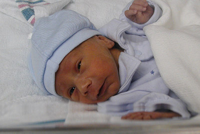
retinopathy of prematurity
All babies born before 31 weeks of pregnancy need monitoring for retinopathy of prematurity. c. Photo credit: National Eye Institute

The telemedicine strategy consisted of electronically sending photos of babies’ eyes to a distant image reading center for evaluation. Staff at the image reading center, who were trained to recognize signs of severe ROP, identified whether infants should be referred to an ophthalmologist for evaluation and potential treatment. The study tested how accurately the telemedicine approach reproduced the conclusions of ophthalmologists who examined the babies onsite.

“This study provides validation for a telemedicine approach to ROP screening and could help save thousands of infants from going blind,” said Graham E. Quinn, MD, professor of ophthalmology at the Children’s Hospital of Philadelphia and the lead investigator for the study, which is reported today in JAMA Ophthalmology. The study was conducted by the e-ROP Cooperative Group, a collaboration that includes 12 clinics in the United States and one in Canada.

Some degree of ROP appears in more than half of all infants born at 30 weeks pregnancy or younger — a full-term pregnancy is 40 weeks — but only about 5 to 8 percent of cases become severe enough to require treatment. In ROP, blood vessels in the tissue in the back of the eye called the retina begin to grow abnormally, which can lead to scarring and detachment of the retina. Treatment involves destroying the abnormal blood vessels with lasers or freezing them using a technique called cryoablation. Early diagnosis and prompt treatment is the best prevention for vision loss from ROP, which is why the American Academy of Ophthalmology recommends routine screening for all babies who are born at gestational age 30 weeks or younger or who weigh less than 3.3 pounds at birth.

The study evaluated telemedicine for ROP screening during the usual care of 1,257 premature infants who were born, on average, 13 weeks early. About every nine days, each infant underwent screening by an ophthalmologist, who assessed whether referral for treatment was warranted. Those who were referred were designated as having referral-warranted ROP (RW-ROP). Either immediately before or after the exam, a non-physician staff member in the neonatal intensive care unit (NICU) took images of the infant’s retinas and uploaded them to a secure server at the University of Oklahoma, Oklahoma City. Trained non-physician image readers at the University of Pennsylvania, Philadelphia, then downloaded the photos, independently evaluated them following a standard protocol, and reported the presence or absence of RW-ROP.

Through the telemedicine approach, non-physician image readers correctly identified 90 percent of the infants deemed to have RW-ROP based on examination by an ophthalmologist. And they were correct 87 percent of the time when presented with images from infants who lacked RW-ROP. The examining ophthalmologists documented 244 infants with RW-ROP on exam. After referral, 162 infants were treated. Of these, non-physician image readers identified RW-ROP in all but three infants (98 percent). “This is the first large clinical investigation of telemedicine to test the ability of non-physicians to recognize ROP at high risk of causing vision loss,” said Eleanor Schron, Ph.D., group leader of NEI Clinical Applications. “The results suggest that telemedicine could improve detection and treatment of ROP for millions of at-risk babies worldwide who lack immediate in-person access to an ophthalmologist,” she said.

About 450,000 (12 percent) of the 3.9 million babies born each year in the United States are premature. The number of preterm infants who survive has surged in middle income countries in Latin America, Asia, and Eastern Europe. In these parts of the world, rates of childhood blindness from ROP are estimated at 15 to 30 percent — compared to 13 percent in the United States.
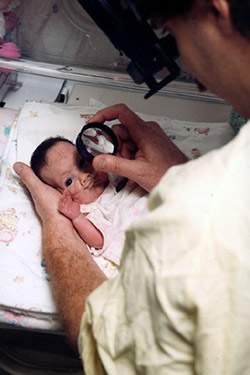
retinopathy of prematurity
NICU care providers take photos of a premature baby’s retinas in the NEI-funded e-ROP study of telemedicine for retinopathy of prematurity. Photo credit: Children’s Hospital of Philadelphia

One advantage of telemedicine ROP screening is that it can be done more frequently than screening by an ophthalmologist. “It’s much easier to examine the retina when not dealing with a wiggling baby,” said Dr. Quinn. “If a baby is too fussy or otherwise unavailable when the ophthalmologist visits the NICU, the exam may be delayed until the ophthalmologist returns — sometimes up to a week later.”

Weekly ROP screening — or even more frequently for high-risk babies — is a realistic goal for telemedicine and could help catch all cases needing treatment, according to the report. In the study, imaging was restricted to occasions when an ophthalmologist examined the baby. In practice, hospital staff could implement an imaging schedule based on the baby’s weight, age at birth, and other risk factors. “With telemedicine, NICU staff can take photos at the convenience of the baby,” said Dr. Quinn.

Telemedicine for evaluating ROP offers several other advantages.

Telemedicine may help detect RW-ROP earlier. In the study, about 43 percent of advanced ROP cases were identified by telemedicine before they were detected by an ophthalmologist — on average, about 15 days earlier.

Telemedicine could save babies and their families the hardship and hazards of being unnecessarily transferred to larger nurseries with greater resources and more on-site ophthalmologists. “Telemedicine potentially gives every hospital access to excellent ROP screening,” Dr. Quinn said.

Telemedicine might also bring down the costs of routine ROP screening by reducing the demands on ophthalmologists, whose time is better allocated to babies who need their attention and expertise. In a separate analysis, the study found that non-physicians and physicians had similar success in assessing photos for RW-ROP. Three physicians evaluated image sets from a random sample of 200 babies (100 with RW-ROP based on the eye exam findings; 100 without) using the standard grading protocol. On average, the physicians correctly identified about 86 percent of RW-ROP cases; the non-physicians were correct 91 percent of the time. The physicians correctly identified about 57 percent of babies without RW-ROP; non-physicians were correct 73 percent of the time.

The cost of establishing a telemedicine ROP screening program includes acquisition of a special camera for taking pictures of the retina, training of NICU personnel to take and transmit quality photos, and establishment and maintenance of an image reading center. “As we move along this road, advances in imaging and grading of images may streamline the process even more,” Dr. Quinn said.

The e-ROP Cooperative Group includes the following clinical sites and resource centers:

  • Children’s Hospital of Philadelphia
  • Johns Hopkins University, Baltimore
  • Boston Children’s Hospital
  • Nationwide Children’s Hospital and Ohio State University Hospital, Columbus
  • Duke University (cost-effectiveness center), Durham, North Carolina
  • University of Louisville, Kentucky
  • University of Minnesota, Minneapolis
  • University of Oklahoma (Inoveon ROP Data Center), Oklahoma City
  • University of Texas Health Science Center at San Antonio
  • University of Utah, Salt Lake City
  • Vanderbilt University, Nashville, Tennessee
  • Hospital of the Foothills Medical Center, Calgary, Alberta
  • University of Pennsylvania (data coordinating center and image reading center), Philadelphia

For more information about ROP.

Click to view a video about e-ROP.

NIH logo without bannerNational Institutes of Health
9000 Rockville Pike
Bethesda, Maryland 20892
301-496-4000,
TTY 301-402-9612

Contact Lens Fitting For Children

8/26/14

Fitting Techniques

The techniques the contact lens professional utilizes to fit an adult with a GP lens must be altered to fit an infant or small child. The ability to capture a reliable image with a topographer or accurate keratometric readings is often impossible to obtain in small children. Keratometric readings obtained at the time of surgery or an exam under anesthesia should only be considered as a starting point or a guide to the initial diagnostic lens. The application and evaluation of a diagnostic lens is the best method to obtain an appropriate fit in small children. I utilize diagnostic lenses that do not have a UV filter.Contact Lens Fitting for Children I find these lenses allow me to better interpret the fluorescein pattern when using a handheld burton lamp or LED cobalt flashlight. Once the appropriate fit has been determined, the lens is remade incorporating a material that provides an ultraviolet filter. I find it easier to determine the approximate corneal shape and curvature initially with a relatively flat fitting lens on the eye. If the diagnostic lens being evaluated vaults the anterior corneal surface, the interpretation and extrapolation of corneal curvature is difficult if not impossible. As in any GP fitting, the goal is to equally distribute lens mass and provide peripheral fulcrums to maintain stability and a central position. This central position of the lens is especially important in higher powers to minimize spherical aberrations. In recent years, I have found myself fitting looser and larger GP diameters. A general rule to follow with small children and GP lenses is that a tight lens will tend to dislodge from the eye and a loose fitting lens will tend to displace off the cornea onto to bulbar conjunctiva.

As with GP fitting on small children, soft lens fitting techniques are also a bit different. In order to determine appropriate movement of a soft lens on a small child, the “spring back” test may be helpful. With the soft lens on the eye, digitally displace the lens off center. If the lens immediately “springs back” into place on the cornea, the lens may fit too tightly on the ocular surface. If the lens stays off center while manually closing the lids to mimic a blink, the lens may be fitted too loosely on the ocular surface. In addition, retinoscopy over the soft lens to determine if the reflex maintains clarity during the blink is a finding seen with a well fitting lens. If the reflex is clearer with a blink, the fit may be too steep. If the reflex is worse with a blink, the fit may be too flat. The reflex seen with a well-fitted soft lens will maintain the same clarity before, during and after a blink. The retinoscope is not only used to determine the final lens power with any type of lens but also an important instrument to guide you to the best cornea lens relationship. Pediatric fitters of contact lens should be proficient with a retinoscope.

Little Lenses for Little People?

The pediatric contact lens professional is not limited to “off the rack” products. In addition to custom GP lenses, there are many lens manufacturers of custom made soft and silicone hydrogel contact lenses that allow us the opportunity to provide any child any parameter. In addition, liberal exchange policies implemented by these manufacturers of custom products allow us to provide these products to the patients who require them in a fair and effective manner. However, the delay in time to deliver the product to a pediatric patient in an urgent situation is a potential problem. Any delay in optical correction and visual rehabilitation with a young pediatric patient may result in permanent loss in vision.

Silicone Hydrogel Custom Products

After many years of anticipation, in 2010 Contamac received FDA approval for Definitive, a latheable silicone hydrogel material. The Definitive material can be manufactured by a limited number of laboratories in the U.S. in virtually any group of parameters. This inherently wettable, high water content and low modules material has a DK of 60. While 60 DK is not as high as other “off the rack” silicone hydrogel materials, the effective DK in many of the parameters utilized in pediatric fitting is higher than the same parameters made in HEMA-GMA materials. While this material is a welcome addition to our armamentarium of contact lens options in our practice, my clinical experience specific to pediatric indications and this material has led me to two conclusions. The application of a lens to the eye of a small child manufactured in the Definitive material is more difficult than HEMA-GMA materials and the time delay of up to ten days is often too long in an urgent case common to the pediatric patient. In time, both of these concerns can be overcome with practice and improved efficiency on the part of the patient, the practitioner and the laboratory.

New News About an Old Lens

Silicone Elastomer (Silsoft) has a long and well-documented history of being the lens of choice for the majority of pediatric professionals to manage small children following cataract surgery. The truth is that there would be many “blind” children if not for this particular lens. Silsoft Super Plus contact lenses for pediatric aphakia (>20 diopters) are available with the following parameters: diameter, 11.3mm, base curves: 7.5 mm (45.00D), 7.7mm (43.75D), and 7.9mm (42.75D), optic zone of 7.0mm and powers ranging from +23.00D to +32.00D in 3D steps. The Silsoft material has an oxygen permeability (Dk) value of 340, with oxygen transmissibility (Dk/t) of 58 at 0.61mm. One of the concerns about Silsoft has been the limited availability of parameters. As a result of the tireless efforts of Joe Barr, O.D., B+L may ultimately decide to expand the parameters of their Silsoft Super Plus product. While this announcement is far from official at the time of this article, I would like to applaud Joe and encourage you to do the same. Whether you are a proponent or opponent of Silsoft, any improved technology to provide children with the opportunity to safely develop better vision is worthy of the efforts. On behalf of the industry, the children and their families, thank you Joe.

Conclusion

As contact lens professionals, we have the responsibility, opportunity and privilege to provide products and service to young patients and their families. These products and associated services are necessary to maintain and or develop possibly the most important gift one may ever possess, the gift of sight. Again I ask you, are you a “healer of children”?

Buddy Russell - pediatric contact lensesBuddy Russell, FCLSA, COMT
Associate, Specialty Contact Lens Service
Emory University Eye Center

Common Pediatric Eye Diseases

8/21/14

In the third of this series, Buddy Russell, from the Emory University Eye Center, provides a great overview of common pediatric eye diseases.

Some Conditions Frequently Seen in Pediatrics

A basic understanding of some of the conditions that may be present in pediatric patients is important to not only know what they are but also understand well enough to explain to the parent or caregiver. The following is intended to be an overview of some of those conditions and not a complete explanation.Girl with eye chart-common pediatric eye diseases

  1. Nystagmus – Nystagmus is a vision condition in which the eyes make repetitive, uncontrolled movements, often resulting in reduced vision. These involuntary eye movements can occur from side to side, up and down, or in a circular pattern. As a result, both eyes are unable to hold steady on objects being viewed. Unusual head positions and head nodding in an attempt to compensate for the condition may accompany nystagmus. Most individuals with nystagmus can reduce the severity of their uncontrolled eye movements and improve vision by positioning their eyes to look to one side. This is called the “null point” where the least amount of nystagmus is evident. To accomplish this they may need to adopt a specific head posture to make the best use of their vision. The direction of nystagmus is defined by the direction of its quick phase (e.g. a right-beating nystagmus is characterized by a rightward-moving quick phase, and a left-beating nystagmus by a leftward-moving quick phase). The oscillations may occur in the vertical, horizontal or torsional planes, or in any combination. The resulting nystagmus is often named as a gross description of the movement, e.g. downbeat nystagmus, upbeat nystagmus, seesaw nystagmus, periodic alternating nystagmus. Having nystagmus affects both vision and self-concept. Most people with nystagmus have some sort of vision limitations because the eyes continually sweep over what they are viewing, making it impossible to obtain a clear image. If a refractive error is found, contact lenses may be the most effective way of obtaining best-corrected vision.
  2. Strabismus – Strabismus is any misalignment of the eyes. It is estimated that 4% of the U.S. population has strabismus. Strabismus is most commonly described by the direction of the eye misalignment. Common types of strabismus are esotropia (turn in), exotropia (turn out), hypotropia (turn down), and hypertropia (turn up). Eye misalignment can cause amblyopia in children. When the eyes are oriented in different directions, the brain receives two different visual images. The brain will ignore the image from the misaligned eye to avoid double vision, resulting in poor vision development of that eye. Also, an eye that sees poorly tends to be misaligned. The goal of strabismus treatment is to improve eye alignment, which allows for better work together (binocular vision). Treatment may involve eyeglasses, contact lenses, eye exercises, prism, and / or eye muscle surgery.
  3. Amblyopia – Amblyopia, sometimes called a “lazy eye,” occurs when one or both eyes do not develop normal vision during early childhood. Babies are not born with 20/20 vision in each eye but must develop it between birth and 6-9 years of age by using each eye regularly with an identical focused image falling on the retina of each eye. If this does not occur in one or both eyes, vision will not develop properly. Instead, vision will be reduced and the affected eye(s) are said to be amblyopic. This common condition, affecting up to 4% of all children, should be diagnosed and treated during infancy or early childhood to obtain optimum three-dimensional vision and to prevent permanent vision loss. What causes amblyopia?
      • Misaligned eyes (strabismus)
        Misaligned eyes are the most common cause of amblyopia. When both eyes are not aimed in exactly the same direction, the developing brain “turns off” the image from the misaligned eye to avoid double vision and the child uses only the better eye — the dominant eye. If this persists for a period even as short as a few weeks, the eye will not connect properly to the visual cortex of the brain and amblyopia will result.
      • Unequal refractive error (anisometropia)
        Unequal refractive error is an eye condition in which each eye has a different refractive error and therefore both eyes cannot be in focus at the same time. Amblyopia occurs when one eye (usually the eye with the greater refractive error) is out of focus because it is more nearsighted, farsighted or astigmatic than the other. Again, the brain “turns off” the image from the less focused eye and this eye will not develop normal vision. Because the eyes often look normal, this can be the most difficult type of amblyopia to detect and requires careful vision screening of acuity measurements at an early age. Treatment with glasses or contact lenses to correct the refractive error of both eyes, sometimes with part-time patching of the better seeing eye, is necessary in early childhood to correct the problem.
      • Obstruction of or cloudiness (deprivation)
        Obstruction of or cloudiness in the normally clear eye tissues may also lead to amblyopia. Any disorder that prevents a clear image from being focused can block the formation of a clear image on the retina and lead to the development of amblyopia in a child. This often results in the most severe form of amblyopia. Examples of disorders that can interfere with getting a clear image on the retina are a cataract or cloudy lens inside the eye, a cloudy and or irregular shaped cornea, or a droopy upper eyelid (ptosis) or eyelid tumor.It is not easy to recognize amblyopia. A child may not be aware of having one normal eye and one with reduced vision. Unless the child has a misaligned eye or other obvious external abnormality, there is often no way for parents to tell that something is wrong. In addition, it is difficult to measure vision in very young children at an age in which treatment is most effective.To treat amblyopia, a child and their caregiver must be encouraged to use the weaker eye. This is usually accomplished by patching the stronger eye. This covering of the stronger eye with an adhesive patch, an cclude contact lens or temporary surgery often proves to be a frustrating and difficult therapy. Patching will often continue for weeks, months, or even years in order to restore normal or near normal vision and maintain the improvement in the amblyopic eye. Occasionally, blurring the vision in the good eye with eye drops or lenses to force the child to use the amblyopic eye treats amblyopia. In some cases, cataract surgery or glaucoma surgery might be necessary to treat form deprivation amblyopia. Patching may be required after surgery to improve vision, and glasses or contact lenses may be required to restore appropriate focusing.Surprising results from a nationwide clinical trial in 2005 show that many children age seven through 17 with amblyopia may benefit from treatments that are more commonly used on younger children.
        Treatment improved the vision of many of the 507 older children with amblyopia studied at 49 eye centers. Previously, eye care professionals often thought that treating amblyopia in older children would be of little benefit. The study results, funded by the National Eye Institute (NEI), appear in the April issue of Archives of Ophthalmology.
  4. Congenital Cataract – A congenital cataract, or clouding of the crystalline lens is present in 2-3 per 10,000 live births of children. The presence of a visually significant cataract in a child is considered an urgent disorder. The resultant form deprivation of vision requires immediate surgery to remove the obstruction, prompt optical correction and amblyopia therapy in unilateral cases. Until the 1970s, it was generally believed that there was no means of restoring the vision in an eye with a unilateral congenital cataract. However, subsequent studies demonstrated that excellent visual results could be obtained with early surgical treatment coupled with optical correction with a contact lens and patching therapy of the fellow eye. However, treatment results continue to be poor in some infants with unilateral congenital cataracts due to a delay in treatment or poor compliance with contact lens wear or patching therapy of the fellow eye. The Infant Aphakia Treatment Study (IATS) was designed to compare the visual outcomes in children 1 to 6 months of age with a unilateral congenital cataract randomized to optical aphakic correction with contact lenses or an intraocular lens (IOL). Children randomized to IOL treatment had their residual refractive error corrected with spectacles. Children randomized to no IOL had their aphakia treated with a contact lens. In previous publications we have shown that the visual results are comparable for these two treatments at 1 year of age, but significantly more of the infants randomized to IOL implantation required additional intraocular surgeries.
  5. Accommodative Esotropia – Accommodative esotropia refers to a crossing of the eyes caused by farsightedness. Accommodative esotropia is a type of strabismus. Children who are farsighted easily and automatically focus on objects at distance and near through accommodation. As a result, a child who is farsighted usually does not have blurred vision. However, in some children who are farsighted, this accommodative effort is associated with a reflex crossing of the eyes. Accommodative esotropia can begin anywhere from 4 months to 6 years of age. The usual age of onset is between 2 and 3 years of age.Full-time use of the appropriate hyperopic glasses prescription or contact lenses will often control the esotropia. When wearing the correction, the child will not need to accommodate and hence the associated eye-crossing reflex will disappear. However, after removing the prescribed correction, the crossing will reappear, perhaps even more than before the child began wearing the correction. Sometimes the correction will only cause the crossing to disappear when the child views a distant object. However, when gazing at near objects, crossing may persist despite the use of the correction. In these circumstances, a bifocal lens is often prescribed to permit the child to have straight eyes at all viewing distances. One potential advantage of contact lenses compared to spectacles when correcting hyperopic powers is the decrease in accommodative demand. The increased effort to converge the eyes with spectacles requires one to over come the resultant base out prism when viewing a near object.


Buddy Russell - pediatric contact lensesBuddy Russell, FCLSA, COMT
Associate, Specialty Contact Lens Service
Emory University Eye Center

Treatment Options For Children

8/19/14

Here is part two in Buddy Russell’s series; this one focusing on contact lenses as a treatment option for children.

We Are Not Born With Good Vision

The human visual system at birth is poorly developed, but rapidly becomes the remarkable combination of nerve tissue, muscles and optics that provide us with the sense of vision. Those babies born with “perfect” eyes have only the opportunity to develop normal vision. The information processed by the eyes is sent directly to the brain and is interpreted as vision.Toddler looking through glasses - treatment options for children During the first few weeks, the child sees shapes, lines and space between objects. The child’s visible world is most usable within 8-14 inches of his/her eyes. During this time, the eyes may appear to wander. After about a month or so, the normal child’s eyes will appear more coordinated and they start to show more interest in looking at objects. It is usually in the third month that a child who has normal eyes can fix and follow on a near object. The growth of the eye is a dynamic process, influenced by genetics and the environment.
Early detection of any eye problem is key to treating the disorder. The prevalence of vision problems in children is higher than you might think. For example:

  • 1 in 10 children are at risk from undiagnosed vision problems
  • 1 in 25 will develop strabismus
  • 1 in 30 will be affected by amblyopia
  • 1 in 33 will show significant refractive error
  • 1 in 100 will exhibit evidence of eye disease
  • 1 in 20,000 children have retinoblastoma

As a result of his granddaughter and her eye problem, former President Jimmy Carter initiated a program in 2002 called InfantSEE. This program allows children to have an eye exam at a very young age at no charge to the family. Participating eye doctors provide a more thorough exam than the busy pediatrician. As a result, there is a greater opportunity to detect and treat eye disorders that may otherwise go undetected.

“Have to” Contact Lenses

Fitting pediatric patients is not usually about routine visits and patients who want to wear contact lenses. It is about critical and often urgent situations and patients who have to wear contact lenses. The more common medical indications for contact lenses can be categorized into three groups; anisometropia, irregular corneal astigmatism and “large” refractive errors.

Anisometropia

One of the more common conditions potentially leading to a permanent loss of vision in a young patient is anisometropia. This difference in the refractive errors of the two eyes can lead to suppression of the less clear image. As a result of the non-focused eye, the brain of a young patient simply turns off the blurred eye. Early detection is key to successful treatment. Following the diagnosis of this problem being present, simply correcting the refractive error may be enough. However, it has been reported that as little as one diopter difference between the two eyes corrected with spectacles and the resultant anisokonia, can lead to foveal suppression impacting stereopsis and depth perception. The use of a contact lens or contact lenses alters the effective image size due to the vertex distance being zero compared to either the magnification or minification of the image size due to the vertex distance with spectacles. One of the most severe examples of this condition would be a child with a unilateral congenital cataract and managed with spectacles postoperatively.

Irregular Corneal Astigmatism

Whether acquired or congenital, the presence of irregular corneal astigmatism of the anterior curve of the cornea is best managed with a contact lens. This condition is to be considered urgent if the patient is of a young age. The eye may forever loose the opportunity to be corrected as the resultant amblyopia develops over a short period of time. By neutralizing the corneal irregularities with a contact lens, the eye of a young child will hopefully gain enough vision improvement to avoid the potential permanent loss.
Obviously, patching the better eye may also be necessary if the treated eye’s vision is not as correctable as the unaffected eye. The length of time the child is to be patched is to be determined by the pediatric ophthalmologist or optometrist, as this area of treatment is sometimes controversial. The factors that are considered include the level of vision obtained, age of the child and the condition of the other eye.

Large Refractive Errors

The optics of spectacle correction in high powers have inherent properties that include distortion, prismatic effect and minification / magnification. For instance, the decrease in image size when one views an object through high minus spectacles may result in less vision. This decrease in image size may impact the opportunity to fully develop normal vision in a young child. The smaller image size that is due to the vertex distance of spectacles may be better managed with a contact lens that has a vertex distance of zero thus providing a larger image. This larger image size often increases best-corrected vision.

“Fitting” the Caregiver

Arguably, the most important factor with young children having a good outcome is the parents / caregivers. The technical challenges that exist in these cases are secondary to the ability the fitter must possess to effectively explain and train the person or persons that will take care of the child outside of the office. They must be your partner in the child’s treatment. They must understand the urgency of the situation, they must understand the seriousness of the problem, they must be trained to properly apply, remove and care for the lens / lenses, they must also follow any and all instructions concerning the child. Many of these parents struggle with feelings of nervousness, guilt and sadness. My strategy is to be sensitive to their feelings but not let them feel sorry for themselves too long as the clock is ticking. I provide verbal instructions, written instructions, videos, my email address and a 24-hour phone number. I welcome the caregiver to ask any question at any time. I do my best to let them know that I do care and that I want them and their child to be successful. I am tough on them. There is no good excuse not to do as I have instructed them to do.

When the child and the parent / caregiver are convinced that I am confident in my ability and they know that I do care, the partnership develops as we walk the path together. I want the child to know that they are coming to see me. I want them to know I will reward their cooperation with all phases of the visit. This positive reinforcement may be in the form of a piece of candy, a small toy or just a sticker when the child allows me to see their eye, measure their cornea or intraocular pressure or they just tell me what they can see. Kids love to please us just like they love to please their parents. Reward them for it. Whether you consider this approach bribery or positive reinforcement, it works.

Buddy Russell - pediatric contact lensesBuddy Russell, FCLSA, COMT
Associate, Specialty Contact Lens Service
Emory University Eye Center

Pediatric Contact Lenses

8/14/14

Because August is Children’s Eye Health Month we are pleased to present a four-part series on pediatric vision issues and contact lenses by Buddy Russell, FCLSA, COMT. With over thirty years experience fitting contact lenses, Buddy is currently an associate of the specialty contact lens service at Emory University Eye Center in Atlanta, Georgia. Buddy is a clinical instructor in Emory’s Ophthalmic Technology Program and teaches students and ophthalmology resident’s contact lens technology. 

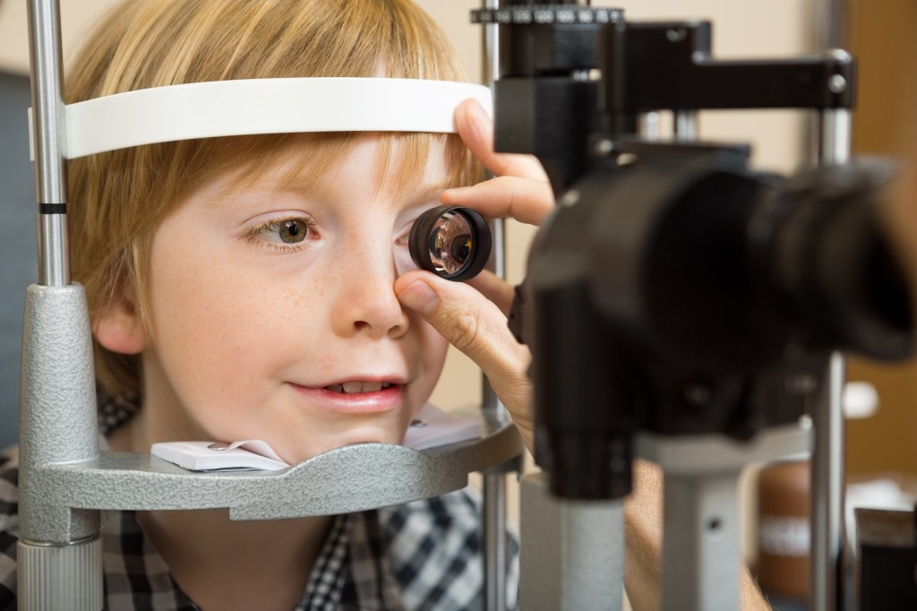
Child refractive exam - pediatric contact lensesHe is a licensed dispensing optician, a Fellow member of CLSA and has been certified by JCAHPO as a Certified Ophthalmic Medical Technologist. He lectures at national and international meetings on contact lens related topics. Buddy has written articles for a number of publications, two chapters for CLSA’s advanced training manual and is a peer reviewer for the Cornea publication. He is also a contributing editor for CLSA’s Eyewitness journal. His current areas of research include pediatric aphakia and keratoconus. He joined the faculty at TVCI in 2006.

The first article will examine that pediatric contact lenses for children go beyond vision correction, the second will explore lenses as a treatment option, the third will look at a variety pediatric eye conditions and the final post will discuss the contact lens fitting challenges you face when you work with children.

Introduction

Working with the pediatric patient and their caregivers / family can be challenging, rewarding, fun, and yet sometimes frustrating. Many of these cases often include factors that are unique to the young patient. In addition to the technical challenges of obtaining the objective data, the fear of uncertainty is often present. The uncertainty of the unknown can either paralyze you or motivate you to step up and simply do what must be done.

The Definition May Vary

The definition of pediatric contact lens fitting can be different to different people. The fitter who works with the occasional twelve-year-old neophyte wearer will define pediatric fitting different from the person that works with babies on a routine basis. Pediatrics is generally defined as a branch of medical care that deals with infants, children and adolescents. The word pediatrics is derived from two Greek words (pais = child and iatros = healer), which means healer of children. Are you a “healer of children” or do you tend to feel better about someone else assuming the challenge and responsibility? This article will discuss some of the conditions, contact lens indications, fitting techniques and challenges that are present with the young patient.

Refractive Indications

What age is “appropriate “ to fit a contact lens on a child? In the absence of a medical indication, Jeff Walline, OD and his colleagues have addressed the answer to this question in the published literature. In addition, the American Academy of Optometry published a position paper in 2004 that stated that by the age of eight, a child was able to handle contact lenses and assume some degree of responsibility. We are all aware that not all eight year olds are capable of dealing with contact lenses. For that matter, not all eighteen year olds are mature enough to assume responsibility for anything. Some of the concerns that a contact lens practitioner may have in fitting these young children include the risk of safety to the child’s health, too much chair time, physical limitations, lack of hygiene, and lack of maturity. These are all legitimate concerns when you consider the child can see well with spectacles.

What does the literature reveal concerning these questions and concerns? Are the answers there?

CLIP Study

The Contact Lens In Pediatrics study compared 169 neophyte wearers in two age groups (children age 8-12 and teens age 13-17) over a period of three months. The summary of the clinical findings in the publication is that adverse events was low and the younger children took a little longer to train application and removal of the contact lenses. The more impressive outcomes from this study was determined by a tool used more frequently in child psychology referred to as the Pediatric Refractive Error Profile (PREP) survey. The PREP survey is a clinically validated quality of life instrument to assess how a child “sees” him or herself. This 26-question survey revealed that contact lenses improved the child’s self image in regards to their appearance, increased confidence in themselves while participating in activities and overall satisfaction of their form of vision correction. These findings were consistent in both age groups. More than 80% of both age groups found contact lenses easy to clean and take care of as all participants were fitted with 2-week disposable soft lenses and used a multipurpose disinfection care system.

The ACHIEVE Study

The Adolescent and Child Health Initiative to Encourage Vision Empowerment (ACHIEVE) were published in 2009. Jeff Walline, OD and his colleagues designed this study to find out the effects that glasses and contacts had on the self-perception of the child. This study examined 484 myopic children 8-11 years. The participants were randomized to spectacles (n=237) or contact lenses (n=247) and followed for three years. The children were evaluated at baseline, 1 month and every 6 months for three years by a validated psychology tool for self-perception referred to as the Self-Perception Profile for Children (SPPC). The SPPC instrument allows a 4 point self-assessment in 6 categories; scholastic competence, social acceptance, athletic competence, physical appearance, behavioral conduct and global self-worth. The participants revealed the most dramatic areas of improvement with contact lenses compared to spectacles in the areas of physical appearance, athletic competence, scholastic competence and social acceptance. Similar to the low occurrence of adverse events with contact lens wear found in the CLIP study, over the three year period there were only 13 adverse events among 9 subjects. In addition, the ACHIEVE study found very similar rates of myopic progression in both groups of patients over the three year period (1.08D spectacle group and 1.27D contact lens group).
What can we conclude from these two studies?

One is that we are in a position to not only help a young person see but we are also in a position to do it safely and assist the child by instilling more confidence in themselves at a young age that may impact them as they mature into an adult who feels good about themselves. Young children are accustomed to following rules. When properly trained, these same young patients may grow into some of the most compliant patients that we have in our practice. There are some practical considerations for prescribing contact lenses to the younger patient. Mary Lou French, O.D. has stated the three M’s are important for success; Maturity (good hygiene, good communication skills, signs of responsibility), Motivation (why do they want contacts? Does the child want them or just the mom or dad? Are they active in activities where freedom from spectacles is important?), Mom (is the mom / dad / older sibling willing to help?). Don’t let age be the deciding factor. Consider your position as one that may positively impact the young patient in how they “see” and feel about themselves.

Buddy Russell - pediatric contact lensesBuddy Russell, FCLSA, COMT
Associate, Specialty Contact Lens Service
Emory University Eye Center

I See You

8/12/14

This is the second article from Kooshay Malek, a blind therapist in Los Angeles. Her first article, The Habit of Seeing, discussed losing her vision and her choice of becoming a marriage and family therapist. Here she discusses at how she uses her vision loss to help her patients.

When I started as a therapist, I was really concerned about my blindness. I had faced prejudice in other jobs. With my first few clients, I gave them this whole spiel at the beginning, explaining about being blind and about why I wear dark glasses.Therapy cloud - blind therapist My supervisor said not to work so hard to explain. He thought it was a nonissue: “If you were blonde and blue-eyed, would you be describing that over the phone to them?” he asked. He was right. It didn’t faze most people. In the 11 years I’ve been practicing, only a few people had a problem with it. To this day, once in a while, it may come out organically that I’m blind. Most of the time, I don’t tell them beforehand.

I think I pick up on certain nuances sighted therapists may miss. I sense shifts in energy in the room. I have very strong attunements: I notice the slightest change in tone of voice — or even in their silences — and I know something’s going on. If necessary, we talk about my blindness, and we process my blindness in the session. I don’t leave it as an elephant in the room. The main concern of everybody who comes to therapy, whether they are seeing a blind person or not, is, “Am I going to be heard and understood?” In this case, they may wonder if my blindness will affect whether I can hear and understand them. I say: “Well, we’ll have to wait and see. If there is something you think I can’t understand, would you be able to tell me?” That makes them self-sufficient in asking for help or expressing a need. Many patients tell me they find it so much easier to talk to me, because I don’t have my eye on them, so to speak, like a microscope. They find it close to the traditional psychoanalyst’s couch, where the therapist would sit behind them and not look at their face. They find out I see them better than anyone else in their life. That’s the reward of it. Especially with clients who have self-image and self-esteem issues. I get to see who is inside, not who is outside, and that’s powerful by itself. People open up more easily. My blindness is a really quiet, subtle intervention in the room at all times. It’s always present. It’s a gift I carry in there with me, and I use it.

Kooshay Malek - seeingKooshay Malek
Marriage and Family Therapist
Los Angeles, CA

Nystagmus In Children

8/7/14

Nystagmus is a condition of uncontrolled eye movements. Patients with nystagmus are unable to maintain their eyes in a fixed position of focus. The movements can be pendular, swaying evenly side to side, or, jerk into one direction and drift toward the opposite direction. It can be present early in life or acquired as an adult. It can occur in eyes with poor vision from other anomalous development, or eyes that appear perfectly normal. In almost all patients the vision is compromised to some degree. In some patients, the eye movement is less, and the vision better, in an eccentric position that causes the patient to adopt a face turn, tilt or head posture so they can use this quieter position (“null point”) to navigate during their daily activities. To date there have been no consistently effective treatments for this condition.

Lingua and Grace - nystagmus
Dr. Lingua and Grace Nassar

Treatment efforts have been either medical (drugs to reduce the amplitude of the nytagmus movement) or surgical (to move the “null point” into straight ahead gaze to eliminate a head turn, or, directed at reducing the effective contracture of all the eye muscles to reduce the amount of movement). In general, surgical treatment of nystagmus has been disappointing.

In 2002, Dr. Robert Sinskey, noted cataract surgeon and phacoemulsification pioneer, proposed a revolutionary concept, that nystagmus could only be truly effectively controlled by removing the forward portion of the eye muscle and detach it completely from the eye. Since the twitching eye muscles were controlled by nerves sending that pulsatile information, any operation that allowed the muscles to remain attached to the eye would never quiet the movement. He performed this novel surgery in 2000 and published the results in 2002. It did not receive attention in the nystagmus surgery community, as most experts worried that the surgery would limit normal eye movements excessively. The operation does remove the forward portion of the eye muscle but, surprisingly, the eyes are still able to move to allow reading, computing, and driving.. In 2012, I had the opportunity to view a patient he operated 10 years prior and was impressed with how successful the results were even after 10 years. Coincidentaly, I was caring for a 17 year-old patient with nystagmus who had already undergone the 2 currently accepted eye muscle procedures for nystagmus without success. His movements remained uncontrolled, he could not maintain eye contact with anyone, and is his vision was less than that needed for a drivers license. In 2013, I offered him the Sinskey procedure and the results were remarkable. His nystagmus was quieted, his vision improved (20/25) enough to qualify for a drivers license and to return to school.

Since 2012, we have adapted, augmented and perfected the procedure and performed the surgery on over 12 patients with similar remarkable results. All patients experience a marked reduction in the amplitude of the nystagmus (60-100%), and all patients demonstrate improved vision (1-8 lines of the acuity chart), especially at the reading position.

Visit the YouTube posting “Meet Grace for an example of how this surgery can impact a child’s life and the hopes of their parents. Visit www.eye.uci.edu for further information, contact information and scientific data on the procedure.

Robert Lingua, MDRobert W. Lingua, MD
Director, Pediatric Ophthalmology and Strabismus
Gavin Herbert Eye Institute, UC Irvine

32 Facts About Animal Eyes

8/5/14

For something different, and a little fun, here some interesting facts about animal eyes that you may not have known.Animal Eyes

  1. Shark corneas are similar to human corneas, which is why they have been used in human transplants.
  2. A worm has no eyes at all.
  3. An owl can see a moving mouse more than 150 feet away.
  4. Guinea pigs are born with their eyes open!
  5. Scorpions can have as many as 12 eyes, but the box jellyfish has 24!
  6. Camels have three eyelids! This is to protect their eyes from sand blowing in the desert.
  7. Most hamsters only blink one eye at a time.
  8. Owls are the only bird which can see the color blue.
  9. Goats have rectangular pupils to give them a wide field of vision.
  10. A scallop has around 100 eyes around the edge of its shell to detect predators.
  11. Snakes have two sets of eyes – one set used to see, and the other to detect heat and movement.  They also don’t have eyelids, just a thin membrane covering the eye.
  12. The four-eyed fish can see both above and below water at the same time.
  13. Owls cannot move their eyeballs – which has led to the distinctive way they turn their heads almost all the way around.
  14. A dragonfly has 30,000 lenses in its eyes, assisting them with motion detection and making them very difficult for predators to kill.
  15. Dolphins sleep with one eye open.
  16. The largest eye on the planet belongs to the Colossal Squid, and measures around 27cm across.
  17. Geckos can see colors around 350 times better than a human, even in dim lighting.
  18. The eyes of a chameleon are independent from each other, allowing it to look in two different directions at once.
  19. A camel’s eyelashes can measure up to 10cm long, to protect its eyelashes from blowing sand and debris in the desert.
  20. An ostrich’s eye is bigger than its brain.
  21. Dogs can’t distinguish between red and green.
  22. Polar bears have a third eyelid that helps filter UV light.
  23. Human eyes are not the most highly evolved. The mantis shrimp has four times as many color receptors as the human eye and some can see ultraviolet light.
  24. Pigeons can see millions of different hues, and have better color vision than most animals on earth.
  25. Cat’s eyes have almost 285 degrees of sight in three dimensions – ideal peripheral vision for hunting.
  26. Although color blind, cuttlefish can perceive light polarization, which enhances their perception of contrast.
  27. A moth’s eyes are covered with a water-repellant, anti-reflective coating.
  28. An ant only has two eyes, but each eye contains lots of smaller eyes, giving it a “compound eye.”
  29. Eagles have 1 million light-sensitive cells per square millimeter of the retina – humans only have 200,000.
  30. A honeybee’s eye is made of thousands of small lenses. A drone may have up to 8,600 and the queen be can have 3,000-4,000 lenses.
  31. The night vision of tigers is 6 times better than humans.
  32. Eyes on horses and zebras point sideways, giving them tremendous peripheral vision, to the point of almost being able to see behind them, but it also means they have a blind spot right in front of their noses.


Susan DeRemerSusan DeRemer, CFRE
Vice President of Development
Discovery Eye Foundation