Pumpkin Season

A sure sign that fall is here is that Starbucks is offering their Pumpkin-Spiced Lattes. Since pumpkins begin to ripen in September, this makes sense. But there are so many other ways to enjoy pumpkins, which can be good for your vision.
pumpkin season
They contain an abundance of antioxidants, vitamins, fiber and phytonutrients that are good for your skin, eyes and heart, and they may also decrease your risk of cancer.

When shopping for your pumpkin you need to look for the sugar or cheese pumpkins varieties that are good for cooking and baking, because of their dense, sweet flesh. A traditional field pumpkin that you use for decoration and carving jack-o’-lanterns has watery, stringy flesh and is not recommended for eating.

You can keep an un-cut pumpkin at room temperature for up to a month. Stored in a cool cellar or refrigerator, they can last up to three months. However, once you cut the pumpkin, pieces should be wrapped tightly and refrigerated and used within five days.

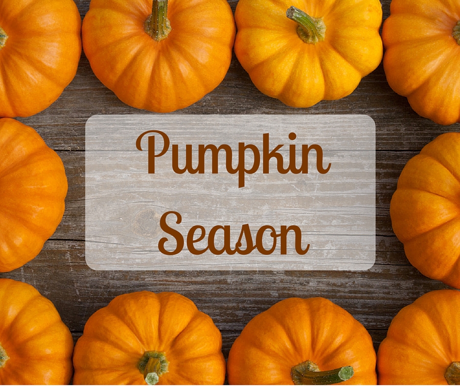
Pumpkin Season Recipes

Here are a variety of tasty recipes that will let you enjoy pumpkins beyond the traditional soup and pie (but we have included those two as well).

Breads and Muffins

pumpkin season
Pumpkin-Cranberry Muffins
Pumpkin-Cranberry Muffins from My Recipes by Heather McRae

Pumpkin Biscuits from Country Living

Pumpkin-Cranberry Breadsticks from Recipe Girl

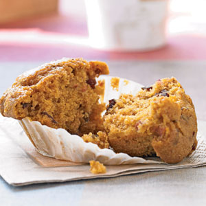
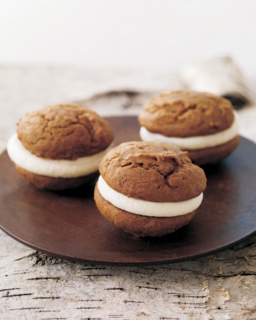
Pumpkin and Cream Cheese Muffins from Country Living

Pastas

pumpkin season
Chicken, Bacon & Pumpkin Gnocchi
Chicken Bacon Pumpkin Gnoochi from Nutmeg Nanny

Ravioli with Pumpkin Alfredo Sauce from Taste and Tell

Soups

pumpkin season
Pumpkin, Beef & Black Bean Chili
Pumpkin, Beef and Black Bean Chili from Country Living

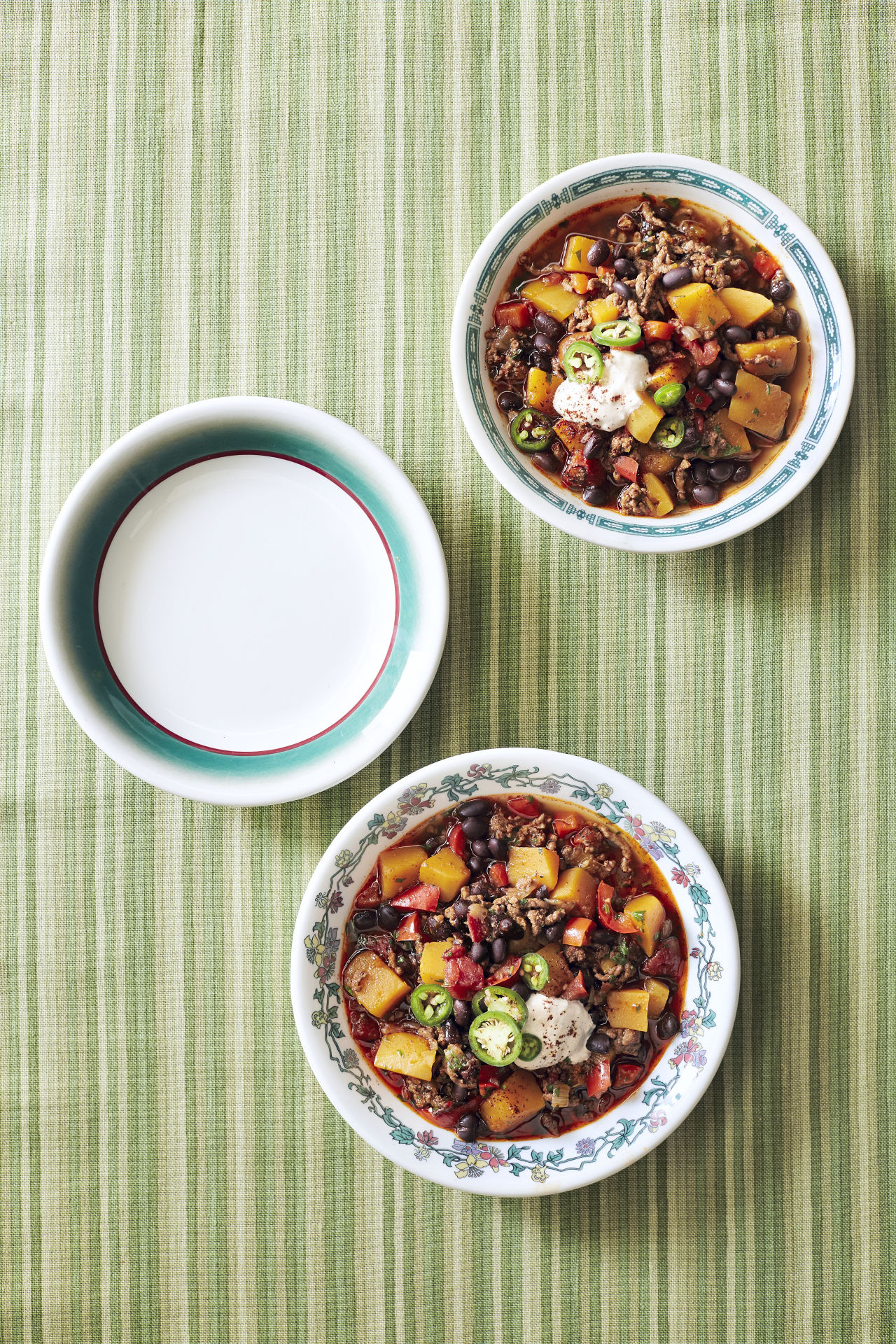
Roasted Pumpkin Soup from Martha Stewart

Breakfast Treats

pumpkin season
Fresh Pumpkin Pancakes
Fresh Pumpkin Pancakes from A Sweet Pea Chef

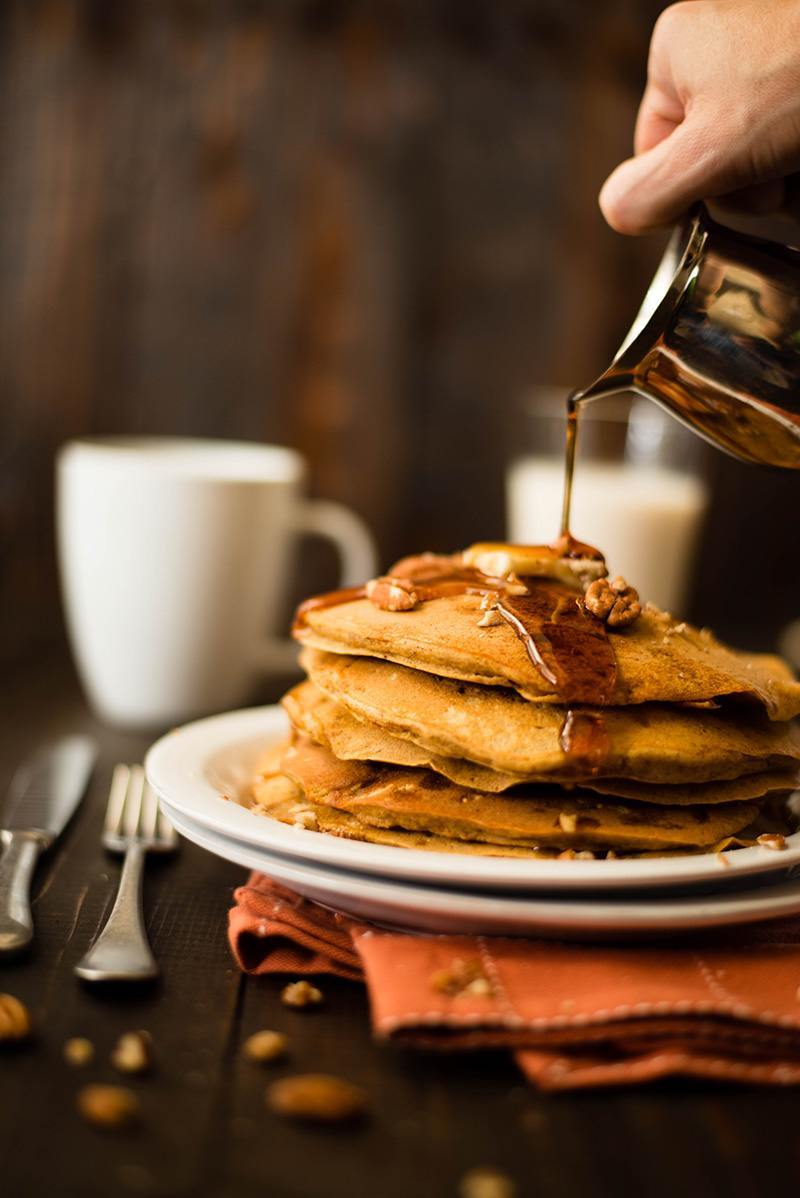
Pumpkin-Ginger Waffles from Country Living

Desserts

pumpkin season
Pumpkin Whoppie Pies with Cream Cheese Filling
Pumpkin Whoopie Pies with Cream-Cheese Filling from Martha Stewart

Ginger Pumpkin Pie with Toasted Coconut from My Recipes by David Bonom

Pumpkin Chiffon Pie with Gingersnap Pecan Crust from Epicurious

Extras

pumpkin season
Pumpkin French Fries
Baked Pumpkin Fries from Kirbie’s Cravings

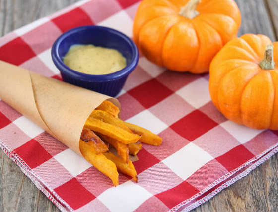
Pumpkin Salsa from Little Figgy

Pumpkin Pie Shake from My Recipes by Vivian Levine

As the days get shorter and the temperatures cool off, these recipes will hopefully get you geared up for autumn, and the holidays that are around the corner. Let us know which recipes are your favorites in the comments below.

9/29/15

Susan DeRemerSusan DeRemer, CFRE
Vice President of Development
Discovery Eye Foundation

Retinal Vascular Occlusions

Just like any other organ in the body, the retina needs a healthy blood supply to function properly. When a blood vessel in the retina becomes blocked, sudden loss of vision can occur. Such blockages, known as retinal vascular occlusions, often occur in older adults. Because there are effective treatments available, seeing a specialist promptly can prevent further problems or even restore vision.

Retinal arterial occlusions

Arteries are the vessels that bring fresh blood containing oxygen and nutrients to different parts of the body. The artery that supplies the retina is known as the central retinal artery. After it enters the eye, the central retinal artery splits up into branch retinal arteries that serve different areas of the retina.

Symptoms
The symptoms of a central retinal artery occlusion include a sudden, painless loss of vision in one eye. Because the circulation to the entire retina is disrupted, the vision loss is profound and encompasses the entire field of vision. In contrast, a branch retinal artery occlusion only leads to loss of vision in a part of the field of vision. This is because only part of the retinal circulation in compromised. Similar to a central retinal artery occlusion, a branch retinal artery occlusion is also typically sudden and painless.

Evaluation
Retinal arterial occlusions can usually be diagnosed simply by examining the retina. Often, the retina is whitened in the area of the occlusion due to the lack of blood flow. Most arterial occlusions occur in patients with diseases such as high blood pressure or diabetes. On occasion, fragments of cholesterol or blood clots can be cause of the occlusion. Therefore, the treating physician will usually order tests looking for the source of these deposits in the carotid arteries or the heart. Another important cause of central retinal artery occlusions is a condition known as temporal arteritis. Temporal arteritis is an inflammatory disorder that can also produce fever, pain, or weight loss. Blood tests are ordered if temporal arteritis is suspected.

Retinal vascular occlusions
Figure 1. Whitening of the retina due to a central retinal artery occlusion
Treatment
Unfortunately, there is no proven, reliable treatment for retinal arterial occlusions at this time. Various methods including hyperbaric oxygen, ocular massage, and anterior chamber paracentesis may or may not be effective if the occlusion is treated right away. However, if the occlusion is due to temporal arteritis, treatment with corticosteroids is effective at preventing loss of vision in the other eye. An unwanted, late complication of retinal arterial occlusions is neovascularization. Neovascularization refers to the growth of new, abnormal blood vessels in different parts of the eye that can result in bleeding, detachment of the retina, or glaucoma. Neovascularization is treated with laser or injections of anti-neovascular medications into the eye.

Retinal venous occlusions

After blood has passed into the eye through the retinal arteries, it leaves through the retinal veins. Blood travels first through the smaller, branch retinal veins and then enters the central retinal vein.

Symptoms
A central retinal vein occlusion usually causes sudden, painless vision loss in one eye. Unlike a central retinal artery occlusion, which causes profound vision loss, the visual deficit in a central retinal vein occlusion can range from mild to severe. More severe occlusions cause more severe deprivation of vital oxygen to retinal tissue. In addition, a central retinal occlusion can result in macular edema, swelling of the retina that also leads to blurred vision. A branch retinal vein occlusion usually leads to blurring of vision in only part of one eye. Like central retinal vein occlusions, branch retinal vein occlusions can vary in severity, but often result in macular edema as well.

Evaluation
Retinal venous occlusions are also diagnosed by examining the retina. The blocked vein will appear dilated and engorged. In addition, the area of the retina served by that vein will contain hemorrhages or become swollen. To assess the amount of oxygen deprivation to the retina, a specialist will often recommend a fluorescein angiogram. The angiogram involves the injection of a yellow dye known as fluorescein intravenously. Subsequent photos allow visualization of the abnormal blood flow within the retina. In addition, optical coherence tomography is often performed to determine the amount of swelling within the retina.

Figure 2. A retinal vein occlusion with hemorrhages seen as the red spots in the bottom half of the image.
Treatment
Treatment of retinal venous occlusions is largely aimed at decreasing swelling in the macula in hopes of improving vision. Currently, the most effective treatments involve injections of medication into the eye. FDA approved medications such as Lucentis and Eylea, that target abnormal levels of growth factors in the eye, can result in significant gains in vision in approximately two thirds of patients with branch retinal vein occlusions and half of patients with central vein occlusions. In addition, injectable steroid medications such as Ozurdex can also be effective, with approximately one third of patients having a significant visual gain. Laser can also be used to treat macular edema due to branch retinal vein occlusions. Keep in mind that these treatments likely need to be repeated on a routine basis or used in combination to achieve maximum benefits. If neovascularization occurs, it is treated as previously described in the section about retinal arterial occlusions.

In conclusion, occlusions of either retinal arteries or veins can cause significant visual impairment. Prompt evaluation and ongoing treatment with a retinal specialist can often improve and maintain vision.

9/24/15

Liao - Macular Pucker and Macular HoleDavid Liao, MD, PhD
Retina-Vitreous Medical Group

Listen Up: Free Low-Vision Audio Recordings

Three million people in the U.S. age 40 and older have low vision. According to 2010 research by the National Eye Institute, the number of Americans with low vision will continue to grow dramatically, from 2.9 million in 2010, to 5 million in 2030, to 8.9 million in 2050, as our population ages. In response, The Hadley School for the Blind, the largest provider of distance education for people who are blind or visually impaired worldwide, has launched a series of 10 free audio recordings designed to help those living with low vision maintain their independence. Available through the new Low Vision Focus @ Hadley program the recordings share practical ways to address daily living skills made difficult by vision loss.

Low-Vision Audio Recordings

The recordings are available on CD, NLS (National Library Service) cartridge and as free mp3 audio downloads from the Low Vision Focus @ Hadley (LVF) website at www.lowvisionfocus.org. Individuals are required to register online to receive access to the free audio recordings or they should call 1-855-830-5355 for the CDs or NLS cartridges. low vision audio recordingsEach recording is approximately 30 minutes long and covers a different aspect of living independently with low vision. This series helps people move forward using step-by-step tips and techniques, along with information and resources to help maximize the vision they have.

Following are the 10 audio lesson topics and a sample tip from each:

  1. Making the Kitchen User Friendly
    It’s a good idea to have two different cutting boards – a light colored one and another that’s darker. This will allow you to choose a background color that contrasts with the color of the food you’re working with. So, slice white onion or mozzarella cheese on the dark surface, and carrots or green peppers on the light colored board.
  2. Low Vision Cooking
    When putting a pan on a burner, make it a habit to move the panhandle over the counter. It’s also a good idea to point it in a consistent direction.
  3. Doing Simple Kitchen Tasks
    When you’re cooking with spices, don’t add spices by shaking them over the mixed ingredients, because once you add too much, they can’t be removed. Instead, shake spices into your palm and pinch the amount that you want. You can always add more.
  4. Basic Tactile Marking
    When marking a microwave keypad with tactile dots, put one dot on each number – but add an extra dot on the number five. Since the five button is in the middle, the double dot will let you identify it, so you can use it to figure out where the other numbers are around it.
  5. Simple Home Modifications
    The direction that the light is coming from is just as important as the source. A lamp with a gooseneck or an adjustable swing arm will help you to position it right where you need the light. Remember, whatever you use, make sure it’s completely shaded, so no light is directed in your eyes making it difficult to see the object.
  6. Getting Around the House
    Every place in your home where sound can be heard is a great landmark for the room. Constant sounds like a ticking clock are a great way to tell where you are. Other less dependable sounds like the traffic on the street can tell you where the windows are, your neighbor’s television can let you know where the living room is, and the intermittent motor hum of your refrigerator can always point you towards your kitchen.
  7. Looking Your Best
    To mark your shampoo and conditioner, think about it this way: when you’re washing your hair, you use the shampoo first, and the conditioner second. To mark them, just put one rubber band around the shampoo, and then two rubber bands around the conditioner. It’s a simple concept – the container used first gets one mark, and the second gets two.
  8. Keeping Prescriptions in Order
    Open your pill bottles over a tray or baking sheet lined with dark colored felt. If you drop a pill, it won’t bounce on the floor and it will be easier to see.
  9. Going Out for a Meal
    When reaching for glassware, approach the glass from above, bringing your hand down to the rim, and then to the bottom to pick it up. Putting it down in a consistent place on the table will help you find it more easily.
  10. Going Out with a Friend
    A sighted guide is someone who has enough vision to help you get from one place to another safely. You will hold onto their upper arm with your hand so both of you maintain physical contact as you walk. When you’re approaching obstacles or changes in your path like doorways, stairs, or sidewalk curbs, it’s the sighted guide’s job to give you verbal and physical information to keep you aware of the surroundings.

While the LVF is geared toward older adults, the program is open to any individual who is experiencing sight loss or caring for someone who may be losing his or her vision. Adult children of seniors living with low vision are encouraged to take advantage of the online resources to assist their parents. Caregivers and professionals, especially those working with low vision support groups in local communities, are also encouraged to utilize the resources available through the LVF website.

In addition to downloadable recordings, the Web site offers links to free low vision webinars, Hadley distance education courses that are relevant to those with low vision, tips and resource lists. In the future, Hadley will provide free, “quick tip” videos through the Web site that complement the audio recordings as well as new monthly webinars.

“We are so excited to offer Low Vision Focus @ Hadley and enable this growing population to retain their independence and live with confidence,” says VP of Education and Training and head of Low Vision Focus @ Hadley, Doug Anzlovar.

For more information or questions, call 855-830-5355 or email lowvisionfocus@hadley.edu.

9/22/15


Sheryl BassSheryl Bass, MA, MSW
The Hadley School for the Blind

Vision Rehabilitation Services

The term “vision rehabilitation” covers a wide range of services that can help you learn how to remain active with vision loss while maintaining your independence and quality of life.
sorting meds - vision rehabilitation

Vision Rehabilitation Services

The following are different vision rehabilitation services you can access in addition to the information and help you will receive from your ophthalmologist or optometrist.

  • Communication skills: These help you with reading, writing and assistive computer technology. In some cases you may even want to learn braille.
  • Counseling: This can be beneficial, not only to you, but and family and/or friends that want to better understand or help you with your vision loss.
  • Independent living skills: These skills can be a simple as learning how to cook safely, take care of your personal appearance, manage your finances, keep exercising, or enjoy hobbies. The people that come to your home to help you keep your independent may also suggest home modifications to make life easier and help ensure your safety.
  • Independent movement and travel skills: These skills are important not only to help with your independence, but also to keep you from isolating yourself. Isolation is a common problem with vision loss as you may not want people to know you are losing your vision, or you are afraid of falling or embarrassing yourself. However, isolation can lead to depression.
  • Low vision evaluations and training: There are a variety of hand-held and desktop magnifiers, special reading glasses, lighting sources and other devices that help you make the most of your remaining vision.
  • Vocational rehabilitation: Loss of vision does not necessarily mean you can no longer work. Vocational evaluation and training, along with job modification and restructuring, can keep you enjoying the work you have enjoyed and depend on.


laptop help - vision rehabilitation

Vision Rehabilitation Service Providers

When looking for vision rehabilitation services, what should you know? You are best looking for a specially trained person including low vision therapists, vision rehabilitation therapists, and orientation and mobility specialists. They each have completed a standardized certification process that will let them help guide you as you work toward your vision goals.

  • Certified Low Vision Therapists (CLVTs): These therapists work with you to use your remaining vision with various devices and assistive technology. They also help you determine what modification you might need at home or work to remain independent and safe.
  • Certified Vision Rehabilitation Therapists (CVRTs): These therapists teach special adaptive skills that will allow you to confidently carry out a range of daily activities.
  • Certified Orientation and Mobility Specialists (COMS or O&Ms): These specialists teach skills that help you navigate safely and confidently when you are home, in the community or traveling far from home. This can include the use of a cane, GPS devices or public transportation. They also include guide dog instructors, but this training is more intense and takes place at a guide dog facility for several days.

To remain active and independent may require learning new skills, but it is important to find the right person to help you learn those skills. By finding the best person to suit your needs, the process can be a fun and positive experience.

9/17/15

Susan DeRemerSusan DeRemer, CFRE
Vice President of Development
Discovery Eye Foundation

Laser Refractive Surgery: LASIK, LASEK, PRK and PTK

The introduction of the excimer laser to eye surgery in the early 1990’s represented a revolutionary innovation in the treatment of refractive errors: nearsightedness, farsightedness, and astigmatism. The development of this technology allows the safe and dependable correction of vision in many patients. For the most part, however, keratoconus (KC) patients are not candidates for such procedures for 2 reasons. First, the inherent biomechanical weakness of the keratoconic cornea could worsen if tissue is removed from the already thin cornea. Second, in addition to standard nearsightedness, farsightedness, and astigmatism, keratoconic vision is also impeded by higher order aberrations, which can be thought of as static in the eye’s optical system. However, recent advances may make variations of such procedures applicable to selected patients with KC.
Laser Refractive Surgery: LASIK, LASEK, PRK and PTK

LASIK, LASEK, PRK and PTK

Today, laser in situ keratomileusis (LASIK) is the most popular method of laser eye surgery. LASIK uses an excimer laser to correct nearsightedness, farsightedness, or astigmatism by removing a thin lenslet of tissue from the surface of the cornea (the clear, front “watch crystal” of the eye). This is analogous to removal of a “tissue contact lens”. In LASIK, which is now an “all-laser” technique, a pancake-like thin flap of the cornea is first prepared with a high speed femtosecond laser. The flap acts to preserve the surface epithelial cells (which are like tiles on a floor) to promote quick healing and recovery of vision. Next, the excimer laser is used to remove a small amount of tissue from the corneal surface beneath the flap. The excimer laser used in LASIK produces a beam of invisible ultraviolet light energy, which when applied via an eye tracking mechanism, results in meticulous removal of this “tissue contact lens”. After corneal reshaping, the LASIK procedure is finished when the corneal flap is repositioned. When the flap is replaced, it lies in the bed of excimer laser removed tissue, causing the surface to change shape with the effect of decreasing nearsightedness, farsightedness, or astigmatism.

Laser vision correction can also be performed without a LASIK flap. These procedures, which are also perfomed with the excimer laser, go by a number of names – PRK (photorefractive keratectomy), LASEK (laser epithelial keratomileusis), epi-LASIK, or ASA (Advanced Surface Ablation). Although LASEK and LASIK sound the same, unlike traditional LASIK, LASEK does not require the preparation of a corneal flap. This has two potential advantages. First, risks of making the corneal flap in LASIK are avoided. This may be important in some patients in whom there is an additional risk in making the flap, such as patients with corneal scars or irregularities. Second, since laser treatment is done on the surface, LASEK/PRK preserves more corneal tissue. In particular, patients who have thinner corneas may be more safely treated with a no flap technique rather than LASIK.

At the beginning of the LASEK / PRK eye surgery procedure, the surface cells of the cornea are loosened and removed. The laser treatment then is applied, just as in LASIK, removing the properly shaped “tissue contact lens” for the desired optical correction. At the end of the procedure, a contact lens bandage is applied. Topical drops are used for a few week afterwards to avoid infection and control wound healing.

Vision after LASEK/PRK takes a little while longer to completely improve and stabilize than after LASIK because the epithelium needs to grow and smooth. Substantial improvement usually is noticed the day after the procedure and fluctuates over the next 2 weeks. The contact lens is removed in 5 days in most patients. Driving vision in the days after the procedure can be variable and take up to 2 weeks in some patients.

For both LASIK and LASEK/PRK, there are two basic types of possible side effects. Because patients may respond and heal differently, it is possible that the entire refractive error may not be fully corrected. In this case, vision will be clearer without glasses, but may not be as good as desired. In these situations, patient often can undergo a re-treatment procedure to further improve their vision. In addition, optical side effects include halos around lights and glare, especially at night, and some patients may experience dry eye sensations. Other, more rare, complications include infection or scarring.
LASIK, LASEK, PRK, PTK
Recently, there has been much talk in the keratoconus community about combining corneal collagen crosslinking with topography-guided LASEK/PRK. Topography-guided PRK uses information gained from your corneal map to program the laser to help make your cornea more optically regular. The goal of topography-guided PRK, like Intacs, is to improve corneal contour in the KC patient to improve glasses corrected vision and contact lens tolerance. In general, you will still need contacts and glasses afterwards.

Typically, LASEK/PRK procedures for keratoconus are combined with corneal collagen crosslinking, which has the goal to strengthen the weak keratoconic cornea and decrease progression of corneal mishapening over time. It is important to note that such treatments are not FDA-approved and are not generally available in the U.S. However, a number of international surgeons have been exploring the potential role of combined LASEK/PRK with crosslinking to improve keratoconus outcomes. In our practice, we have also had the opportunity to use Intacs and other procedures to further improve corneal shape in patients who have undergone topography-guided treatments with crosslinking with encouraging results.

In addition to LASIK and LASEK/PRK, the excimer laser may provide a novel therapeutic modality in the treatment of a number of superficial corneal disorders. This treatment is known a phototherapeutic keratectomy or PTK. Whether PTK eye surgery is used alone or as an adjunctive strategy in traditional corneal surgical techniques, a number of disorders affecting the corneal surface may be successfully treated by taking advantage of the excimer laser’s ability to meticulously remove superficial corneal tissue. These include a variety of corneal degenerations and dystrophies, corneal irregularities, and superficial scars, such as surface nodules found at the apex of the keratoconic cone. While some of these conditions, heretofore, could be treated by mechanical superficial keratectomy techniques, PTK may minimize tissue removal and surgical trauma.

So, for patients with keratoconus, it is important to know that, although LASIK type procedures are generally not indicated, research using these advanced technologies continues. A tailored therapeutic approach over time may combine a variety of procedures to optimize the corneal shape and ultimate visual outcome for the patient with keratoconus.

9/15/15


Peter Hersh, MD - Laser Refractive Surgery: LASIK, LASEK, PRK and PTKPeter S. Hersh, MD
Cornea and Laser Eye Institute – Hersh Vision Group
CLEI Center for Keratoconus

Using Andragogy to Help People with AMD

Adults and children do not learn in the same way. The concept of “andragogy” was widely introduced to the world of educational theory by Malcolm Knowles, PhD, in the latter part of the 20th century. He contrasted “pedagogy,” which is generally used to describe the method or practice of teaching — and comes from the Greek for “child learning” — with the practice of “andragogy,” which applies specifically to adult learning. While Knowles did not invent the term, he advanced the argument that andragogy should be very different from pedagogy.
Andragogy to Help People with AMD
Maureen Duffy, CVRT, LVT, an editor for Journal of Visual Impairment & Blindness, a writer and blog editor for VisionAware, and author of Making Life More Livable: Simple Adaptations for Living at Home After Vision Loss, relies heavily on andragogy in her work with vision-impaired adults. Loved ones — especially adult children of older people — have a tendency to push care and “help” on grown-ups as if they were children, she says. Not surprisingly, this method seldom works.

Using Andragogy to Help People with AMD

From her work as a certified vision-rehabilitation therapist (CVRT) and low-vision therapist (LVT), Duffy calls out three concepts of andragogy to keep in mind when trying to help an adult who is coping with vision loss:

1. Adults have life experience.
Children don’t have a lot of life experience. And while adults have an incredible amount of life experience, we don’t always treat them as if they do. They already know how to problem-solve. If they have been cooking for their entire lives, they don’t need to learn to learn how to cook all over again; they just need to make adaptations.

“We treat adults as if they have to forget everything they’ve learned, and try to teach them something new,” Duffy says. “That doesn’t usually work. I say, ‘Show me how you’ve always done this. There’s probably just a little adaptation you need to make, and you can keep doing things the way you’ve always done them.’ You can’t just go in, and tell someone you’re going to change everything they do. Just figure out an adaptation.”

2. Adults are reactive learners.
Children are ready to learn when they are told they are ready to learn: We send them to school, and they know it’s time to learn. Adults are ready to learn when they decide they need to know or do something to improve their daily lives — not before. You cannot make an adult learn until they are ready to learn. That’s a big difference.

“Adults tend not to say, ‘I’m going to learn this now because I might need it later.’ They say, ‘I have this problem right now; I need to learn this thing right now,’” Duffy says. “It’s common for family members to tell a person with macular degeneration, ‘You need to learn braille in preparation for when you go blind in the future.’ That’s not true. They can read larger and larger print for as long as they are able to do so. It creates additional anxiety to tell an adult they have to start now to prepare for when they can’t see. You can’t prepare for that. It’s a process, and you make changes as you go.”

Andragogy to Help People with AMD
3. Adults are self-directed.
Adults want to be independent and determine, for themselves, what it is they need to learn. And that won’t necessarily match what family members think or want.

“Adults will decide what they want to learn, usually based on a hierarchy of needs and what they really need to know now,” Duffy says. “That hierarchy starts with survival needs: making food, getting food, reading medications, etc. And the only way to know what an adult wants and needs to learn is to ask them.”

At the heart of andragogy, it seems, is respect for the fact that an adult is an adult — and should be treated as such. “Andragogy is more focused on teaching problem-solving skills, which can be used over and over again by the adult in a variety of situations,” Duffy says.

Another difference between the way children and adults learn is the need for adults to maintain a sense of autonomy, points out Judi Delgado, director of the Macular Degeneration Partnership (MDP). “People with AMD may need to adjust the way they do some things, but they can still do them,” she says. “It doesn’t help them if others try to take over or do things for them. It’s important to understand that the person is already losing so much; if loved ones try to take over their lives, it just adds to the loss of independence.”

9/10/15


Maureen Duffy, CVRTMaureen A. Duffy, CVRT, LVT
Social Media Specialist, visionaware.org
Associate Editor, Journal of Visual Impairment & Blindness
Adjunct Faculty, Salus University/College of Education and Rehabilitation

3 Tips for Caregivers Helping People With Low Vision

Receiving a diagnosis of age-related macular degeneration (AMD), diabetic retinopathy or glaucoma can be a shock. Loved ones naturally want to help, but they don’t always know what to do or how to do it. Here are 3 tips for caregivers helping people with low vision.

We asked vision-rehabilitation expert Maureen Duffy, CVRT, for advice. She suggests turning to local low-vision agencies, trained low-vision professionals and online resources, such as the Macular Degeneration Partnership and the VisionAware services guide. Perhaps most importantly, she says, look for a support group.
3 tips for caregivers
“I have found that most adults with whom I’ve worked turn to their peers, and they get the most guidance and help in vision-loss support groups,” says Duffy, an editor for Journal of Visual Impairment & Blindness, a writer and blog editor for VisionAware, and author of Making Life More Livable: Simple Adaptations for Living at Home After Vision Loss.

Duffy shared the three most important things to ask a loved one after they find out they have AMD:

1. What do you understand about what the doctor said and about what’s going on with your vision? What don’t you understand? What do we need to clear up?
If they don’t understand completely, ask if you can go to doctor with them to be a note-taker and information-gatherer. Ask the doctor for explanations. Be clear and concrete about the information you need — and ask the doctor for next steps.

The Macular Degeneration Partnership website has a downloadable list of questions to take along, as well as suggestions on how to be an advocate during a visit to the doctor on its “Be an Advocate” page.

2. What is the one thing you are most afraid of RIGHT NOW?
At first, their biggest fear is of going totally blind: “I won’t be able to do anything; I’ll be all alone; I’ll be totally helpless.” With AMD, they won’t go totally blind, and they can learn to make the most of their peripheral vision. There are services that can help, but it’s tough in the beginning: Go slowly.

Vision-rehabilitation services can help teach them to function safely and independently in critical day-today activities, such as:

• Independent movement and travel:

  • getting around indoors
  • walking with a guide
  • using a long white cane
  • crossing streets
  • using public transportation
  • using electronic travel devices

• Independent living and personal management:

  • preparing meals
  • managing money
  • labeling medications
  • making home repairs
  • enjoying crafts and hobbies
  • shopping

• Communication and technology:

  • telling time with an adapted clock or watch
  • signing their name
  • using tablets and smartphones
  • using computers with speech or screen magnification
  • learning braille

3. What is the ONE thing you are most afraid you can’t do?
Don’t start talking about everything that may need to go on; it’s just too much and is overwhelming. Start with the one thing. “I can’t aim for the toilet”; “I can’t keep food on the fork”; “I can’t make my coffee in the morning.” Help them find solutions for simple things. Figure out alternatives. That little bit of accomplishment encourages self-analysis.

“Many people have difficulty telling currency bills apart,” Duffy says. She shares a simple, effective way to do this by folding each bill differently:

  • Keep the $1 bill flat and unfolded.
  • Fold the $5 bill in half crosswise (with the short ends together).
  • Fold the $10 bill in half lengthwise (with the long sides together).
  • Fold the $20 bill like a $10 bill lengthwise, and then in half again crosswise, like the $5 bill.

It’s important to remember that no matter how much you may want to help, your loved one may not be ready to accept assistance. Pushing too much too soon isn’t helpful. Once you ascertain that your loved one is ready to be receptive, offer your help gently, slowly and with empathy.

9/8/15


Maureen Duffy, CVRTMaureen A. Duffy, CVRT
Social Media Specialist, visionaware.org
Associate Editor, Journal of Visual Impairment & Blindness
Adjunct Faculty, Salus University/College of Education and Rehabilitation

What You Need to Know About AMD

Age-related macular degeneration (AMD) is the leading cause of vision loss in people over 60. And as we continue to live longer, our chances of being affected, either by being diagnosed, or being a caregiver for someone close who is diagnosed, increases considerably. This infographic from the National Eye Institute clearly defines who is at risk, what the risk factors are and how to reduce your risk – what you need to know about AMD.

need to know about amd

9/3/15

NEI Logo


National Eye Institute
National Institute of Health

Cortical Visual Impairment: What Is It?

What Is It?

Cortical Visual Impairment (CVI) refers to decreased vision resulting from the visual processing parts of the brain (e.g., the posterior visual pathways and/or the occipital lobes) rather than from the eyes themselves. For example, individuals with CVI typically have normal eye exam findings. However, vision loss from CVI can range from mild to total blindness. It is also one of the more frequent causes of visual impairment in children. Regardless, children with CVI often have some level of vision that may improve over time, particularly if they receive therapy to teach them how to integrate the visual signals their brains are receiving.
cortical visual impairment
CVI may be caused by a number of different conditions that damage the visual parts of the brain. Examples include stroke, decreased blood supply to the brain, decreased oxygenation in the brain, brain malformation or infection, hydrocephalus, seizure, metabolic diseases, head trauma, and other neurologic disorders. Conditions such as these make it difficult for the brain to understand and interpret what the eyes see.

In most cases, individuals with CVI do not have other neurological problems, although epilepsy and cerebral palsy are not uncommon. The presence of CVI is not an indicator of the cognitive abilities of the individual; therefore, CVI should be distinguished from vision loss secondary to global neurological damage, where other functional deficits are also present in motor, cognitive, and physical abilities.

Indicators of Cortical Vision Loss

In children, one of the most common indicators of CVI is their poor attention to visual targets, particularly to more complex targets, such as a person’s face. Other indicators are that children with CVI often prefer to look at lights for long periods of time and that when reaching for an object, they will often look away from the object before grabbing it. This is because children with CVI have difficulty integrating visual stimuli (looking at an object) with their motor ability (grabbing the object). The diagnosis of CVI is given based on the combined results of magnetic resonance imaging (also known as an MRI) and an eye evaluation by a pediatric ophthalmologist.

Treatments

If a child is suspected of having CVI, he or she should be evaluated by a pediatric ophthalmologist as part of the initial evaluation. The pediatric ophthalmologist will assess the child’s eye health as well as the need for glasses to make sure there are no additional factors that may be limiting vision.

Often, there is concern that a child with CVI has little to no vision early in life. However, vision is a learned sense, so as the child matures, he or she may have improved visual responses. As such, early intervention is important for improved visual responses over time, as well as because the treatment period for visual development is limited to the early years of life. State and local educational agencies and early intervention programs should be contacted as soon as a visual concern is noted so that an organized plan of visual stimulation activities can be developed and implemented, based on the specific needs of each child. The professionals involved in the evaluation of a child with vision loss from CVI can include teachers of students who are blind or visually impaired, physical therapists, occupational therapists, speech therapists, and certified orientation and mobility specialists. It is important to note that although the vision of an individual with CVI may improve with intervention, rarely does the vision become totally normal.

The realization and acceptance that a child is visually impaired can be a difficult adjustment for the child’s parents. Fortunately, there are many things that can enhance the functional abilities of individuals with vision loss at any age. To learn about available resources for individuals with vision loss, visit the National Eye Health Education Program low vision program page.

9/1/15


Dr. Wilkinson - driving with vision lossMark Wilkinson, OD
University of Iowa Carver College of Medicine
Director, Vision Rehabilitation Service, UI Carver Family Center for Macular Degeneration
Medical Director, UI Optical
Chair of the National Eye Health Education Program Low Vision Subcommittee