What You Should Know About Eye Color

One of the most common identifying factors of a person is their eye color. Here is what you should know about eye color.

eye color
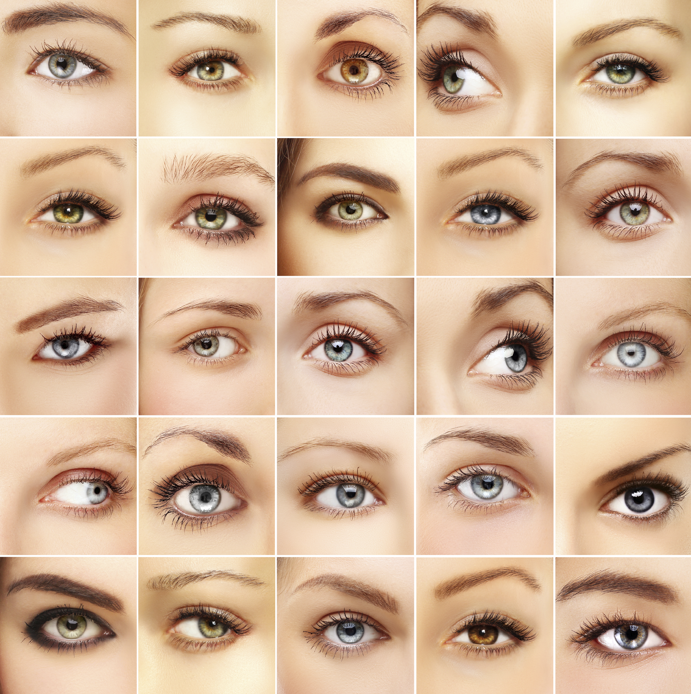
We all know that eye color is genetic, depending on the genes we inherit from our parents. We used to think that brown eyes were “dominant” and blue eyes were “recessive,” but modern science has shown that eye color is not at all that simple. Human eye color originates with three genes, but there may be as many as 50 genes that have influence over eye color. Most people in the world have brown eyes, with the second most common colors being blue and grey. Green is the rarest color.

What gives you your eye color?

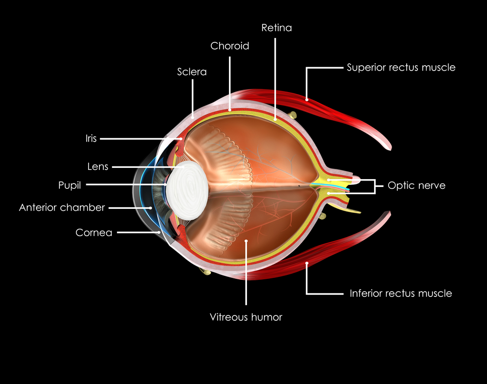
The color of your eyes depends on how much of the pigment melanin you have in your iris. The more pigment you have, the darker your eyes will be. Blue, grey, and green eyes are lighter because there is less melanin in the iris. Most Caucasian babies are born with blue eyes because melanin is not present at birth, but develops with age. However, by the age of three their eye color is determined.

A contributing factor is also your stroma, the front layer of the iris that contains fibers that scatter the light that is reflected outward. This helps to account for why you your eyes might appear to change color or intensify depending on what color you are wearing, amplifying the scattered light reflecting back.

Some children are born with eyes that are not the same color. This can be caused by faulty developmental pigment transport, trauma either in the womb or shortly after birth or a benign genetic disorder. However, because there could be a small chance of it indicating eye disease, such as Horner’s syndrome, it is suggested you have an early eye exam to make sure nothing serious is going on.

Can your eyes change color?

Contrary to popular opinion, your eyes do not change color based on your emotions. The iris is a muscle that expands and contracts to control the size of your pupil. It gets bigger in dim lighting and gets smaller in bright lighting. It also shrinks when you are focusing on close tasks, such as reading a book. Certain emotions can also change the pupil size, such as anger, grief or happiness. This can cause the pigments in the iris compress or spread apart, slightly changing the appearance of your eye color. Also because the pupil is black, your eyes appear darker. There are times when your eye color might darken slightly, such as puberty, pregnancy, and aging for 10%-15% of Caucasians with light-colored eyes.

What else does melanin do?

Besides giving our eyes color, melanin helps to protect them from the sun. Because they have less pigment, light eyes are much more sensitive to harmful UV rays from the sun and electronic devices than brown or black eyes. This makes UV protection even more important for babies and people with light-colored eyes.

Finally, because some people believe that the eyes are the windows to the soul, there are some superstitions about eye color. People with blue eyes are thought to have rich imaginations, people with green eyes have sharp minds, hazel eyes indicate a passionate soul and people with brown eyes are calm, but have underlying passion. What does your eye color say about you?

3/17/16


Susan DeRemerSusan DeRemer, CFRE
Vice President of Development
Discovery Eye Foundation

Women’s Eye Health

With this being Women’s History Month, we thought it appropriate to discuss women’s eye heath. Your vision is one of the most important things in your life. Vision loss can make everyday tasks more difficult, impede your work and lead to depression.
women's eye health
According to Prevent Blindness America (PBA), 66% of people who are blind or visually impaired are women. This is primarily due to the fact that women have more risk factors for vision loss than men. Sadly the same study also revealed that only 9% of women realize this. Early intervention can help prevent blindness in many cases. Many blinding eye diseases can be treated to prevent blindness and almost all eye injuries can be prevented.

Here is a closer look at why women are affected more than men, and what condition they need to be aware of for early diagnosis and sight-saving treatments.

Why Women Lose Vision

  • They live longer than men and many eye diseases are age-related. Examples are cataracts, macular degeneration and diabetic retinopathy. The rates of these diseases are increasing as the baby boomer population ages.
  •  

  • Some eye diseases are intrinsically more prevalent among women, like dry eye syndrome which is believed to be linked to hormones. It is 2-3 times more likely in women than men. Hormonal changes can influence vision changes across the life span of a woman, from pregnancy to post-menopause.
  •  

  • Women have higher rates of autoimmune diseases such as lupus, rheumatoid arthritis and multiple sclerosis. The serious side effects of these conditions can affect your eyes, causing vision loss.
  •  

  • Social and economic factors can access to health care for women restricting early detection and treatment which could prevent or limit vision loss.
  •  

  • While behavioral and environmental factors are not restricted to women, poor nutrition and obesity can cause diabetes and subsequent diabetic retinopathy; while smoking is also a proven risk factor for eye diseases such as cataracts and macular degeneration. Women under 23 are the fastest growing segment of new smokers.

women's eye health
Eye Conditions More Prevalent in Women

  • Cataracts are the world’s leading cause of blindness. They are the result of a clouding of your eye’s normally clear lens. They can be treated with cataract surgery, where the cloudy lens is replaced with a clear synthetic lens.
  •  

  • Glaucoma is the second-leading cause of blindness in the Us. It is called the silent thief of sight because there is no warning. It happens when pressure in the eye — intraocular pressure or IOP — is too high, damaging the optic nerve which sends vision signals to the brain. Open-angle glaucoma, the most common type, affects men and women equally. But women are 2-4 times more likely than men to get closed-angle glaucoma. One of the possible reasons for this is that the front chamber between the iris and cornea is shallower in women than men and can block fluids from draining out of the eye, thus increasing pressure. Glaucoma is also genetic, meaning you are at higher risk if someone in your family has had glaucoma. When caught early, there are treatments that can help control your IOP. If it is not controlled early, blindness can result and it is irreversible.
  •  

  • Age-related macular degeneration (AMD) is the number one cause of vision loss in people over 40 in the US. One of the reasons women are at higher risk is because they tend to live 5-7 years longer than men. AMD gradually destroys the central part of your sight that helps you read or drive. There are two types of AMD. Dry AMD, which occurs when drusen accumulate under the retina. There is no treatment for dry, yet accounts for 90% of the cases. Wet AMD occurs when new blood vessels grow and leak between the retina and eye’s outer layer. There is a treatment of anti-VEGF injections for this version of AMD. The earlier the disease is diagnosed and treated, the better the results. Like glaucoma, it is an inherited eye disease.
  •  

  • Diabetic retinopathy is a result of diabetes. When blood sugar levels are too high, the vessels that feed the retina weaken and eventually leak, which cause the macula to swell. In its most dangerous form the retina may detach from the back of the eye leading to blindness. Diabetes is also hereditary, but controllable with early diagnosis and treatment.
  •  

  • Dry eye syndrome occurs when your eyes produce too few tears. Your eyes may burn, sting or feel gritty, making your vision blurry or you may blink more. Contrary to the name, you may appear weepy as the dryness may stimulates more tear production. Thanks to hormones, women are more susceptible. In postmenopausal women, the shift in balance between estrogen and progesterone can be responsible.
  •  

  • Pregnancy-related eye changes, like menopause, are caused by hormone shifts and can cause several temporary eye conditions, such as dry eye and corneal swelling.

3/10/16

Susan DeRemerSusan DeRemer, CFRE
Vice President of Development
Discovery Eye Foundation

The Brain and the Eye – How They Work Together

The Brain and the Eye

The eye works like a camera. The iris and the pupil control how much light to let into the back of the eye, much like the shutter of a camera. When it is very dark, our pupils get bigger, letting in more light; when it is very bright our irises constrict, letting in very little light.

The lens of the eye, like the lens of a camera, helps us to focus. But just as a camera uses mirrors and other mechanical devices to focus, we rely on eyeglasses and contact lenses to help us to see more clearly.

The focus light rays are then directed to the back of the eye, on to the retina, which acts like the film in a camera. The cells in the retina absorb and convert the light to electrochemical impulses which are transferred along the optic nerve to the brain. The brain is instrumental in helping us see as it translates the image into something we can understand.

The Brain and the Eye

The eye may be small, but it is one of the most amazing parts of your body. To better understand it, it helps to understand the different parts and what they do.

Choroid
A layer with blood vessels that lines the back of the eye and is between the retina (the inner light-sensitive layer that acts like film) and the sclera (the outer white part of the eyeball).

Ciliary Body
The muscle structure behind the iris, which focuses the lens.

Cornea
The very front of the eye that is clear to help focus light into the eye. Corrective laser surgery reshapes the cornea, changing the focus to increase sharpness and/or clarity.

Fovea
The center of the macula which provides the sharp vision.

Iris
The colored part of the eye used to regulate the amount of light entering the eye. Lens focuses light rays onto the retina at the back of the eye. The lens is transparent, and can deteriorate as we age, resulting in the need for reading glasses. Intraocular lenses are used to replace lenses clouded by cataracts.

Macula
The area in the center of retina that contains special light-sensitive cells, allowing us to see fine details clearly in the center of our visual field. The deterioration of the macula can be common as we age, resulting in age related macular degeneration.

Optic Nerve
A bundle of more than a million nerve fibers carrying visual messages from the retina to the brain. Your brain actually controls what you see, since it combines images. Also the images focused on the retina are upside down, so the brain turns images right side up. This reversal of the images Is a lot like what a mirror does in a camera. Glaucoma can result when increase pressure in the eye restricts the flow of impulses to the brain, causing optic nerve damage and makes it difficult to see.

Pupil
The dark center opening in the middle of the iris changes size to adjust for the amount of light available to focus on the retina.

Retina
The nerve layer lining the back of the eye that senses light and creates electrical impulses that are sent through the optic nerve to the brain.

Sclera
The white outer coating of the eyeball.

Vitreous Humor
The clear, gelatinous substance filling the central cavity of the eye.

3/3/16

Susan DeRemerSusan DeRemer, CFRE
Vice President of Development
Discovery Eye Foundation

Pink Eye Tips and Prevention

pink eye
Pink eye is an inflammation or infection of the thin, clear covering of the white of your eyeball (the conjunctiva) and the inside of your eyelids. When the small blood vessels in the conjunctiva become inflamed they are more visible making the whites of your eye to appear pink. Also called conjunctivitis, it can affect one or both eyes.

Common symptoms of pink eye include:

  • Redness in the white of the eyeball(s) and or inner eye lid(s)
  • Increased tearing or discharge
  • Slightly blurred vision from discharge
  • Crusting of the eyelashes from the discharge that may prevent eyes from opening after sleep
  • Mild eyelid swelling
  • Itching or burning sensation
  • Increased sensitivity to light
  • Irritation or gritty feeling in your eye(s)

Make an appointment with your eye doctor if you notice and of the symptoms of pink eye. Some forms are highly contagious for as long as two weeks, so an early diagnosis could protect those around you from contacting the disease. If you were contact lenses, stop using them until directed by your doctor.

There are four general types of pink eye.

Allergic Conjunctivitis
This form is caused by eye irritants such as pollen, dust, animal dander and other environmental factors. It is not contagious. Treatment often includes applying a cool compress to your eyes and using allergy eye drops and artificial tears. In severe cases non-steroidal and anti-inflammatory medications may be prescribed.

Bacterial Conjunctivitis
This type is most often caused by staphylococcal or streptococcal bacteria, is highly contagious and can cause serious damage to the eye if left untreated. This is treated with antibiotic eye drops or ointments to speed up the healing process that can take one to two weeks. While you may see improvement after three to four days, the entire course of treatment needs to be used to prevent a recurrence.

Because this is so highly contagious here are a few things to remember so you don’t spread it to others or re-infect yourself:

  • Don’t touch your eye with your hands
  • Wash your hands frequently and thoroughly
  • Change towels and washcloths daily – and don’t share them
  • Change pillowcases often
  • Get rid of all eye cosmetics and personal care items such as eye creams – and don’t share them
  • Avoid swimming
  • Don’t reuse tissues when wiping your eyes, and throw them out immediately
  • Follow your eye doctor’s instructions related to your contact lens usage and care

Viral Conjunctivitis
This is the same type of virus associated with the common cold. Antibiotics will not work on a viral infection. Like a cold, the infection just needs to run its course which could take anywhere from a few days up to 2-3 weeks. It is also contagious like a cold, so follow the same instructions as listed above to not spread the infection.

Chemical Conjunctivitis
This can be caused by irritants like air pollutions, chorine in swimming pools or exposure to noxious chemicals. To treat this type of pink eye requires a doctor to carefully flush your eyes with saline and may require topical steroids. Acute chemical injuries are very serious and need prompt medical attention to avoid corneal scarring, intraocular damage, vision loss or the loss of an eye.

Of course the best way to deal with pink eye is not to get it. Here are some ways to protect yourself and others.
pink eye

2/24/16

Susan DeRemerSusan DeRemer, CFRE
Vice President of Development
Discovery Eye Foundation

4 Super Greens for Better Sight

A healthy diet not only helps your heart, but also your eyes. Your diet should include lots of fruits and vegetables to provide you with a natural source of nutrients to help protect your sight. As wonderful as supplements are, eating the actual foods is always better. Some of the best vegetables for you are the dark, leafy greens that are rich in valuable vitamins and nutrients. These are the super greens for better sight.

With the US experiencing bitter, freezing temperatures on the East Coast, while the West Coast is having summer in February, with record-breaking hot temperatures, I thought it would be interesting to see how you could enjoy those super greens, no matter what the temperature is outside. Below is a quick look at four dark leafy greens that are a great addition to a healthy diet, watercress, arugula, spinach and kale. For each vegetable I have included a recipe that is served hot, along with one that is served cold.

Here is what you need to know about super greens for better sight.
super greens
WATERCRESS

Watercress is a cruciferous plant and part of the brassica family, like arugula and kale. It contains vitamins A, B6, B12, C, K, iron, magnesium, calcium, phosphorus, lutein and zeaxanthin. In fact, weight for weight, watercress contains more vitamin C than an orange, more calcium than milk, more iron than spinach and more folate than bananas. Watercress has the highest amount of nutrients for the smallest amount of calories.

The health benefits of watercress include boosting immunity, cancer & diabetes prevention, reducing cognitive decline, thyroid support, better cardiovascular health and stronger bones. As for your eyesight, it can help prevent or slow the onset of age-related macular degeneration and possibly cataracts.

Watercress is most commonly enjoyed fresh in salads, but can also be use in pastas, casseroles, soups and sauces. Choose watercress with deep green, crisp leaves, with no signs of wilting. Trim the stems, rinse the greens in cold water and dry. It is best if used immediately, but can be store for up to four days in the refrigerator.

Watercress Soup by William Anatooskin

Watercress and Grapefruit Salad by Martha Stewart
super greens
ARUGULA

Arugula is also known as a salad or garden rocket. It is a small low growing herb that is packed with vitamins, minerals and antioxidants. It is rich in folates, vitamin A, B-complex, C and K and has copper, iron, calcium, potassium, manganese and phosphorus.

The health benefits of arugula include a lowered risk of cancer, healthy bones, strengthened brain function, improved mineral absorption and it boosts the immune system. Because of being a source of carotenoids, it also helps to slow the progression of age-related macular degeneration.

Arugula is popular in salads, used with milder greens to add a peppery flavor. But it can also be used in pastas, casseroles, pizzas, soups and sauces. Choose arugula that is crisp with green young leaves. Avoid the flowered harvest as those leaves are tough and have a bitter taste. Wash leaves in a bowl of water, swishing thoroughly to get rid of all sand and soil. Drain and pat dry before storing in the vegetable bin of your refrigerator for no more than a few days.

Grilled Stuffed Swordfish by Stormy Scarlett

Pasta Salad with Goat Cheese and Arugula by Martha Stewart
super greens
SPINACH

Spinach is a very popular leafy green vegetable, with two common varieties cultivated for food; the savory-type with dark green crinkled leaves and the flat-leaf type with smooth surfaced leaves. Spinach contains vitamin A, B-complex, C and K, along with lutein, zeaxanthin beta-carotene, potassium manganese, magnesium, copper and zinc.

The nutrients in spinach help improve blood glucose control in diabetes, lower the risk of certain cancers, reduce blood pressure, increase bone health and help iron deficiency. The lutein, zeaxanthin and beta-carotene help to potentially prevent and slow the progression of age-related macular degeneration. Lutein also works to protect the eye from free radical damage by helping filter out damaging blue and ultraviolet light.

Spinach is a universally popular vegetable, used around the world in a variety of ways, including salads, soups, noodles, pies, casseroles, dips, sauces, etc. Look for leaves that are dark green in color, crisp and not dull or yellow and spotted. Wash thoroughly to remove sand and soil, dry, trim away tough stems and store in the refrigerator for up to a week.

Turkey-Spinach Meatballs from Bon Appètit

Spinach Salad with Dates from Bon Appètit
super greens
KALE

Kale is a member of the mustard and cabbage families and has more nutrients than spinach. Less than ½ cup has 333% of the recommended daily allowance (RDA) of vitamin A, 587% RDA of vitamin K and 200% RDA of vitamin C. This frilly-leafed vegetable also has vitamin B-complex, lutein, zeaxanthin, beta-carotene, copper, calcium, sodium, potassium, iron, manganese, and phosphorus.

The health benefits of kale include healthy muscles and skin, improved blood glucose control, lower colon and prostate cancer risk, better cardiovascular health, stronger bone health, reduced neuronal brain damage and support for red blood cell formation. The advantage for your eyes comes from the lutein, zeaxanthin, beta-carotene and vitamin A, all which work to support a healthy retina. They help protect against blue and ultraviolet light as well as the early onset and progression of age-related macular degeneration. Because of the positive impact on diabetes it also reduces the onset and progression of diabetic retinopathy.

Kale is very versatile and can be served in a variety of ways including salads, soups and casseroles. It can also be braised, broiled, sautéed and even made into kale chips by tossing them in extra-virgin olive oil and sprinkled with your choice of cumin, curry powder, chili powder, red pepper flakes or garlic powder and baking at 275 degrees for 15-30 minutes depending on how crisp you want them to be. When shopping for kale look for leaves that are crispy and crunchy with a brilliant dark blue-green color. Wash thoroughly to remove soil and sand, dry well, and remove all tough stems. It is extremely perishable, so use it as quickly as possible.

Kale and Chicken Casserole by Martha Stewart

Kale with Pomegranate Dressing and Ricotta Salata from Bon Appètit

All of these dark green leafy vegetables are not only healthy for you, but can be used in many ways to make it easy to incorporate them into your diet. Here are a few ideas:

  • Throw a small handful into your blender when making your favorite smoothie
  • Add them to your next omelet or egg scramble
  • Use them for making pesto or adding to pasta sauce
  • Sauté with a small amount of extra-virgin olive and season with freshly ground black pepper and freshly grated Parmesan cheese to serve at a topping for your baked potato
  • Add it to your wrap, sandwich or flatbread

2/17/16

Susan DeRemerSusan DeRemer, CFRE
Vice President of Development
Discovery Eye Foundation

Understanding Your Eyeglass Prescription

If you need eyeglasses it is because you were diagnosed with a refractive error during your eye exam. This means your eyes have trouble focusing and images can appear blurry. Types of refractive errors include being near-sighted (myopia), far-sighted (presbyopia) or having an astigmatism (your cornea is an irregular shape).
Understanding Your Eyeglass Prescription

Understanding What 20/20 Vision Means

The number is based on you standing a distance of 20 feet (considered to be the norm) in front of a Snellen or Tumbling E eye chart to test your visual acuity. If during an eye test you can read the big E at the top of the eye chart, but none of the letters below that, your vision is considered 20/200. That means you can read a letter at 20 feet that people with “normal” vision can read at 200 feet, meaning you have very poor visual acuity. If you can read the fourth line from the bottom of most charts, you vision is 20/20. Any line below that would be 20/15 or below and indicates that you have exceptional visual acuity. Each eye is tested separately as your eyes are designed to compensate for each other and would not give an accurate reading.

Understanding Your Eyeglass Prescription

The prescription you receive may be a chart with headings that are filled in, or it may just be written out. In any case, it is written in a specific order, with the results for each eye listed first.

  • OD is an abbreviation for oculus dexter, meaning right eye, while OS is oculus sinister, meaning left eye. If both eyes are being referred to the abbreviation you will see is OU (oculus uterque) meaning both eyes.
  • The next term on your prescription is Sphere (SPH) and is are measured in diopters (D). This number tells you how strong your lenses need to be to correct your vision. If this number has a minus sign (–), you are nearsighted; if the number has a plus sign (+) or is not preceded by any sign, you are farsighted.
  • Next may be cylinder (CYL), an indicator of the lens power if you have an astigmatism. The number for the CYL has the same format as the SPH, a minus sign (for the correction of nearsighted astigmatism) or a plus sign (for the correction of farsighted astigmatism). If there is no number given you either have no astigmatism, or your astigmatism is so slight that it is not really necessary to correct it.
  • If you do have a CYL number you will also have an axis number, based on a protractor scale that tells you where on the eye the astigmatism is found. For example, SPH –2.50 D CYL +5.00 Axis 40 means you have a nearsightedness of -2.50 dioptics with an astigmatism of +5.00 along the 40 degree axis.
  • If you are getting bifocals or progressive lenses, there is another number called an “Add.” This number is the amount of additional correction your eyes need to focus at close distance and will always be a positive number, whether or not the + sign is there.

Needless to say, eyeglasses are expensive, but understanding a prescription should help you understand why. The stronger the lens the more expensive it is and then with each adjustment, the cost also increases. Also don’t forget that anti-glare or UV protective coatings add to the costs, but may be well worth it. Eye glasses are an investment in your safety, job, independence and letting you enjoy any leisure-time activities you enjoy.

2/9/16


Susan DeRemerSusan DeRemer, CFRE
Discovery Eye Foundation

Having Trouble with Your Scleral Lenses?

Scleral lens have become very popular and while many enjoy the comfort and vision correction they provide, some find it difficult to apply (insert) and remove these large diameter RGP lenses. If you are one of the many who are having difficulty managing to get your scleral lenses in or out, there are a number of tools available to help.

The Scleral Lens Education Society website provides a wealth of information about the care and handling of these lenses trouble with your Scleral Lenses as well as an excellent video. There are photos demonstrating various ways to hold the lens while applying it …some you may not have tried! If you have trouble with your schleral lenses, see below.

Troubleshooting tips and tricks:

    • If you are unable to maintain fluid in the bowl of the lens as you bring it towards your eye, make sure that your face is fully parallel to the floor. It may seem like you are nearly standing on your head when you’re in the correct position to apply the lens.
    • Lid control is essential; use one hand to hold lids completely out of the way, and don’t release the lids until the lens is actually fully in place and the plunger (or your finger tripod) has been removed.
    • If you are unable to successfully apply a solid lens with saline, you could practice applying the lens after filling the bowl of the lens with Celluvisc™ or another non-preserved viscous lubricant. These viscous lubricants will blur your vision compared to saline, however, so you may simply want to use them to practice lens application. Once you’ve mastered this step, switch to saline to give you better vision.
    • Try to keep both eyes open as you apply your lenses. This may also help you to position the lenses correctly.
    • If you are using a bulbed (DVM) plunger, and can see the opening in the center of the suction cup, look directly at the hole as you bring the lens into position. This will help you to position the lens correctly.

    Many find the DVM plungers helpful. They are readily available at your doctor’s office and online. These are just a few of the places to find them: DMV Corp, Dry Eye Zone, and Amazon.

    trouble with your schleral lensesAnother variation to the standard lens inserters is a ring-style lens applicator by EZI Scleral Lens. It was designed by a post-transplant patient who like so many, had trouble inserting his scleral lens without getting a bubble. Read Tim’s story.

    If you have tried the above techniques and still have trouble applying scleral lenses there are a number of devices available that may help. Dalsey Adaptives has developed the See-Green devicetrouble with your schleral lenses that can be used to help successfully apply scleral contact lenses. The See-Green system comes with a stand that holds a lighted plunger (Figure 2). Using this system, you don’t hold the lens, you lower your eye onto it, which leaves both hands free for improved lid control. The light at the center of the plunger is used as a target to help you position the lens centrally on the eye. Click here to see the detailed instruction sheet.

    Scleral lenses offer good vision and comfort but can be challenging to manage. Discuss these options with your eye care professional to get his or her recommendation for your specific situation.

    1/28/16


    CathyW headshotCathy Warren, RN
    Executive Director
    National Keratoconus Foundation

Layers of the Retina

The retina at the back of the eye is essential for all vision. Each layer of cells in this tissue serves a specific purpose. As we prepare for Age-Related Macular Degeneration Awareness Month in February, a closer look at the layers of the retina and their function.

layers of the retina

Layers of the Retina

Choroid – This is made up of a layer of blood vessels that supply oxygen and nutrients to the retina. Defect in the CHM gene can cause choroideremia, leaky blood vessels can expand in the retina causing wet age-related macular degeneration (AMD) and diabetic retinopathy.

Retinal pigment epithelium – This is a single layer of cells that provide essential nutrition and waste removal for the photoreceptor cells. Accumulation of waste can lead to AMD and Stargardt disease.

Photorecptors – This is where the rods and cones are located that convert light into electrical signals. Rods help you with night and peripheral vision. Cones are more concentrated in the macula (the central part of the retina) and proved central and color vision. Death of the rods can cause vision loss called retinitis pigmentosa, while AMD is the loss of central vision.

Horizontal cells – These cells are connect to the photoreceptors that surround the bipolar connected photoreceptor cells and help the help integrate and regulate the input from multiple photoreceptor cells, increasing your visual acuity.

Bipolar cells – The dependence of each layer of the retina on each other is exemplified here. These cells take the electrical information from the photoreceptor cells and pass it along to other retinal cells.

Ganglion cells – These cells extend to form an optic nerve that conveys information to the brain and take the electrical information from the bipolar cells and process it to determine shapes, contrast and color. Glaucoma vision loss results from high intraocular pressure that affects the optic nerve, interrupting the signals to the brain.


Wearing Contact Lenses in Winter

Wearing Contact Lenses in Winter This has been a cold winter so far, and since it is only January, it is bound to get colder. The extreme cold, combined with winds, snow, rain and other environmental factors, can really take a toll on your eyes. And while it may be snowing or raining, winter air is actually drier than any other season. This can be especially difficult if you wear contact lenses. Here is what you should know about wearing contact lenses in winter.

  • Wear sunglasses for protection from UV rays and wind. Your eyes can become sunburned which cause blurry vision and can make your eyes feel like they are burning (think of your sunburned skin feels) for 24 to 72 hours. It will also protect your eyes from snow, rain or anything else the wind can send your way.
  • Avoid direct sources of heat such as heating vents and fireplaces. Indoor heating can draw the moisture out of the air, so consider a humidifier to help maintain the correct amount of moisture in the air to help keep eyes moist. Cool-air humidifiers have less of a tendency toward mold and bacteria.
  • Speaking of hydration, we also tend to drink less water in the winter months, so make a concentrated effort to keep up your water intake.
  • If it is so dry, why are my eyes watering? This is a common question and the answer may be a bit counter-intuitive. Anything that irritates your eyes, including dryness, causes a tearing reflex. Your tear glands go into overdrive trying to replace the moisture to your cornea. To try and reduce the tearing, you can use eye drops or artificial tears specifically designed for use with contact lenses.
  • Your eyes are not the only thing that dries out in the winter, so does your skin. Try to put in your contacts before moisturizing your skin, especially your hands. So wash your hands, put in your lenses and then use your creams and lotions.
  • Change out your contact lenses regularly in cold weather according to the recommended schedule, be it daily, every two weeks or monthly. This will allow them to better conduct oxygen, reduce irritation and increase comfort.
  • Take a break from your contacts and wear your eyeglasses. Putting them on when you get home from work can make a big difference. Contact lenses dry your eyes out on their own, when you add cold weather it gets that much worse.
  • Get plenty of sleep, which also helps with the dryness and fatigue. This will help you start the day with your eye refreshed and ready for the many things you will put them through throughout the day ahead.

Do you have any other suggestions that have helped you cope when wearing contact lenses in winter?

1/15/16


  Susan DeRemerSusan DeRemer, CFRE Vice President of Development Discovery Eye Foundation

7 Tips for Living With Glaucoma

living with glaucoma
Did you know January is Glaucoma Awareness Month?  Although no cure exists, the eye disease may be treated with medication and surgery.  If left undiagnosed and untreated, however, glaucoma can cause permanent damage to vision.  According to the National Eye Institute, glaucoma affects more than three million people in the United States. The population that is at highest risk is adults over the age of 60. Glaucoma affects vision by damaging the optic nerve and typical vision loss occurs in the peripheral visual field. This type of loss can create difficulty performing everyday tasks. To mark the importance of Glaucoma Awareness Month, Low Vision Focus @ Hadley offers these tips to improve independence and safety for people with glaucoma-related vision impairments.

Tips for Living With Glaucoma

  1. Loss of peripheral vision makes it difficult to see steps and stairways.  Marking treads and handrails with contrasting colored paint or tape can improve navigation and reduce the risk of falling.
  2. Persons with glaucoma often experience difficulty adjusting to darkness or darkened rooms. Increase illumination in dark closets or hallways by installing additional lighting fixtures. When outdoors at night, carry a strong flashlight.
  3. Area rugs can pose a hazard for persons with visual field loss.  It’s best to keep home pathways and work areas free of extra floor coverings.
  4. In all locations where you might be a frequent visitor, ask someone to give you a tour.  It goes without saying you need to know where the bathroom is located, as well as how to exit the building from a variety of locations.
  5. Some persons with glaucoma prefer to use a human guide when traveling in unfamiliar places.  To use a human guide effectively, grasp the guide’s arm firmly just above the elbow and walk a half a step behind. This positioning gives the most protection from potential obstacles and allows both people to function as a team.
  6. Get in the habit of consistently closing kitchen and bathroom cabinets, especially those above countertops.  Make sure doors are either all the way open or shut.  Practicing both of these safety techniques can greatly reduce the risk of head injury.
  7. Before reaching down to pick up dropped objects, place your hand, palm out, about 12 inches in front your face.  This way, you’ll make sure you don’t hit the edges of tables or countertops with your forehead.

Remember, although glaucoma cannot be cured, it can be treated.  Regular eye exams, especially for adults over 60, are critical to control the spread of the disease.  For those who are affected by vision loss, it’s important to remember that the acquisition of some simple adaptive skills and techniques can help maintain safety and independence.  Low Vision Focus@Hadley is dedicated to assisting persons with low vision to live life to the fullest all year long. For more information on how Low Vision Focus@Hadley can help you or someone you know, please visit our website www.lowvisionfocus.org, or call (855) 830-5355 to find out about our free programs and materials.

1/12/16

Ed Haines - living with glaucomaEd Haines
The Hadley School for the Blind
Low Vision Focus @ Hadley