Help for Computer Users

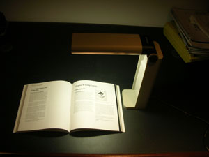
Working long hours in front of the computer requires a fairly unchanging body, head and eye position which can cause discomfort.  Correct working position, periodic stretch breaks, frequent eye blinking, artificial tears for lubrication are all very important.  However, it’s not always easy to remember this when you are engrossed in work. Here are a few fun, free and easy-to-install “break reminders” to help:

WorkSafe Sam - break reminder
WorkSafe Sam
WorkSafe Sam is a desktop tool that provides stretching tips to help reduce eye and muscle strain for office workers (clicking on this link will open a file on your computer because this is a zip file).

Workrave is break reminder program that alerts you to take “micro-pauses” and stretch breaks.

Take Your Break is another break reminder designed to prevent or minimize repetitive strain injury, computer eye strain and other computer related health problems.  It has a friendly interface and a tray icon status indicator.  It runs quietly in the background, monitoring your activity and reminding you to take regular breaks.

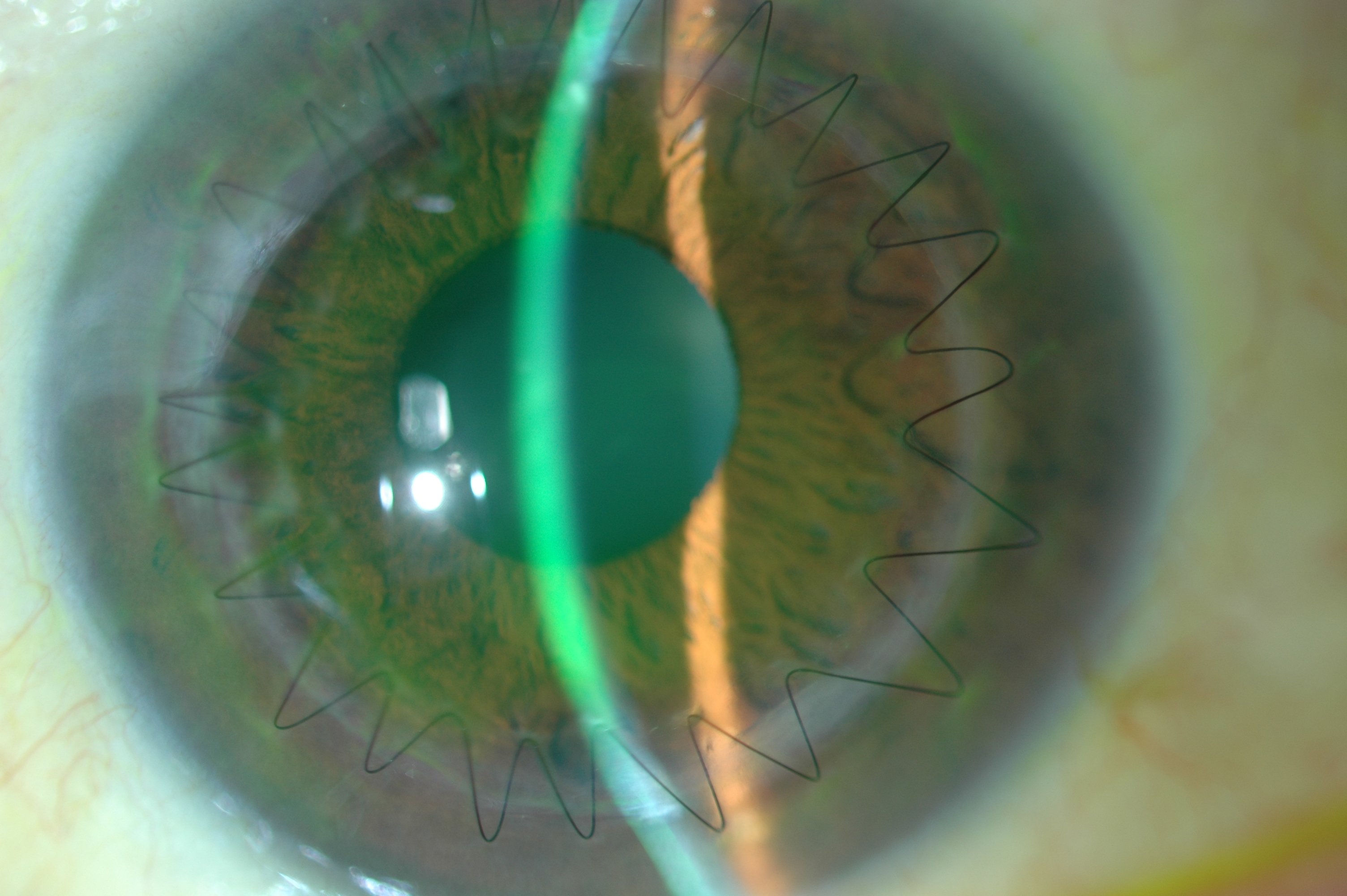
And remember to blink.  Blinking cleans the ocular surface of debris and flushes fresh tears over the ocular surface. Each blink brings nutrients to the eye surface structures keeping them healthy. The flow of tears is responsible for wetting the lower third of the cornea. This is very important in KC, since this area is generally below the bulge of the cone and in many cases irritated by wobbly RGP lenses.  Maybe your job requires hours of work at a computer. Maybe you like to spend your free time surfing the internet. Whatever the reason, your body is probably feeling the effects of spending too much time staring at a computer monitor, which could result in Computer Vision Syndrome (CVS).  The most common symptoms are: eye strain, dry or irritated eyes,redness in eyes,difficulty in refocusing eye,neck pain,double vision,blurred vision, fatigue, and headaches.

Please join us on Thursday when Dr. Bezalel Schendowich will be providing a detailed insight into the importance of blinking, going beyond computer usage.

CathyW headshotCathy Warren, RN
Executive Director
National Keratoconus Foundation

What Are The Differences In The Immune System of An Age-related Macular Degeneration Patient?

A symposium was held in Bethesda, Maryland at the National Institutes of Health on March 6, 2014.  The purpose was to bring together clinicians and researchers from a wide variety of background to discuss the recently discovered differences in the function of the immune system in patients with age-related macular degeneration (AMD) and how it relates to aging.  These differences are important to understand because they may lead to new therapeutic approaches to treat people which are high risk for AMD.

Courtesy of National Eye Institute, National Institutes of Health - immune system
Courtesy of National Eye Institute, National Institutes of Health

  • Three researchers associated with the Discovery Eye Foundation attended the one-day conference.  Cristina Kenney, MD, PhD, has worked in the field of AMD for over twelve years and is a leading expert on the genetics and molecular changes in the mitochondria as it related to the retinal cell death associated with AMD.
  • Lbachir BenMohamed, PhD, is an expert immunologist with a tremendous understanding of how the immune system responds to infections and stress.
  • Anthony Nesburn, MD, has been involved in AMD research at both the clinical and research levels.

By attending this meeting, all three researchers gained insight into the importance of the immune system with respect to maintaining a healthy retina and slowing the progression of AMD.

The highlights of the meeting were the following:

1.  Emily Chew, MD (National Eye Institute) reviewed the clinical aspects of the disease stressing that there are many stages of AMD.

The early stage of AMD is diagnosed based upon the medium-size drusen (about the width of an average human hair) that can be seen underneath the retina.  There may not be any visual changes in these patients.

The intermediate stage of AMD is when subjects have larger drusen and some degree of retinal pigment epithelial cell drop out.  These patients may not have vision loss or other symptoms.

The late stages of AMD which can be categorized into the dry form (geographic atrophy) which has significant loss of the retinal pigment epithelial cells and overlying photoreceptor cells.  Presently there in no treatment for this type of AMD.

These individuals can have changes that cause decreased vision.  The second form of late AMD is the wet form (neovascular), which has growth of abnormal blood vessels beneath the retina that can cause significant loss of vision.  The treatments for this type of AMD are anti-VEGF medications that block the growth of these vessels and help maintain good visual acuity.

2.  Anand Swaroop, PhD (National Eye Institute) reviewed the genetics of AMD and summarized the work of numerous laboratories.  It is now recognized that there are over 20 different genes associated with AMD.  These genes fall into the categories of those involved with Complement Activation, Cholesterol Pathway, Angiogenesis, Extracellular Matrix and Signaling Pathways.  Many of these genes have additive effects, meaning that if a patient has more than one high risk gene, then the likelihood of developing AMD increases.  While we have learned a lot about the genes that are important, we still do not have any gene therapies that can be used to treat AMD.

3.  Six different speakers presented their data related to animal models of AMD and it was agreed that there is not a “perfect” model because most of the animals do not have a macula, the region of the retina that is affected the most by AMD.  However, there is still a lot to be learned by using the models that we do have because if we can better understand the basic pathways involved, then we can block or modify the pathways to prevent the damage.

4.  Jayakrishna Ambati, PhD (University of Kentucky) presented data showing that there is a deficiency of an enzyme called DICER1 in the retinal pigment epithelial cells which leads to increased activation of inflammation via a protein complex called the inflammasome.  He described some of the signaling pathways which are involved in the inflammasome activation.  This is important because these pathways can become targets for treatment of the dry form of AMD.

5.  Jae Jin Chae, PhD (National Human Genome Research Institute) also talked about the role that inflammation plays in the development of AMD.  The data presented reviewed the pathways involved with activation of the inflammasomes which is the first step in a cascade of events that result in inflammatory diseases.  They have identified a calcium-sensing receptor (CASR) which triggers the activation of the NLRP3, a key component of the inflammasome.  Understanding how this series of events works allows researchers to develop medications to block or interfere with the pathway and therefore decrease the levels of inflammation.  

Dr. M. Cristina KenneyM. Cristina Kenney, MD, PhD
Professor and Director of Ophthalmology Research
School of Medicine, Dept. of Ophthalmology
University of California, Irvine

Is Omega-3 Important to Your Diet?

Last week a study that appeared in the Annals of Internal Medicine (AIM) showed that fish oils such as omega-3 don’t reduce the risk of heart disease.  It caused quite a stir, as we have been told for years, by organizations like the American Heart Association that we needed to increase our intake of omega-3.  For some people that meant taking supplements instead of eating flax seeds, walnuts, sardines or salmon, just a few of the sources of omega-3. omega-3 sources

In May of 2013 the results of the AREDS II study on eye supplements found that while lutein and zeaxanthin may be helpful in helping vision, omega-3 did not have a positive effect over five years.

In both of these studies they are referring to omega-3 supplements.  This does not mean you should give up eating fish, or other sources of omega-3.  To derive the benefits of omega-3 you need to get it from the source –  directly from the food you eat, so it is still advised you eat fish as part of a healthy diet.

If you do use supplements check with your doctor to make sure they are right for you.  A doctor can help you determine what, if anything you might need, making a decision based on your diet, medical history and any medications you might be taking.

If you want to learn more about dietary supplements, here is a fact sheet from the National Institute of Health.

Remember – supplements are what the name implies – something to supplement a well-rounded diet – they are not a substitution.

Susan DeRemerSusan DeRemer, CFRE
Vice President of Development
Discovery Eye Foundation

Low Vision Resources

What to do when “There’s nothing more that can be done.”

“I’m sorry, but there’s nothing more that can be done. There is no cure for your eye condition.”

In your work as healthcare professionals and health educators, it’s likely you’ve encountered a significant number of adults and older adults who have been on the receiving end of this devastating news.

When an eye care provider says, “There’s nothing more that can be done,” what he or she likely means is, “There’s nothing more I can do for you surgically.” But instead of saying, “There’s nothing more that I can do,” the discouraging message delivered to the patient is, “There’s nothing more that can be done.”

Thus, in many cases, the discussion ends there. Patients will either exhaust their resources searching for an elusive cure or become resigned to a life that is restricted and defined by incurable vision loss.

When receiving a diagnosis of vision loss, many adults who have managed to overcome a host of obstacles in their lives may now believe they are facing an obstacle with no viable solution. A natural, and understandable, initial reaction is to focus instead on the devastating losses that are seen as an inevitable accompaniment to blindness and low vision such as:

  • Loss of independence: “How will I prepare meals, clean my home, or shop? Will I become a burden to my family and friends?” 
  • Loss of confidence and self-worth: “All my life I’ve been physically active and self-reliant. Has my life as an independent person come to an end?” 
  • Loss of privacy: “I won’t be able to handle my finances independently. Will I have to surrender control of my life to someone else?” 
  • Loss of employment: “I’ll have to quit my job. How will I earn a living?” 

It’s important to let your clients and patients know that there is indeed hope—and life—after vision loss. A wide range of vision rehabilitation services enable adults who are blind or have low vision to continue living independently. The term “vision rehabilitation” includes highly trained professionals and comprehensive services that can restore function after vision loss, just as physical therapy restores function after a stroke or other injury.

Patient working with a low vision therapist

Vision rehabilitation professionals include:

Additional vision rehabilitation services can include:

  • Peer support and counseling: talking with peers, sharing common concerns and frustrations, and finding solutions to vision-related problems. 
  • Vocational rehabilitation: vocational evaluation and training, job training, job modification and restructuring, and job placement. 
  • Veterans’ services: vision rehabilitation and related support services for blinded veterans of all ages. 

There are many resources available to help your patients and clients locate vision rehabilitation services. For example, the VisionAware Directory of Services allows you to browse by state and type of service, including counseling resources, support groups, low vision services, independent living skills, and orientation and mobility. The VisionAware “Getting Started” Kit provides tip sheets on specialized services and products that can assist with everyday life after vision loss.

The National Eye Institute’s National Eye Health Education Program (NEHEP) also has low vision education resources. The video, Living with Low Vision: Stories of Hope and Independence, explains how, as a health professional, you can help your patients make the most of their remaining vision and improve their quality of life by referring them for vision rehabilitation services. Share it with your colleagues, too. You can find additional resources and ideas for promoting vision rehabilitation on the NEHEP Low Vision Program page.


Maureen Duffy, CVRTMaureen A. Duffy, CVRT
Social Media Specialist, visionaware.org
Associate Editor, Journal of Visual Impairment & Blindness
Adjunct Faculty, Salus University/College of Education and Rehabilitation


Implantable Miniature Telescope Update

The Macular Degeneration Partnership, a program of the Discovery Eye Foundation,  has received numerous questions about the implantable miniature telescope (IMT) since it was approved by the FDA in 2010.

Implantable Miniature Telescope
The IMT is becoming more widely available now. The IMT is a tiny telescope implanted inside the eye that may benefit older adults with advanced AMD. Smaller than a pea, this device is proven to restore sight and quality of life in eligible candidates. Unfortunately, the inclusion criteria to be eligible for the device are narrow.  Most importantly, the IMT can only be implanted into an eye that has not had a cataract removed yet.   We encourage you to review the below information to see if you or a loved one might be a candidate for this procedure. Approximately 2 million Americans have advanced forms of AMD, which is the leading cause of blindness in people over the age of 65. When an individual has severe wet macular degeneration, or dry AMD with geographic atrophy, it is sometimes called “end-stage AMD”. Patients with end-stage AMD have a central blind spot or missing area in their vision. But, despite the availability of drug treatments that slow the progression of AMD, the number of people with end-stage AMD is expected to double by the year 2050.

Specifically, the telescope implant uses micro-optical technology to magnify images which would normally be seen in your “straight ahead,” or central, vision. The images are projected onto the healthy portion of the retina not affected by the disease, making it possible for patients to see straight ahead. The procedure is performed on one eye only, and involves removing the eye’s natural lens and replacing it with the tiny telescope implant. This is similar to the surgery performed to remove a cataract, which is a clouding of the natural lens. The other eye remains as is to preserve peripheral vision, which is important for balance and orientation. The surgery is done in an outpatient setting by a specially-trained ophthalmologist called a cornea/cataract surgeon. The telescope implant is FDA approved and available through Medicare.

Although the telescope implant is not a cure for AMD, studies showed that in general patients were able to see 3 to 4 lines better on the eye test chart and demonstrated improved quality of life on the National Eye Institute Visual Functioning Questionnaire. Two multi-year clinical studies enrolled over 225 patients to evaluate the safety and efficacy of the telescope implant used in the CentraSight treatment program.   To be considered a candidate for the telescope implant, an ophthalmologist must first confirm that you:

• Have irreversible, End-Stage AMD resulting from either dry or wet AMD
• Are no longer a candidate for drug treatment of your AMD
• Have not had cataract surgery in the eye in which the telescope will be implanted
• Meet age, vision, and cornea health requirements

Some people with end-stage AMD may not be a candidate for a telescope implant. Patients and their physicians will assess if the benefits of the procedure outweigh the potential risks to decide if this treatment option is right for them.

CentraSight is the program that guides people with end-stage AMD through the telescope implant evaluation, surgery and rehabilitation process.  While the out-patient procedure is quick, patients also must commit to a comprehensive occupational therapy program to learn how to use their new vision (and way of seeing) in daily life. Click here to watch a video that shows how the implantable telescope works.

“After surgery, one of the most important aspects of the telescope implant procedure is the rehabilitation,” said Dr. Marjan Farid, Associate Clinical Professor of Ophthalmology at the University of California-Irvine School of Medicine. “Specially trained optometrists and occupational therapists work with patients to teach them how to use their new vision because there are different techniques involved when you are sitting still (for example, reading or watching TV) than when you are moving around, such as walking or cooking.”

The CentraSight treatment program is coordinated by retina specialists who treat macular degeneration and other back-of-the-eye disorders.  Before deciding to have the surgery, a special vision test is given in the office.  A device simulates what a person may expect to see once the telescope is implanted to determine if the potential improvement will meet the patient’s expectations. Once the telescope has been implanted by an eye surgeon, the patient will need to work with vision rehabilitation specialists (approximately 6 to 12 weeks) to learn how to use their new vision in their everyday activities. Risks include all those associated with cataract surgery, such as postoperative inflammation, raised intraocular pressure, corneal swelling, and the potential for comprised corneal health.

“The first patient whom I implanted with this telescope over a year and a half ago states that she can now recognize the faces of her children and grandchildren,” said Dr. Farid. “For patients with AMD, face recognition of loved ones is a major improvement in the overall quality of their life.”

CentraSight treatment centers are available across the nation. Patients can call 1-877-99-SIGHT to find one in their area.

Judi Delgado headshotJudith Delgado
Executive Director
Macular Degeneration Partnership

Better Lighting for Better Vision

Lighting is the most essential element for vision. Without light, we cannot see. Conversely, too much light causes glare and eye discomfort. People with macular degeneration, diabetic retinopathy, glaucoma, retinitis pigmentosa, and other vision conditions require specific levels and colors of light to maximize their vision. For each person, the lighting requirements are specific and one should consult with a low vision optometrist or ophthalmologist and find out the best color temperature of light, the best brightness of light, and the best type of light bulb to maximize vision.

There are many different forms of lighting for indoor use. The incandescent light bulb has been the most popular light bulb for decades but it will soon be discontinued because there are new light bulbs that use less energy, produce less heat, and produce a brighter light of different colors.

OTT task lamp - lighting
OTT task lamp

Compact Fluorescent Lights

The most frequent replacement for the incandescent light bulb is the compact fluorescent light bulb, sometimes abbreviated as CFL. The CFL bulb consists of a tube slighter larger than the diameter of a pencil that is swirled to produce a bulb approximately the same size as a conventional light bulb. The base of the bulb has threads to allow the bulb to be screwed into most all light fixtures and this will enable people to keep their older lamps.

LED Bulbs

Another common replacement for the incandescent light bulb is the LED bulb, also called the light emitting diode. LED bulbs are very efficient and use less energy that the CFL bulbs. There are many varieties of LED bulbs with various color temperatures. Although the price of an LED bulb is higher than the CFL, the overall cost savings makes them an excellent choice if your eyes respond best to the LED bulb.

How to shop for replacement bulbs
One of the easiest ways to purchase replacement light bulbs for your lamps at home is to go to a light bulb store or to a home improvement store. Take the information provided by your low vision eye doctor and ask a clerk to help you to find the bulb that you need. The following information is the features to search for on the package:

 Lumens: Lumens inform you about how much light the bulb will produce. The higher the lumens, the brighter the light.

 Temperature: The package of the bulb will describe the Kelvin temperature of the bulb. 2800 degrees will produce a white light with a reddish tint. 3500 degrees will be a whiter light while the 5000 degree bulb will be white with a tint of blue. Ask your doctor about the best temperature color because some bulbs with a temperature over 5000 degrees contain blue light which can be hazardous to the retina of some people’s eyes.

 Watts: The watts only tell you how much energy the bulb uses. The lower the wattage, the less energy it consumes.

 Base: A medium base is the standard base of the typical incandescent light.

Task Lighting

Task lights are designed to provide the recommended lighting for the specific activity that you are performing. A desk lamp is the most popular task light for reading, writing, paying bills, arts and crafts, and eating. The lamps are small and easy to move from one location to another. The table lamp has a cover that will direct the light from the light bulb directly on the items you want to see. This is very important because light will not shine into the eyes of the user and cause glare. Desk lamps are also positioned close to the table and this increases the amount of light that illuminates the item. The closer the light is to the reading material, the brighter the illumination. Too many times, people with low vision attempt to improve their lighting by installing a brighter bulb in the ceiling. However, the distance between the bulb and the reading materials is too large and one will not be able to see maximally.  Some of the most popular desk lamps for people with low vision are the Ott desk lamp and Veralux desk lamps. These lamps are very easy to turn on and off and they provide a wide area of illumination for reading and desk-work.

Track lighting
Track lighting
Some people require task lighting at different locations in the home, such as above the stove, kitchen counter, or in the living room. The most effective solutions for this type of task lighting are track lights. A track light consists of a metal strip that is mounted to the ceiling. Light fixtures can then be connected to the metal strip. Low voltage halogen light fixtures with a MR-16 bulb provide users with the largest variety of lighting needs. The MR-16 bulbs do not use much energy, they are available in different color temperatures, and they are very bright. Many people will install a track light above their lounge chair to read in the living room. Similarly, they can be installed over the stove or kitchen counter. The bulbs come in a spot light design and a flood design to spread the width of the light to a level that works best for the person. Track lights are very elegant and work very well in dining rooms and in conference rooms.

Bill Takeshita - July 2011Bill Takeshita, OD, FAAO, FCOVD
Chief of Optometry, Center for the Partially Sighted
Consulting director of low vision education, Braille Institute

10 Ways to Save Your Vision

More than half of the people responding to a 2012 survey from the American Optometric Association said they valued their eyesight more than their memory or ability to walk. In honor of Save Your Vision Month, here are some everyday things you can control to help “save your vision.”

© Tammy Mcallister - save your vision
© Tammy Mcallister | Dreamstime Stock Photos

 1. Enjoy a cup of tea – Green tea contains antioxidants, like vitamins C and E, lutein, and zeaxanthin that help protect against AMD and cataracts.  It is it hydrating, helping you produce tears.

 2. Take time to blink – On an average you blink about 15 – 20 times a minute. However, that rate drops by half when viewing text on a screen. Try using the 20/20/20 rule when staring at a screen: Every 20 minutes, look 20 feet away for 20 seconds so you can blink naturally and give your eyes time to relax.

 3. Wear sunglasses and a hat – Exposure to ultraviolet (UV) rays can deteriorate vision over time, leading to cataracts and age-related macular degeneration (AMD). The American Optometric Association recommends sunglasses that block at least 99 percent of UVA and UVB radiation and that screen out 75 – 90% of visible light. And if you plan to spend a lot of time outdoors, it’s a good idea to get sunglasses with lenses that are polarized, which means that they’ve been treated to reduce glare.  Since the sun doesn’t just affect your eyes from the front, try wearing a large brimmed hat to further protect your eyes.

4. Increase the seafood in your diet – Omega-3 fatty acids have been shown to bolster heart and brain health, as well as decrease your risk of eye disease. According to a study published in the 2011 Archives of Ophthalmology, women who ate canned tuna and dark-fish meat (mackerel, salmon, sardines, bluefish, swordfish) just once a week reduced their risk for AMD by 42%, as opposed to people that ate the same fish less than once a month.

 5. Eat a rainbow – Eating a full rainbow of fruits and vegetables regularly helps give your body the nutrients it needs.  In addition to fiber, vitamins and minerals, naturally colored foods contain what are known as phytochemicals, which are disease-fighting substances that also give fruits and vegetable their array of colors.  Star nutrients are lutein and zeaxanthin—pigments found in such foods as dark, leafy greens, broccoli, zucchini, peas, and Brussels sprouts. Also important are antioxidants found in red foods such as strawberries, cherries, red peppers and raspberries,  Orange foods have beta-carotene and include carrots, pumpkins, sweet potatoes and yams.

6. Use digital screens at a safe distance – The brightness and glare from computers, tablets, smartphones and televisions can lead to eyestrain after prolonged use. Recent studies have also shown ill-effects from the UV rays from these devices.  Symptoms can include headaches, blurred vision, dry or red eyes and difficulty refocusing. Experts recommend keeping the computer screen at least an arm’s length away and that you hold a handheld device at least 16 inches from your eyes.

 7. Contact lens solutions serve a purpose – While approximately 85% of contact lens wearers claim that they’re caring for their lenses properly, only 2% are according to a study out of Texas. The most harmful but common problem is moistening contacts with saliva instead of saline solution.

 8. Make-up makeover – Replace tubes of mascara after three months, as it is a breeding ground for bacteria. Sharpen liner pencils regularly and while it is okay to line the base of your lashes, using the liner inside the lash line can block oil glands. Replace eye shadows yearly and don’t share your eye cosmetics.

 9. Use protective goggles –   According to a 2008 study from the American Academy of Ophthalmology and the American Society of Ocular Trauma, of the 2.5 million eye injuries in the US annually, nearly half happen at home.  Sports activities are another cause of eye injuries, from contact sports to sports that use balls that could catch you unaware.  When snowboarding or skiing remember to protect your eyes from the sun and wind with tinted goggles that have UV protection.

 10. Have a yearly eye exam – Even if you don’t wear corrective lenses, adults should get a comprehensive eye exam (which includes dilating your pupils with drops) by age 40. After that a yearly eye exam is recommended to keep your eyes healthy and catch any changes in your eyes that may be indicators of eye disease.  If you have a family history of glaucoma or age-related macular degeneration, or you have diabetes, you are at a higher risk for vision-related issues and your doctor may elect to see you more often.  If you have symptoms such as persistent pain inside or behind your eyes, redness, or gradual loss of vision, make an appointment with your doctor immediately.

Susan DeRemerSusan DeRemer, CFRE

Corneal Transplant Surgery Options

In this day and age of advancing technology, corneal transplants have changed from a long arduous ordeal to a more simple and precise procedure that offers faster visual recovery.  Instead of replacing the entire cornea for any and all corneal diseases, we now perform disease targeted partial corneal transplants.  If the disease involves the back layer of the cornea, we perform endothelial keratoplasty and replace only the diseased inner layer of the cornea.  Conversely, if the problematic portions are the front layers of the cornea, we perform anterior lamellar keratoplasty.  The co-morbidity and risk of rejection from partial corneal transplants are significantly less than the traditional full thickness transplants.

With endothelial keratoplasty, a small incision, about 4-5 mm is made and a sheet of donor endothelial cells are placed into the anterior chamber of the eye.  A large air bubble is then used to float this sheet up so that it opposes the posterior or back portion of the cornea.  The patient is asked to position face up for 24 hours.  Over this period of time, the cells will “stick” on their own and thus no sutures are required to keep the graft in place.,/span>

Figure 1 - corneal transplant
Figure 1

Anterior lamellar keratoplasty is done for superficial scars and opacities of the cornea or for keratoconus, a genetic degeneration of the cornea that is seen in younger individuals.  In this case, the native endothelial cells of the patient are healthy and therefore are left intact while the remainder of the cornea is transplanted.  This significantly lowers the risk of rejection, which is traditionally a much higher risk in young patients.  Multiple sutures are required to maintain this graft in place however, with the advent of femtosecond laser technology, the wound configuration is made in such a way as to promote rapid healing and visual recovery. (Figure 1)  Sutures are removed at an earlier time than with traditional surgery and the eye is able to undergo visual rehabilitation with glasses or contact lenses in 3-6 months’ time.

Corneal transplantation does not require waiting on a list for a donor to become available like it once did.  There are now multiple excellent eye banks across America that harvest, screen, and distribute donor tissue to surgeons.  This way, tissue is readily available and patients only need to schedule a time based on their own and their surgeon’s time schedule.  Post operatively, patients are asked to return to regular activity with the exception of no heavy lifting or bending for a period of 2 months.  Antibiotic and anti-rejection drops are started immediately after surgery and continued for several months after.  No oral medications aside from the patient’s regular medications are required.

Farid 3.6.14Marjan Farid, MD
Director of Cornea, Cataract, and Refractive Surgery
Vice-Chair of Ophthalmic Faculty
Director of the Cornea Fellowship Program
Associate Professor of Ophthalmology
Gavin Herbert Eye Institute, University of California, Irvine

Welcome to the Discovery Eye Foundation Blog

discovery eye foundation_logoWelcome to your blog, where you will find the most important information about eye health and sight-threatening eye diseases.

The Discovery Eye Foundation team has more than four decades of knowledge, experience and relationships to help you better understand your eye disease and discuss it with your eye doctor — so you can make more informed decisions and lifestyle choices.

You will find information from leading ophthalmologists, optometrists, eye researchers, low-vision specialists, nutritionists and other professionals who are interested in helping you keep your eyes as healthy as possible, and/or deal with eye disease and vision loss.

We will be posting twice a week, with a guest post from an eye-care professional every Thursday addressing an issue important to our constituents.

We want to hear from you. What information do you find useful? What topics are important to you? And while we will respond to general questions about eye diseases, we will not be able to address patient-specific questions that can best be answered by your own eye doctor.

For more in-depth information, please visit one of our three websites to learn about specific eye diseases, view videos, listen to recorded presentations and interviews, and discover disease-specific research and resources.

•   DiscoveryEye.org offers information on age-related macular degeneration (AMD), cataracts, diabetic retinopathy, glaucoma, keratoconus (KC), ocular herpes and retinitis pigmentosa, along with other eye conditions. For help with positive lifestyle choices, check out Eye Cook for delicious eye-healthy recipes.
•   AMD.org was one of the first online sources for information focused solely on age-related macular degeneration (AMD). This site is continually updated with the latest news on potential treatments. It fully explains the role of vitamins in eye health and offers insight into AREDS II. It has helpful information for caregivers and a national directory of resources.
•   NKCF.org is your one-stop site for in-depth information on keratoconus (KC). In addition to providing you with the latest on KC treatments and clinical trials, it has two free online support groups for people diagnosed with KC and their families.

Susan DeRemerSusan DeRemer, CFRE
Vice President of Development
Discovery Eye Foundation