No one wants to loose their eyesight, so Keeping your eyes healthy is a number one priority. Here are the top 10 tips for saving your vision.

8/4/15
 Susan DeRemer, CFRE
Vice President of Development
Discovery Eye Foundation

No one wants to loose their eyesight, so Keeping your eyes healthy is a number one priority. Here are the top 10 tips for saving your vision.
8/4/15
Susan DeRemer, CFRE
Vice President of Development
Discovery Eye Foundation
The stories that people share about their vision loss help remind everyone not to take your vision for granted. The following article is from the Keratoconus Group Blog, and is used with their permission. It reveals the emotional toll of keratoconus, while trying to find the most comfortable treatment option that will allow you to see.
My keratoconus story begins just as one major life event ended and another was just starting.
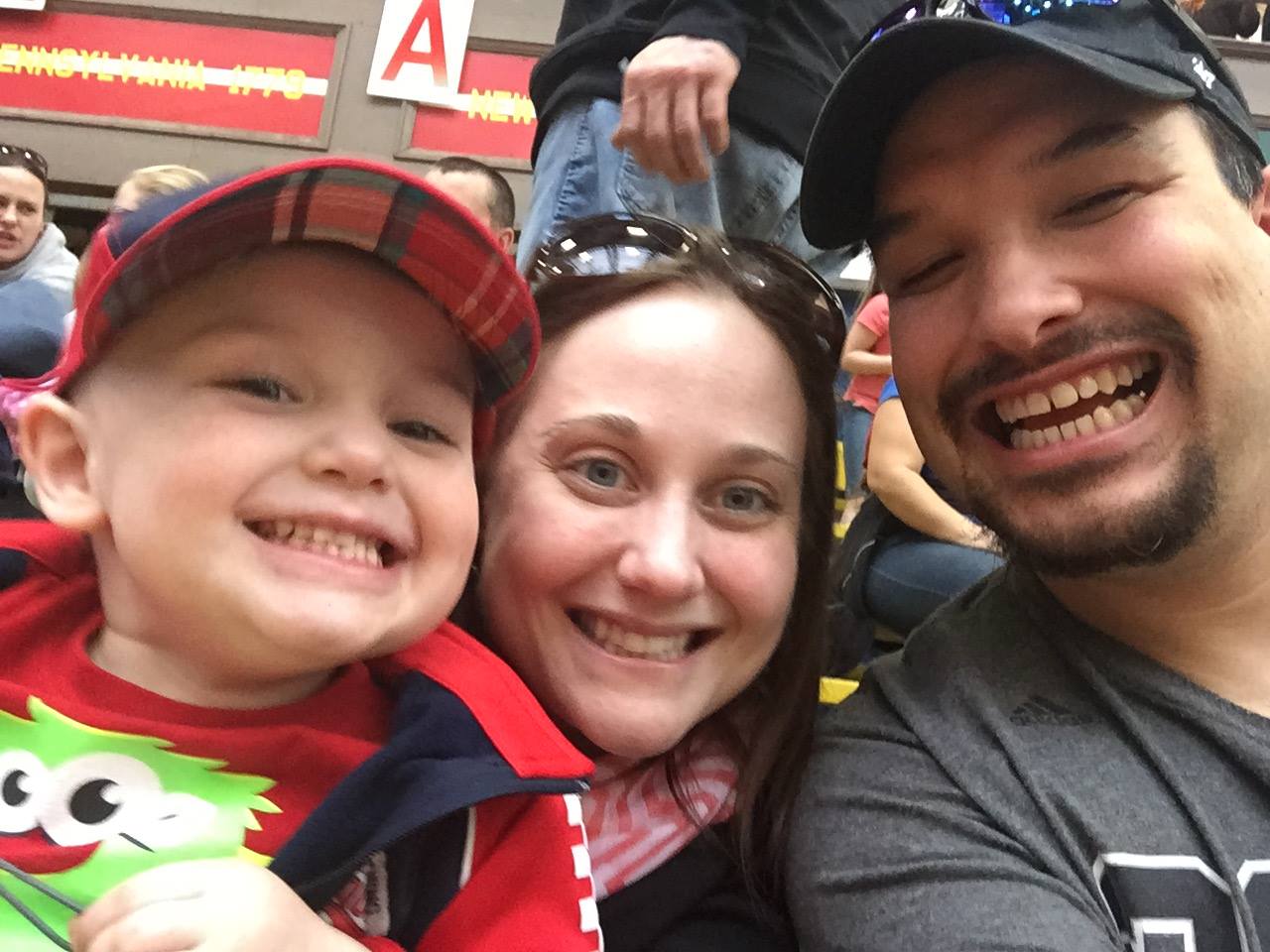
I finished my dissertation and successfully defended it in June of 2007 then set off across the country with my then girlfriend (now wife) to start new jobs. Everything was on the upswing and all appeared normal. However, it had been about a year since my last visit to the optometrist and being in a new place, I had to go through the dubious task of finding one.
I ended up getting an appointment with the optometrist at our local Walmart as I had broken my only pair of glasses and needed a quick replacement. The visit was going normal (or so I thought) until he came back into the exam room after checking on something. It was with a grave expression that he told me that he noted some concerning findings and I should probably speak with someone who had more experience with keratoconus.
For the next hour or so, I was in a panic that I was going blind. That is until I got online to get more details on what this was all about. After reading into the late evening, a great many eye-related issues over the past several years suddenly made sense. I immediately recalled my many complaints about my night time driving becoming more bothersome because oncoming headlights were blinding. It was on the many forums where I first learned of the terms halos and ghosting, which I would become all too familiar with in subsequent years. Lastly, I had tried to switch from glasses to soft contact lenses roughly a year before my move and was not able to do it. I didn’t know it then but my left contact kept sliding off because of my enlarged cone. In retrospect, I still cannot fathom how my optometrist did not recognize the symptoms.
After a little research, I found a local optometrist who had experience with KC. He basically wanted to put me in RGP lenses and call it a day. This was a horrible experience and I will save everyone the gory details. Suffice it to say, I would stay in glasses until February of this year–nearly 7.5 years since my initial diagnosis.
Little by little I was becoming aware that my vision was getting worse. Night time driving was becoming near impossible, reading on the computer (which is a huge bulk of my job) was increasingly difficult and the photophobia was simply impossible to ignore–especially at stores and restaurants that used brutal florescent lighting. I did have tomography scans done yearly and thankfully the progression was slow, but it was still progression. Something had to change.

After leaving my optometrist for several philosophical reasons, I was able to find a group outside of my area, but still within driving distance, who were expert with patients with KC. It was there that I learned about scleral lenses and how they could be of benefit. My ophthalmologist and I also discussed cross-linking, but decided that since FDA approval is around the corner, we have the luxury of time to wait. However, he strongly encouraged me to switch to the scleral lenses.
I am ever thankful that he did. My life, in just a few short weeks, has been irrevocably changed. We are still working out the fine tuning, but the vision restored is unbelievable. I never thought I would see this well again. Overall, vision is 20/25 and I’ve also noted that my peripheral vision is back to normal. Also, and most thankfully, my night time vision and ability to drive safely have been restored. It is still unbelievable what these lenses have done for me and my overall quality of life.
There is the old cliché that you don’t know what you have until it is lost. I am still amazed how I didn’t truly realize how bad my vision had gotten until I got the sclerals. For those with KC and are on the fence as to how best to deal with it, please don’t hesitate to talk with your doctor to find the best solution for you.
7/30/15
 Rene Vasquez
Keratoconus Advocate
Vision, while it is important to humans to carry out daily tasks, is a matter of life and death to animals in the wild. Predators need powerful, accurate vision to stalk their targets, while prey animals have developed a wide field of vision that is sensitive to movement, alerting them of danger. For each animal, their vision is a product of their environment and may have evolved over time. Here are some animals with unique eyes.

The tarsier is a small mammal about the size of a squirrel with eyes that are the largest of any mammal relative to body size, and weighing more than its brain. For a human to have eyes in the same proportion to body size, our eyes would be the size of grapefruits! The other unusual thing about a tarsier’s eyes is that they do NOT rotate in the eye socket. The tarsier must use his very flexible neck to rotate his head 180 degrees to see around him. He has great night vision, but very poor color vision, which is common with most nocturnal animals.
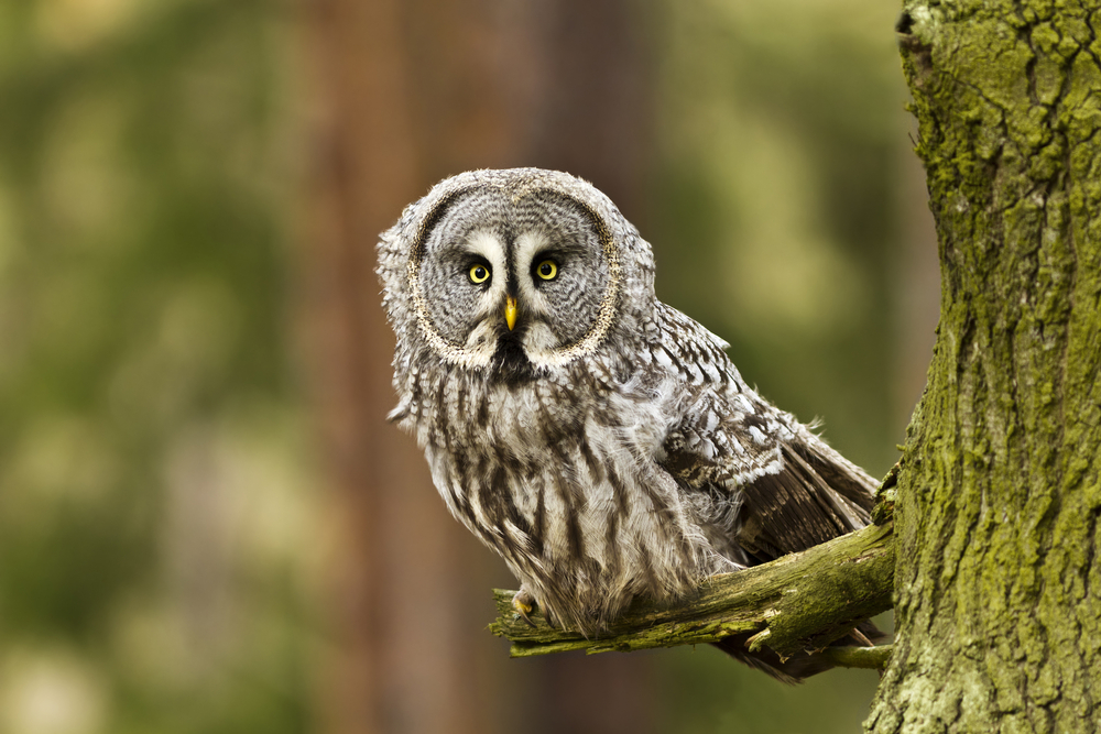
Another animal with relatively large fixed eyes is the owl. He also has a layer of tissue in the eye called the tapetum lucidum. This tissue is commonly found in nocturnal animals and deep sea animals in the back of the eye immediately behind the retina. It reflects visible light back through the retina, increasing the light available to see. Also because the eyes are fixed, owls can rotate their heads up to 270 degrees in both directions and 90 degrees vertically to look around.
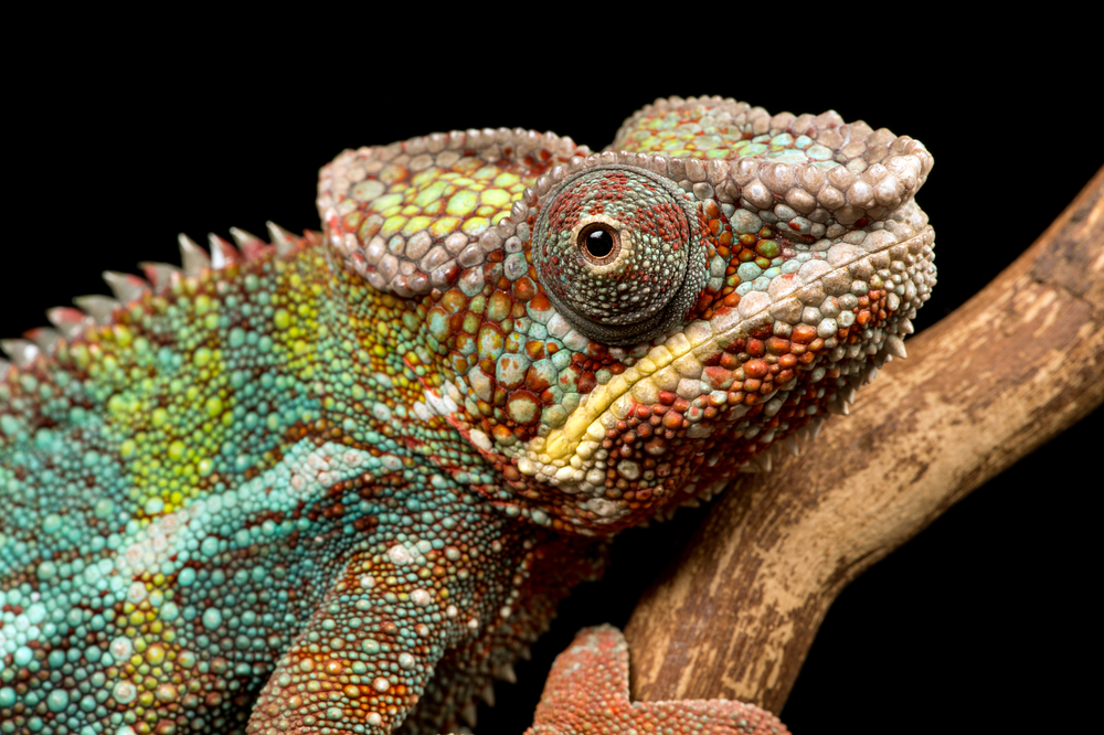
A chameleon’s eyelids are fused, covering almost the entire eyeball except for the pupil. What is more unique is that the eyes work independently of each other and the chameleon can process the completely different images at the same time. However, when the chameleon spots potential prey, it focuses both eyes in the same direction, using stereoscopic vision for precise distance and depth perception. They have a full 360 degree field of vision and can see ultraviolet light.

Goats, along with most other hooved animals, have horizontal rectangular pupils. These pupils give them a field of vision of 320-340 degrees. Because of the special shape and large size of these pupils, the goat is able to have more control on how much light enters the eye, so they can see more easily at night.

Dragonflies have compound eyes that almost cover their heads and give them a full 360 degree filed of vision. The large part of their eyes is made up of 30,000 visual units called ommatidia (a cluster of light-sensing photoreceptor cells with a lens). 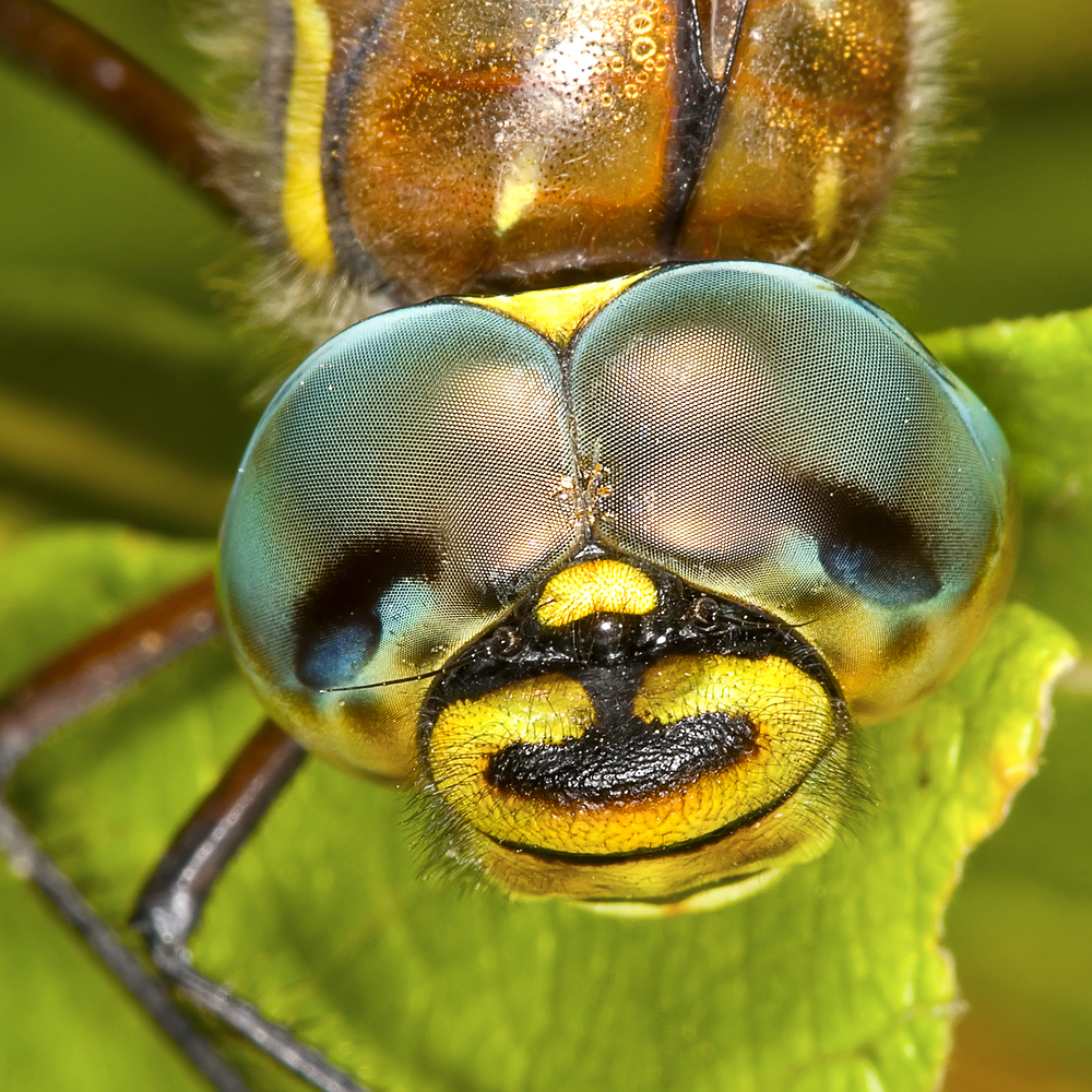 This gives them very acute vision, even in low light. What makes their vision even more remarkable is that they also have three smaller eyes call ocelli which can detect movement faster than the larger eyes, sending the information to the dragonfly’s motor center allowing for spilt second reaction times.
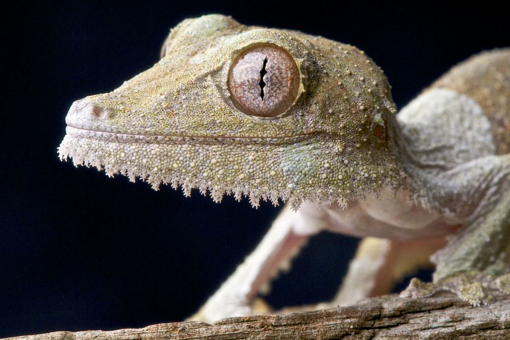
One of the most unusual eyes is that of the leaf-tailed gecko. The pupils are vertical with a series of “pinholes” that widen at night, allowing as much light as possible into the eye. Since they also have a more photoreceptor cells than most animals, they have incredible night vision. The eyes also have a series of intricate eye patterns to help with camouflage since there is no eyelid. Their eyes are protected by a transparent membrane, which they clean with their tongues.
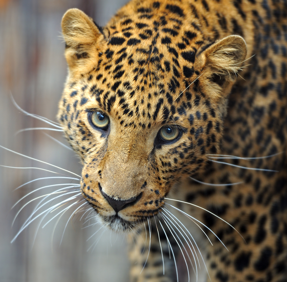
Cats such as lions, tigers jaguars and leopards have extremely sharp night vision due to the fact they have the tapetum lucidum reflective tissue and far more rods (the light sensors of the eye) than cones (the color sensors of the eye) in their eyes compared to humans. However, because of the reduced number of cones, they can only distinguish a very limited range of colors.
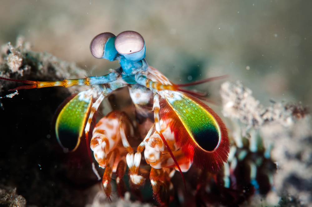
The mantis shrimp has compound eyes like the dragonfly but with only 10,000 ommatidia per eye. However, each row of ommatidia has a particular function – some are for detecting light, others for detecting colors, etc. Mantis shrimp (who are not actually shrimp) have remarkable color vision with 12 types of color receptors (humans have three) as well as ultraviolet, infrared and polarized light vision. This means they have the most complex eyesight of any animal. Each of the manits shrimp’s eyes sit at the end of stalks allowing them to move independently from each other and the ability to rotate up to 70 degrees. Finally, unlike humans, the visual information is processed by the eyes themselves instead of the brain.
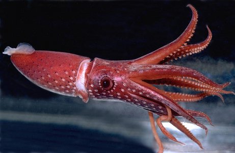
While the tarsier has the largest eyes relative to its size, the colossal squid has the largest eyes in the animal kingdom. Each of the colossal squid’s eyes can be as large as a foot in diameter. These exceptionally large eyes allow them to see well in dim light conditions, 2000 meters below the ocean surface. Also each eye has a built-in “flashlight” which can produce light so that whenever the squid focuses it eyes to the front, there is enough light for it to see its prey in the dark.
These are just a few of the animals with varied and unique vision. If you know of any others, please share them in the comments section below.
7/14/15
Susan DeRemer, CFRE
Vice President of Development
Discovery Eye Foundation
When it comes to our health, we often visit our doctor or nurse regularly to make sure our bodies are healthy. But what about our eyes? They’re not always top of mind, but they’re just as important.
During Healthy Vision Month, held each May, the National Eye Institute (NEI) reminds you to make your eye health a priority and encourages you to take important steps to protect your sight.
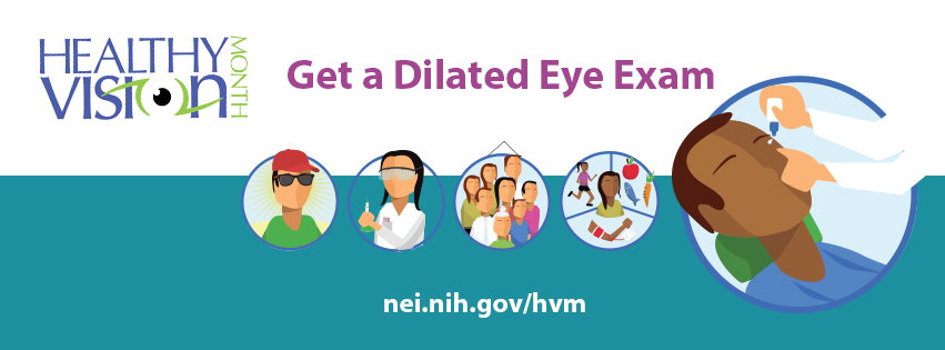
Get a dilated eye exam. Getting a dilated eye exam is the only way to detect eye diseases early, because with many, there are no warning signs. Talk to your eye care professional about how often you should have one. If you want to see what your eye care professional sees during a dilated eye exam, check out NEI’s new eye exam animation!

Live a healthy lifestyle. Eating healthy foods, maintaining a healthy weight, managing chronic conditions, and not smoking can lower your risk of eye disease.
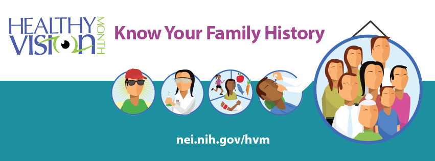
Know your family history. Talk to your family members about their eye health history. It’s important to know if anyone has been diagnosed with an eye disease, since many are hereditary. This will help to determine if you are at higher risk for developing an eye disease yourself.
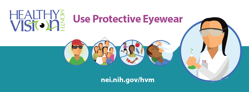
Use protective eyewear. Protect your eyes when doing chores around the house, playing sports, or on the job to prevent eye injuries from happening. This includes wearing safety glasses, goggles, safety shields, and eye guards that are made of polycarbonate.

Wear sunglasses. When purchasing sunglasses, look for ones that block out 99 to 100% of both UVA and UVB radiation, so you can keep your eyes healthy. Prolonged exposure to sunlight can increase your risk for getting an eye disease. A wide-brimmed hat offers great protection, too!
These steps can help you keep your eyes healthy and prevent vision loss and blindness from eye disease.
To learn more about Healthy Vision Month and find additional eye health information, visit www.nei.nih.gov/hvm.
5/5/15
 National Eye Institute
National Institute of Health
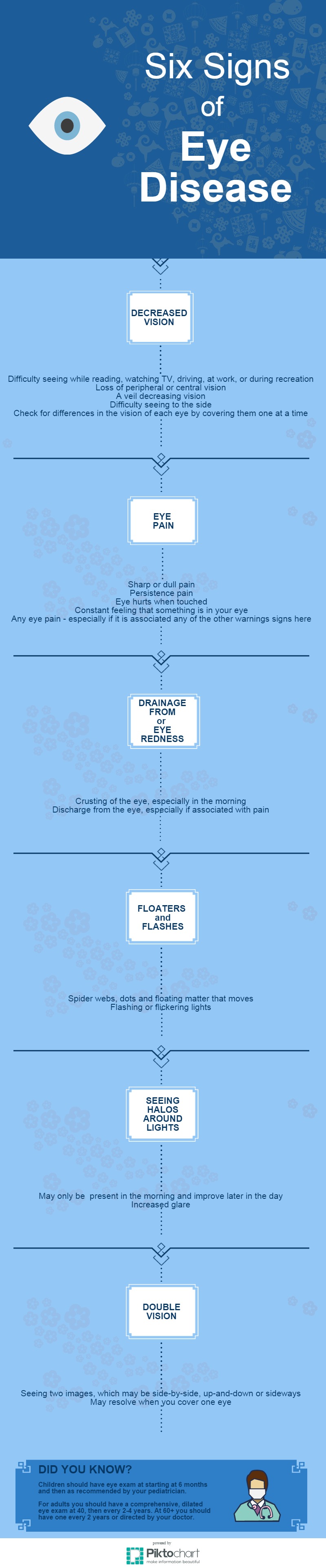
Signs of eye disease that you should be aware of for early treatment.
4/9/15
Susan DeRemer, CFRE
Vice President of Development
Discovery Eye Foundation
The National Keratoconus Foundation, a program of the Discovery Eye Foundation, has been serving the keratoconus (KC) community for over 27 years. They have been a leading source of information on KC, while also providing support and educational programs, such as the OC Keratoconus Seminar this Saturday, March 14th. One of the reasons for the timely, quality information they are able to share, are their relationships with leading eye care professionals around the world.
We are happy to report that a new professional organization for eye care professionals treating KC and other forms of corneal ectasia has been formed, the International Keratoconus Academy of Eye Care Professionals. It is our hope that we can join together in in providing accurate information on KC as well as find future treatments and cures, defining the future of keratoconus. Here is more information about the new organization.
The International Keratoconus Academy of Eye Care Professionals (IKA) was recently established to promote ongoing professional education and scientific development in the area of keratoconus and other forms of corneal ectasia. Its mission is to promote and develop the knowledge base and awareness of the state of the art pertaining to the diagnosis and management of keratoconus and other forms of corneal ectasia. And further to promote the awareness and understanding of the most appropriate and effective treatment strategies for the management of these diseases. According to S. Barry Eiden, OD, FAAO, co-founder and president,” I.K.A. is dedicated to providing comprehensive education to the eye care professions and to foster ongoing clinical research in order to improve the lives of patients suffering from these diseases. “
I.K.A. will accomplish its mission by providing an array of educational initiatives which will include live events, web-based education, social media activities and publications in the professional literature. It will also be dedicated to supporting ongoing clinical research. I.K.A. will function as a complementary entity to other organizations that support patients with these diseases such as the National Keratoconus Foundation (NKCF). These organizations will work cooperatively to establish a comprehensive effort to advance knowledge, awareness and quality of care.
The founding executive board of the International Keratoconus Academy of Eye Care Professionals is comprised of a group of highly regarded experts in the field including:
An international physician’s advisory board comprised of expert optometrists and ophthalmologists is being formed. For more information or questions about IKA, please contact them at info@keratoconusacademy.com.
3/10/15
Susan DeRemer
Vice President of Development
Discovery Eye Foundation
12/23/14
On behalf of Discovery Eye Foundation, we would like to wish you the best this holiday season!

Because this is such a busy time of the year, we will not be posting to the blog, giving you more time to enjoy your family and friends.
We will resume posting on Tuesday, January 6th.
Susan DeRemer, CFRE
Vice President of Development
Discovery Eye Foundation
9/2/14
“The theater of the mind could be generated by the machinery of the brain.”
For more information on Charles Bonnet Syndrome, please go to the Macular Degeneration Partnership website.
*Testing for functional brain imagery as individuals hallucinate can find different parts of the brain are activated.
Your brain has a particular area or lobe where vision is interpreted. The light energy that bounces off objects enters the eye and is converted to chemical energy by the retinal cells. That energy is sent through the optic nerve where processing of the vision starts to occur. When it reaches the visual cortex, it is sent to very specific areas of the brain and specific areas of the brain see specific things.
The fusiform gyrus processes faces, but different areas process the parts of faces. While damage in the fusiform gyrus causes you to lose the ability to recognize faces, abnormal activity in that area will cause you to hallucinate faces. An area in the anterior part of this gyrus is where teeth and eyes are recognized. There are other areas that specifically sees cartoons and another part for buildings and landscapes.
 Judith Delgado
Executive Director
Macular Degeneration Partnership
A Program of Discovery Eye Foundation
8/21/14
In the third of this series, Buddy Russell, from the Emory University Eye Center, provides a great overview of common pediatric eye diseases.
A basic understanding of some of the conditions that may be present in pediatric patients is important to not only know what they are but also understand well enough to explain to the parent or caregiver. The following is intended to be an overview of some of those conditions and not a complete explanation.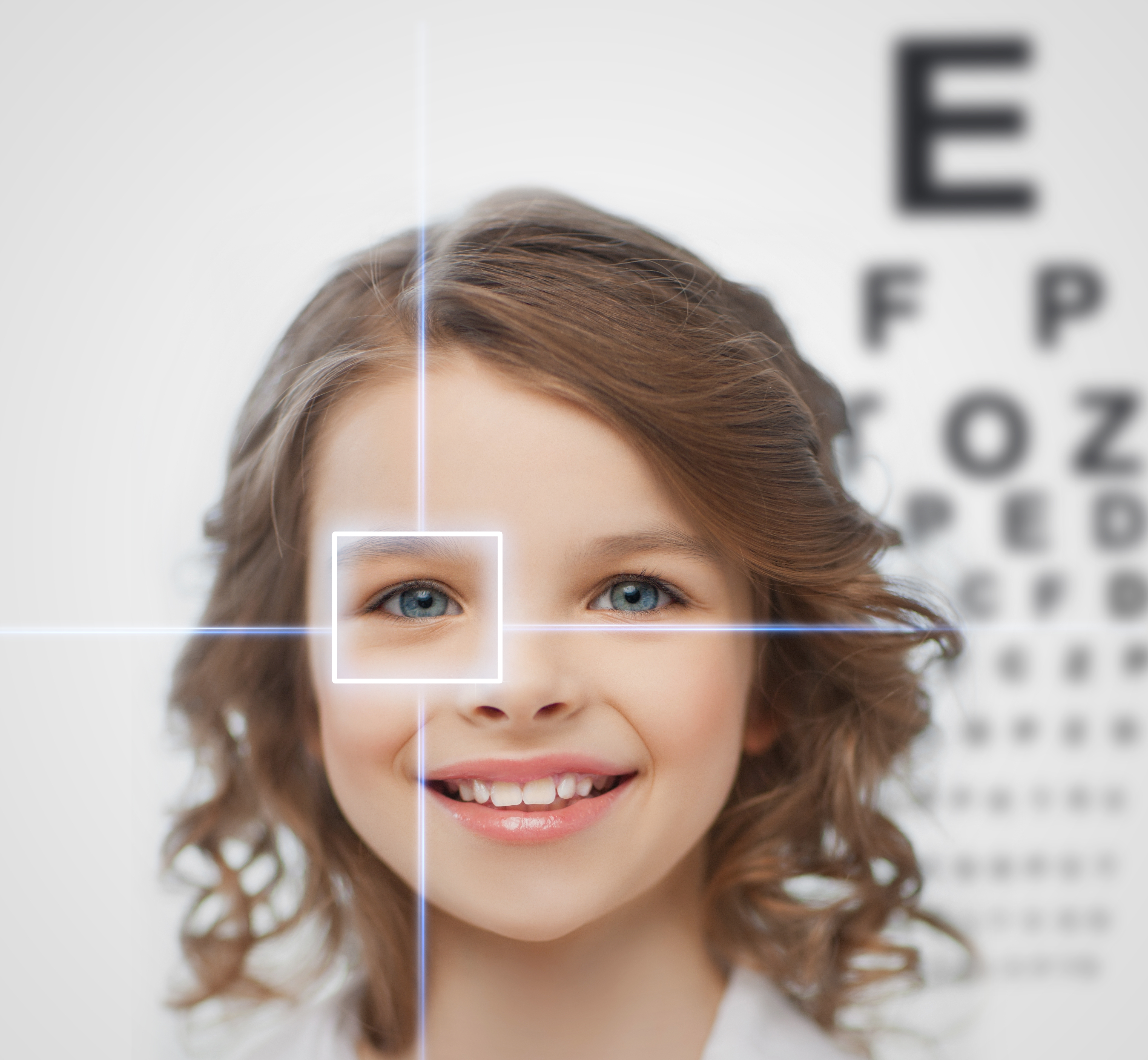
 Buddy Russell, FCLSA, COMT
Associate, Specialty Contact Lens Service
Emory University Eye Center
8/19/14
Here is part two in Buddy Russell’s series; this one focusing on contact lenses as a treatment option for children.
The human visual system at birth is poorly developed, but rapidly becomes the remarkable combination of nerve tissue, muscles and optics that provide us with the sense of vision. Those babies born with “perfect” eyes have only the opportunity to develop normal vision. The information processed by the eyes is sent directly to the brain and is interpreted as vision.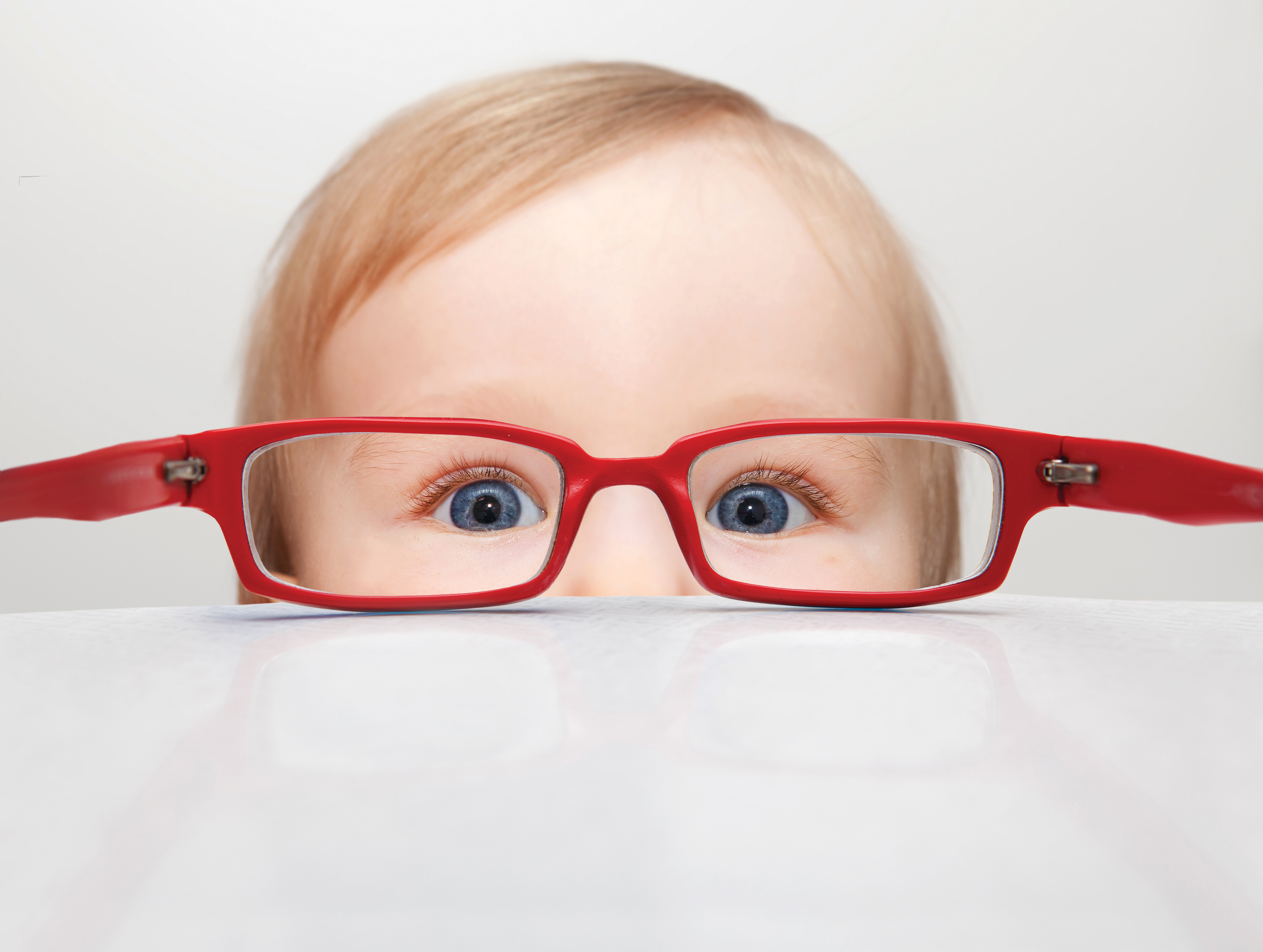 During the first few weeks, the child sees shapes, lines and space between objects. The child’s visible world is most usable within 8-14 inches of his/her eyes. During this time, the eyes may appear to wander. After about a month or so, the normal child’s eyes will appear more coordinated and they start to show more interest in looking at objects. It is usually in the third month that a child who has normal eyes can fix and follow on a near object. The growth of the eye is a dynamic process, influenced by genetics and the environment.
Early detection of any eye problem is key to treating the disorder. The prevalence of vision problems in children is higher than you might think. For example:
As a result of his granddaughter and her eye problem, former President Jimmy Carter initiated a program in 2002 called InfantSEE. This program allows children to have an eye exam at a very young age at no charge to the family. Participating eye doctors provide a more thorough exam than the busy pediatrician. As a result, there is a greater opportunity to detect and treat eye disorders that may otherwise go undetected.
Fitting pediatric patients is not usually about routine visits and patients who want to wear contact lenses. It is about critical and often urgent situations and patients who have to wear contact lenses. The more common medical indications for contact lenses can be categorized into three groups; anisometropia, irregular corneal astigmatism and “large” refractive errors.
One of the more common conditions potentially leading to a permanent loss of vision in a young patient is anisometropia. This difference in the refractive errors of the two eyes can lead to suppression of the less clear image. As a result of the non-focused eye, the brain of a young patient simply turns off the blurred eye. Early detection is key to successful treatment. Following the diagnosis of this problem being present, simply correcting the refractive error may be enough. However, it has been reported that as little as one diopter difference between the two eyes corrected with spectacles and the resultant anisokonia, can lead to foveal suppression impacting stereopsis and depth perception. The use of a contact lens or contact lenses alters the effective image size due to the vertex distance being zero compared to either the magnification or minification of the image size due to the vertex distance with spectacles. One of the most severe examples of this condition would be a child with a unilateral congenital cataract and managed with spectacles postoperatively.
Whether acquired or congenital, the presence of irregular corneal astigmatism of the anterior curve of the cornea is best managed with a contact lens. This condition is to be considered urgent if the patient is of a young age. The eye may forever loose the opportunity to be corrected as the resultant amblyopia develops over a short period of time. By neutralizing the corneal irregularities with a contact lens, the eye of a young child will hopefully gain enough vision improvement to avoid the potential permanent loss.
Obviously, patching the better eye may also be necessary if the treated eye’s vision is not as correctable as the unaffected eye. The length of time the child is to be patched is to be determined by the pediatric ophthalmologist or optometrist, as this area of treatment is sometimes controversial. The factors that are considered include the level of vision obtained, age of the child and the condition of the other eye.
The optics of spectacle correction in high powers have inherent properties that include distortion, prismatic effect and minification / magnification. For instance, the decrease in image size when one views an object through high minus spectacles may result in less vision. This decrease in image size may impact the opportunity to fully develop normal vision in a young child. The smaller image size that is due to the vertex distance of spectacles may be better managed with a contact lens that has a vertex distance of zero thus providing a larger image. This larger image size often increases best-corrected vision.
Arguably, the most important factor with young children having a good outcome is the parents / caregivers. The technical challenges that exist in these cases are secondary to the ability the fitter must possess to effectively explain and train the person or persons that will take care of the child outside of the office. They must be your partner in the child’s treatment. They must understand the urgency of the situation, they must understand the seriousness of the problem, they must be trained to properly apply, remove and care for the lens / lenses, they must also follow any and all instructions concerning the child. Many of these parents struggle with feelings of nervousness, guilt and sadness. My strategy is to be sensitive to their feelings but not let them feel sorry for themselves too long as the clock is ticking. I provide verbal instructions, written instructions, videos, my email address and a 24-hour phone number. I welcome the caregiver to ask any question at any time. I do my best to let them know that I do care and that I want them and their child to be successful. I am tough on them. There is no good excuse not to do as I have instructed them to do.
When the child and the parent / caregiver are convinced that I am confident in my ability and they know that I do care, the partnership develops as we walk the path together. I want the child to know that they are coming to see me. I want them to know I will reward their cooperation with all phases of the visit. This positive reinforcement may be in the form of a piece of candy, a small toy or just a sticker when the child allows me to see their eye, measure their cornea or intraocular pressure or they just tell me what they can see. Kids love to please us just like they love to please their parents. Reward them for it. Whether you consider this approach bribery or positive reinforcement, it works.
Buddy Russell, FCLSA, COMT
Associate, Specialty Contact Lens Service
Emory University Eye Center