
Ways to Reduce the Harmful Effects of Sun Glare

During the height of summer sunshine (and heat!), it’s helpful to discuss the importance of eye protection, including ways to reduce the harmful effects of sun glare.

Fundamentally, we need light to see. Approximately 80% of all information we take in is received through the sense of sight. However, too much light – and the wrong kind of light – can create glare, which can affect our ability to take in information, analyze it, and make sense of our surroundings.

Facts about Sunlight

Every type of light has advantages and disadvantages, and sunlight is no exception:

Advantages:

• Sunlight is the best, most natural light for most daily living needs.
• Sunlight is continuous and full-spectrum: the sun’s energy at all wavelengths is equal and it contains all wavelengths of light (explained below).

Disadvantages:

• It is difficult to control the brightness and intensity of sunlight.
• Sunlight can create glare, which can be problematic for many people who have low vision.
• Sunlight is not always consistent or reliable, such as on cloudy or overcast days.

Visible Light and Light Rays

An important factor to consider is the measurement of visible light and light rays, beginning with the definition of a nanometer:

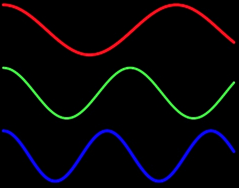
• A nanometer (nm) is the measurement of a wavelength of light.
• A wavelength is the distance between two successive wave crests or troughs:
Wavelength - glare

• A nanometer = 1/1,000,000,000 of a meter, or one-billionth of a meter. It’s very small!

The human visual system is not uniformly sensitive to all light rays. Visible light rays range from 400 nm (shorter, higher-energy wavelengths) ? 700 nm (longer, lower-energy wavelengths).

Visible Light Spectrum - glare
The visible light spectrum occupies just one portion of the electromagnetic spectrum, however:

• Below blue-violet (400 nm and below), is ultraviolet (UV) light.
• Above red (700 nm and above), is infrared (IR) light.
• Neither UV nor IR light is visible to the human eye.

Ultraviolet Light and Blue Light

Ultraviolet (UV) light has several components:

• Ultraviolet A, or UVA (320 nm to 400 nm): UVA rays age us.
• Ultraviolet B, or UVB (290 nm to 320 nm): UVB rays burn us.
• Ultraviolet C, or UVC (100 nm to 290 nm): UVC rays are filtered by the atmosphere before they reach us.

Blue light rays (400 nm to 470 nm) are adjacent to the invisible band of UV light rays:

• There is increasing evidence that blue light is harmful to the eye and can amplify damage to retinal cells.
• You can read more about the effects of blue light at Artificial Lighting and the Blue Light Hazard at Prevent Blindness.

A new study from the National Eye Institute confirms that sunlight can increase the risk of cataracts and establishes a link between ultraviolet (UV) rays and oxidative stress, the harmful chemical reactions that occur when cells consume oxygen and other fuels to produce energy.

Sunlight and Glare

Glare is light that does not help to create a clear image on the retina; instead, it has an adverse effect on visual comfort and clarity. Glare is sunlight that hinders instead of helps. There are two primary types of glare.

Disability glare

• Disability (or veiling) glare is sunlight that interferes with the clarity of a visual image and reduces contrast.
• Sources of disability glare include reflective surfaces (chrome fixtures, computer monitors, highly polished floors) and windows that are not covered with curtains or shades.

Discomfort glare

• Discomfort glare is sunlight that causes headaches and eye pain. It does not interfere with the clarity of a visual image.
• Sources of disability glare include the morning and evening positions of the sun; snow and ice; and large bodies of water, (including swimming pools).

Controlling Glare

You can protect your eyes from harmful sunlight and minimize the effects of glare by using a brimmed hat or visor in combination with absorptive lenses.

• Absorptive lenses are sunglasses that filter out ultraviolet and infrared light, reduce glare, and increase contrast. They are recommended for people who have low vision and are also helpful for people with regular vision.
• Lens colors include yellow, pink, plum, amber, green, gray, and brown. Ultra-dark lenses are not the only choice for sun protection.
• Lens tints in yellow or amber are recommended for controlling blue light.
NoIR Medical Technologies: NoIR (No Infra-Red) filters absorb UVA/UVB radiation and also offer IR light protection.
Solar Shields: Solar Shields absorb UVA/UVB radiation and are available in prescription lenses.
• You can find absorptive lenses at a specialty products store, an “aids and appliances store” at an agency for the visually impaired, or a low vision practice in your area. Before you purchase, it’s always best to try on several different tints and styles to determine what works best for you.

More Recommendations

• Always wear sunglasses outside, and make sure they conform to current UVA/UVB standards.
• Be aware that UV and blue light are still present even when it is cloudy or overcast.
• Make sure that children and older family members are always protected with UVA/UVB-blocking sunglasses and brimmed hats or visors.

Maureen Duffy-editedMaureen A. Duffy, CVRT
Social Media Specialist, visionaware.org
Associate Editor, Journal of Visual Impairment & Blindness
Adjunct Faculty, Salus University/College of Education and Rehabilitation


What Are Floaters? | Causes and Treatment

6/12/14

Floaters can be anything seen in your vision that moves “to and fro” with your eye movement. The movement is not stationary compared to a blind spot which is fixed or stationary in your field of vision.

Example of floaters
Example of floaters
The key is that the position changes with eye movement. Size, shape, color, etc. don’t matter. Anything that moves in your vision is called a floater.

Remember, if you experience new floaters; please see an eye doctor for an examination.

Here are some common causes of floaters:

  • Posterior Vitreous Detachment (PVD)
  • Retinal Tears
  • Blood (Vitreous Hemorrhage) can be caused by;
  • Advanced Diabetic Retinopathy
  • Retinal Tears
  • Retinal Vascular Occlusions
  • Inflammation or Infection
  • Asteroid Hyalosis

Posterior Vitreous Detachment
A posterior vitreous detachment (PVD) is a common cause of floaters. A PVD occurs when the vitreous separates from the retinal surface. This is a normal event and will eventually occur in everyone. Retinal tears are more likely to occur right after a PVD has started.

The vitreous is usually a clear watery gel. As we age, enough of the proteins in the vitreous liquefy and degenerate. Eventually, there is a physical separation of the vitreous from the retina due to this liquefaction.

“Floaters” can develop due to opacities/haziness due to the aging gel which is no longer absolutely clear due to changes in the optical properties of the gel or the interface between the vitreous and water inside the eye.

Tears in the Retina
Retinal tears can cause floaters by either causing a small vitreous hemorrhage, or by dislodging cells into the vitreous which are normally located underneath the retina.

Vitreous Hemorrhage
Blood in the vitreous can also cause floaters. A so-called vitreous hemorrhage may result from either a retinal tear or advanced diabetic retinopathy. Occasionally, retinal vascular occlusions can also lead to bleeding in the vitreous.

Blood in the eye often absorbs, but only your doctor can determine the exact nature of the floaters and if they are associated with a retinal tear. It’s impossible for you to tell the cause of floaters without a proper dilated eye exam.

Inflammation
Certain types of inflammation or infection can cause significant floaters. Actually, these are white cells which migrate to the retina and vitreous and can be seen a floaters.

Asteroid Hyalosis
This is a common entity where lots of fine white opacities are suspended in the vitreous. In most cases, these are not noticed by the patient. Sometimes, however, the so-called asteroid bodies are so dense they prevent good examination of the retina. Other times, especially after a PVD, the asteroid bodies are noticed and patients may complain of floaters.

New Floaters: Evaluation
All new floaters must be examined by your eye doctor within 24-72 hours after occurring. Even if the floaters disappear in that time frame, you should be examined to look for a possible tear in the retina.

Again, you, the patient, can NOT tell the cause of the floaters or if you’ve sustained a retinal tears. Retinal tears can cause a retinal detachment – a potentially blinding problem.

Treatment of Floaters
There are no medicines or eye drops to treat floaters. Most doctors advise simply putting up with the floaters.

For patients who have chronic problems with floaters, I recommend a vitrectomy. A vitrectomy is an eye operation, performed by a retinal specialist. The operation is comparable to a cataract operation in terms of safety and possible complications.

There are a handful of doctors who perform Yag laser to break up floaters.

Randall E. Wong MDRandall V. Wong, MD
Retina Specialist
Fairfax, Virginia

The Costs of Eye Care

5/6/14

**Update 6/16/14 – New estimate on the cost of vision problems just came out in the new Prevent Blindness America report showing they could reach $717 billion by 2050.

This past week I attended a dinner where a topic of discussion was the cost of chronic disease on healthcare – 75% of healthcare dollars go to the treatment of chronic diseases. This led me to explore the costs chronic eye diseases such as age-related macular degeneration, cataract, glaucoma and diabetic retinopathy.
PBA 2012 booklet
In 2007 Prevent Blindness America (PBA) published a report, The Economic Impact of Vision Problems that was updated in 2012, which explored the economic burden related to people 40 and older and the conditions of, age-related macular degeneration (AMD), cataract, diabetic retinopathy, primary open-angle glaucoma and refractive error, as well as the economic impact of vision problems and blindness.

They looked at direct medical costs including inpatient, outpatient and prescription costs; direct non-medical costs, which include nursing home care, guide dogs, and government programs for the blind and visually impaired; and loss of productivity, which includes lower labor force participation and lower wages for those with vision problems as opposed to the same age group with normal vision.

Direct Medical Costs $16.2 billion
Direct Non-medical Costs $11.2 billion
Productivity Losses $8.0 billion
TOTAL ECONOMIC IMPACT $35.4 billion

Looking more closely at the direct medical cost with regards to age-related eye diseases, the costs per patient broke down as follows:

AMD Cataract Diabetic Retinopathy Glaucoma
Medical Costs $797 $12,625 $6,012 $3,552

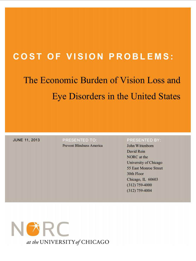
A year later, the NORC at the University of Chicago presented their findings, but also included the age groups of children 0-17 and adults 18-39 in a publication titled, Cost of Vision Problems: The Economic Burden of Vision Loss and Eye Disorders in the United States. It was interesting to see how the estimates varied in the two approaches when I only focused on the group of age 40 and older, as they did in the PBA study.
Noca booklet
The reclassification of the costs into two line items, from the previous three of the PBA report, presented a challenge and is one reason for discrepancies.  They went into more depth and were broken down as follows: direct costs included medical care, medical vision aids, low vision aids, special education, school screenings and Federal assistance programs; and indirect costs included loss of productivity for adults and caregivers, long-term care and transfer payments.

Direct Costs $52.58 billion
Indirect Costs $58.49 billion
TOTAL ECONOMIC IMPACT $111.07 billion

And when I looked more closely at the medical cost with regards to age-related eye diseases, the costs per patient broke down as follows:

AMD Cataract Diabetic Retinopathy Glaucoma
Medical Costs $4,457 $10,570 $4,005 $5,617

Regardless of the data sources and reporting classifications, one thing is clear – the direct medical costs of eye disease is high and growing each year.

Both reports contained a wide variety of information that is very interesting.  Links for both studies have been included so you can review the numbers and draw your own conclusions.

Susan DeRemerSusan DeRemer, CFRE
Vice President of Development
Discovery Eye Foundation

Welcome to the Discovery Eye Foundation Blog

discovery eye foundation_logoWelcome to your blog, where you will find the most important information about eye health and sight-threatening eye diseases.

The Discovery Eye Foundation team has more than four decades of knowledge, experience and relationships to help you better understand your eye disease and discuss it with your eye doctor — so you can make more informed decisions and lifestyle choices.

You will find information from leading ophthalmologists, optometrists, eye researchers, low-vision specialists, nutritionists and other professionals who are interested in helping you keep your eyes as healthy as possible, and/or deal with eye disease and vision loss.

We will be posting twice a week, with a guest post from an eye-care professional every Thursday addressing an issue important to our constituents.

We want to hear from you. What information do you find useful? What topics are important to you? And while we will respond to general questions about eye diseases, we will not be able to address patient-specific questions that can best be answered by your own eye doctor.

For more in-depth information, please visit one of our three websites to learn about specific eye diseases, view videos, listen to recorded presentations and interviews, and discover disease-specific research and resources.

•   DiscoveryEye.org offers information on age-related macular degeneration (AMD), cataracts, diabetic retinopathy, glaucoma, keratoconus (KC), ocular herpes and retinitis pigmentosa, along with other eye conditions. For help with positive lifestyle choices, check out Eye Cook for delicious eye-healthy recipes.
•   AMD.org was one of the first online sources for information focused solely on age-related macular degeneration (AMD). This site is continually updated with the latest news on potential treatments. It fully explains the role of vitamins in eye health and offers insight into AREDS II. It has helpful information for caregivers and a national directory of resources.
•   NKCF.org is your one-stop site for in-depth information on keratoconus (KC). In addition to providing you with the latest on KC treatments and clinical trials, it has two free online support groups for people diagnosed with KC and their families.

Susan DeRemerSusan DeRemer, CFRE
Vice President of Development
Discovery Eye Foundation