How We Get Them & How We Get Rid Of Them
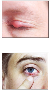
Do you have something that looks like a pimple on the outside or inside your eyelid? Is it terribly uncomfortable and unsightly? You either have a stye or a chalazion on your eye.
 Chalazion is a non-infected swelling of the lid caused by a blocked lipid gland. Lipid from the blocked gland leaks into the eye lid tissue causing an inflammation which eventually clears the irritating lipid. This can take 2 to 4 weeks or more.
Styes are caused by bacteria from your skin that gets into and irritates the oil glands in the eyelids. These bacteria, which normally exist harmlessly on the skin of the eye, can sometimes get trapped along with dead skin cells on the edge of the eyelid. The result is a swollen, red, and painful bump that can develop over the course of a few days.
While most styes or chalazions are harmless and will heal on their own in about a week or two, they can still be thoroughly unpleasant. Fortunately, there are a few remedies that may help you get rid of a stye or chalazion a little faster — or at least reduce some of the discomfort and swelling that often accompany them.
Keep Your Eyelids Clean
- The first thing you should do if you develop a stye is cleanse your eyelids. You can use diluted tear-free baby shampoo on a cotton ball, washcloth, or makeup remover pad. Then rinse your eyelids with warm water and gently pat them dry.
- Also, be sure to wash your hands before and after touching the stye, and don’t share your towels or washcloths with others.
- Pre-moistened eyelid cleansing pads are another option. You can find these non-prescription items in most drugstores.
- It’s wise to stop wearing eye makeup temporarily when you have a stye or chalazion, because covering it up can delay the healing process. Also, discard old makeup or applicators that could be contaminated.
- And if you need vision correction, wear glasses rather than contact lenses until your stye heals.
Apply Warm, Moist Compresses
- You can encourage a stye or chalazion to heal faster by applying hot compresses for 10 to 15 minutes, three or four times a day.
- Some people use teabags for this purpose, but a basic clean washcloth dipped in warm (not hot) water will do the trick and is easy to prepare. Wring the cloth so it’s not dripping, then place it over your closed eyes.
- The goal of this therapy is to bring the stye or chalazionto a head, like you see on a pimple. But whatever you do, don’t get anxious and try to pop it! The warmth from the compress often will allow it to open, drain and heal on its own without causing trauma to the eyelid or possibly spreading an infection by squeezing it.
Ease the Discomfort
- Over-the-counter painkillers like acetaminophen and ibuprofen probably won’t do much to speed healing, but these medications may ease discomfort if a stye is particularly bothersome.
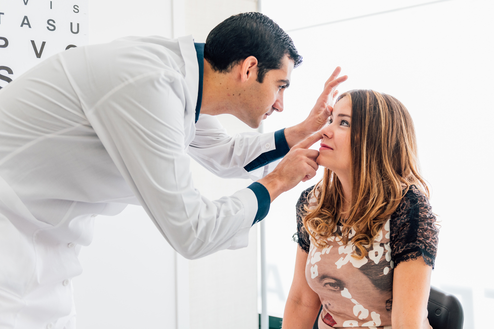
- Your eye doctor can also address pain associated with styes or chalazions. Sometimes, your eye doctor may choose to surgically open a large stye to relieve discomfort and prevent a serious infection.
Always consult a medical professional if you have concerns, pain, or if an external stye that does not clear up within one week or an internal one (on the inside of the eyelid) in three weeks.

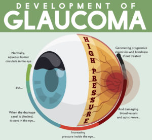 tic nerve, a part of the central nervous system that carries visual information from the eye to the brain.
 For example, lutein and zeaxanthin are important antioxidants that help prevent degeneration in the lens and retina. Eating a diet rich in these carotenoids helps reduce the risk of AMD by fighting oxidation in the retinal cells of the eye.
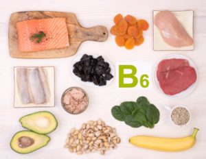
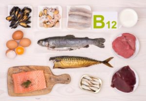
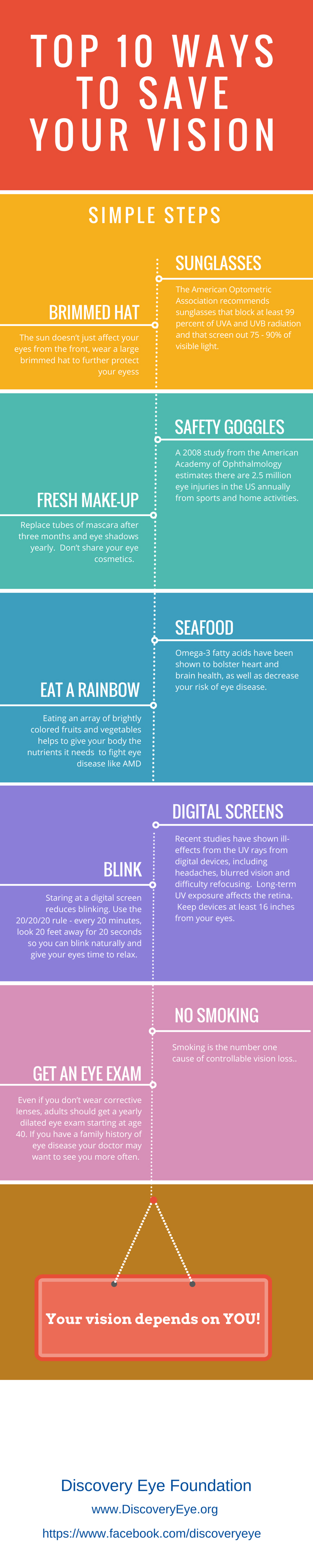
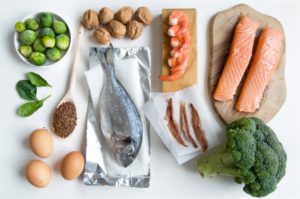 Eating fatty fish, such as salmon, tuna, mackerel, and sardines, that are rich in omega-3 fatty acids also helps lower the risk of AMD. Omega-3 fatty acids are rich in docosahexaenoic acid (DHA), which is important for eye health and visual function. People with dry eye syndrome (i.e., low tear production) can benefit from a diet rich in omega-3 fatty acids because dry eye is linked to low levels of DHA.