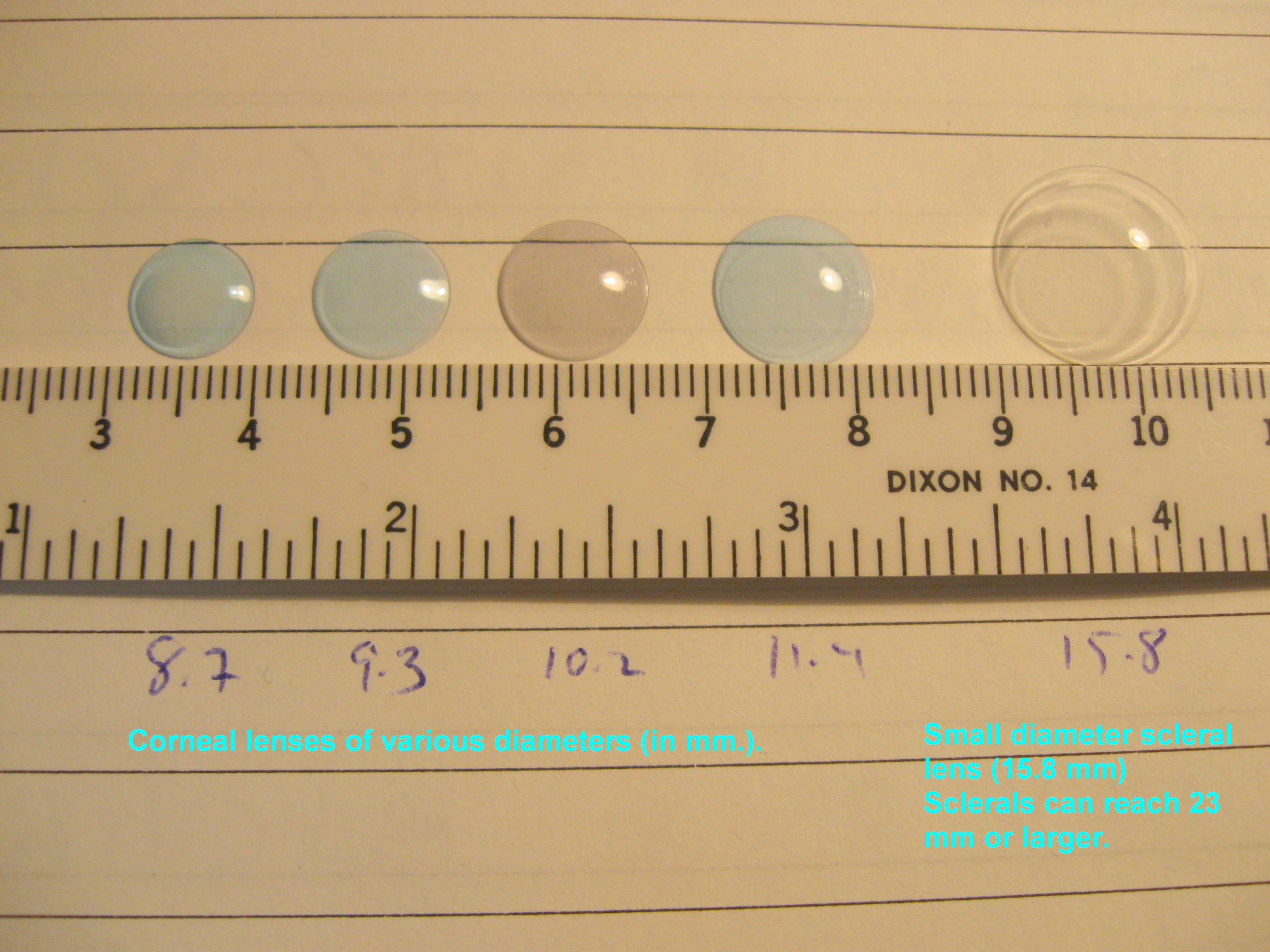
Do I Need Vision Insurance?

With the rising costs of health insurance, many people are looking for ways to reduce their costs. Since not all insurance packages include vision insurance, many people wonder, do I need vision insurance?
vision insurance

Standard Vision Insurance

Vision insurance is a type of health insurance that entitles you to specific eye care benefits such as routine eye exams and other procedures, as well as a specified dollar mount or discount for the purchase of eyeglasses and contact lenses. It only supplements regular health insurance and is designed to help reduce your costs for routine preventative eye care and eyewear.

You can get vision insurance as part of a group, such as your employer, an association, etc., through a government program such as Medicare or Medicaid, or as an individual. It is often a benefit linked to your regular HMO (health maintenance organization) or PPO (preferred provider organization) health insurance.

There are two primary vision insurance plans available:

  • Vision Benefits Package – provides free eye care services and eyewear within a fixed dollar amount for which you pay an annual premium or membership fee and a small co-pay. It may also include a deductible.
  • Discount Vision Plan – provides eye care and eyewear at a discounted rate after you pay an annual premium or membership fee.

Both insurance plans generally include:

  • Annual eye exams
  • Eyeglass frames (usually once every 24 months)
  • Eyeglass lenses (usually once every 24 months)
  • Contact lenses (usually once every 24 months)
  • Discounted rates for LASIK and PRK

Here is where you can check for a list of some vision insurance providers.

Medicare and Medicaid

Different kinds of vision care are included in the US government programs, Medicare and Medicaid. These programs are for qualifying American age 65 and older, individuals with specific disabilities and people with low income.

The Types of Medicare For Vision:

    • Medicare Part A (Hospital Insurance) –Medical eye problems that require a hospital emergency room attention, but routine eye exams are NOT covered.
    • Medicare Part B (Medical Insurance) – Visits to an eye doctor that are related to an eye disease, but routine eye exams are NOT covered.
    • Medicare Part D (Prescription Drug Coverage) – Will help pay for prescription medications for eye diseases.

If you have Medicare Parts A & B you are generally eligible for the following vision coverage, however, there is a deductible before Medicare will start to pay, at which point you will still be paying a percentage of the remaining costs.

  • Cataract surgery – covers many of the cost including a standard intraocular lens (IOL). If you chose a premium IOL to correct your eyesight and reduce your need for glasses, you must pay for this added cost out-of-pocket.
  • Eyewear after cataract surgery – one pair of standard eyeglasses OR contact lenses.
  • Glaucoma screening – an annual screening for people at high risk for glaucoma, including people with diabetes or a family history, and African-Americans whom are 50 or older.
  • Ocular prostheses – costs related to the replacement and maintenance of an artificial eye.

There is also Medicare Supplement Insurance (Medigap) which is sold by private insurance companies to supplement only Medicare Parts A & B. It is intended to cover your share of the costs of Medicare-covered services including coinsurance, co-payments and deductibles. For more details about Medicare plans and coverage check their website or call 800-633-4227.

Medicaid is the US health program that gives medical benefits to low-income people who may have no or inadequate medical insurance. A person eligible for Medicaid may be asked to make a co-payment at the time medical service is provided. Vision benefits for children under the age of 21 include eye exams, eyeglass frames and lenses. Each state determines how often these services are provided and some states offer similar vision services to adults. To learn more about Medicaid eligibility requirements and vision benefits call your state’s Medicaid agency or visit their website.
vision dial - vision insurance

Defined Contribution Health Plans

A way to lower your vison care costs is to take part in a defined contribution health plan (DCHP). You are given a menu of health care benefits to choose from where a portion of the fees you receive for health coverage come from money that is deducted from our paycheck before federal, state and social security taxes are calculated. Four types of DCHP are:

Cafeteria Plans – your employer takes a portion of your salary and deposits it into a non-taxable account for health care spending. The amount taken depends on the number and costs of the benefits you select.

Flexible Spending Accounts (FSA) – your employer takes a predetermined portion of your pre-tax salary and deposits it into health care account for you to pay medical expenses. But generally preventative care such as routine eye exams and are not reimbursable. Nor are eyeglasses and contact lenses reimbursable. You would need to verify with your employer. If you do not use all the money at the end of a 12 month period, the money goes back to your employer.

Health Reimbursement Arrangement (HRA) – this is similar to an FSA except you can use it for preventative care like eye exams and you do not lose the money if it isn’t spent within a certain time period as it can be carried from year to year.

Health Savings Account (HSA) – it can be employer-sponsored of you can set up one independently; however you must purchase a high-deductible health insurance plan to open an HSA and you cannot exceed the annual deductible of your health insurance plan. You cannot be enrolled in Medicare of be a depended on someone else’s tax return. You can use it for preventive care such as eye exams. You can learn more about HSAs by visiting the US Treasury’s website.

There are a variety of options when it comes to vision insurance. You just need to determine your needs and ask providers the correct questions.

4/14/15


Susan DeRemerSusan DeRemer, CFRE
Vice President of Development
Discovery Eye Foundation

Medical Research Funding Needs Individual Donors

The Need for Medical Research Funding

About 1.75 million U.S. residents currently have advanced age-related macular degeneration with associated vision loss, with that number expected to grow to almost 3 million by the year 2020.

About 8.4 million individuals worldwide are blind from primary open-angle glaucoma, with that number expected to grow to almost 11 million by the year 2020.

About 22 million Americans have cataracts affecting their vision, with that number expected to grow to more than 30 million by the year 2020.

The economic impact of this increase of people with vision loss will be tremendous.  But right now scientist are working on ways to treat and eventually cure many eye diseases.  The only problem is the funding necessary to support this sight-saving research. Here is a look at the decline of medical research funding in the US and what you can do to help.
medical research funding

3/17/15


Susan DeRemerSusan DeRemer, CFRE
Vice President of Development
Discovery Eye Foundation

Imagination and KC

Imagination is a powerful thing. It can take you to great heights or take you into a downward spiral. I know. It’s done both to me. Thirty years ago I was diagnosed with keratoconus (KC) in both eyes. Then, I wasn’t sure what it was all about and my imagination took over spinning out all kinds of scenarios. Was I going to go totally blind? Would I be able to continue working? Would I still be able to drive? Was I scared then? Yes!

Imagination and KC
Over the years I’ve been through many of the ups and downs KC’ers face – uncomfortable lenses, vision changes, cornea abrasions, the piggyback system and pushing the limits of lens wear-time. The KC in my left eye deteriorated and a cornea transplant was the only option left. In the early 80s, my surgeon performed the transplant while listening to tracks from Michael Jackson’s album, Thriller. The transplant gave me the vision I needed stay in the workforce. I travelled for business, spent hours in front of a computer, belonged to a bowling league, walked on a glacier, climbed a fraction of the Great Wall, shed inhibitions in an acting class, answered crisis hotline calls, took “artsy” out-of-focus photos and gazed into the innocent, perfect eyes of my grandsons.

Everything wasn’t all rosy. There were highs and lows throughout those years because my other eye with KC kept going downhill before it stabilized. I still encountered all those difficulties KC contact lens wearers face when lenses are critical to functioning. But, I never let KC take over my life. Sometimes after I tried something new, I had to concede that vision challenges lessened the enjoyment and I labeled it “not for me” and moved on to something else.
Juror 1389 - Imagination and KC
I’m retired now and am sixty-nine. Now, my transplant cornea has filamentary keratitis and chronic dry eye so wearing a RGP lens is out. I’m at 20/200 in that eye but am still thankful for the good vision years. My other eye fluctuates between 20/50-60 with a RGP lens but comfortable wear-time is down to 3-4 hrs. Old anxieties have resurfaced. I live alone. Is driving my golf cart over? What am I going to do? “Explore your options,” my inner voiced commanded. So, I tackled the worst-case scenario first – what if I can’t wear any kind of lenses even scleral? I researched tools and services available to those with all kinds of vision problems. I visited the Southeastern Guide Dog Campus in Palmetto, FL in the US and learned all about Seeing Eye guide dogs. They’re amazing! My doctor started conversations about scleral lenses but that got put on hold.

Why? I was in the middle of a huge project. My imagination was taking me to great heights in this project. I was at the critical stages of writing a novel. It required my full attention. Fitting sessions and lens adjustment time would derail my momentum or even force me to take a detour off my route to my destination of having my novel catalogued in Books in Print. I didn’t need high functional vision to imagine scenes and characters. What I did need was a soft contact to act as a bandage to alleviate the pain of filamentary keratitis in my left eye. The soft lens worked! I published Juror 1389 – Dorsie Raines Renninger! Did vision challenges hinder me? Yes, at times. But, I pushed on and worked with what vision I had. I adapted – I bumped up MS Word font size way beyond 200%. I set an alarm clock to signal a stop after two hours of screen time. I removed my RGP lens and took eye-soothing breaks. I used various colors of paper for my research subjects so I could find notes easily. Thera® Tears were constant buddies. And, I asked for help! I formed a 1389 project team with good vision. They read. They highlighted mistakes to correct. I’m 100% certain any reader of Juror 1389 would never guess the author had vision challenges. Why would they? It’s of no importance to them. They’re only interested in what my imagination produced – a good story, a good read.

My message to all KCers is this – KC is a life altering condition not a life threatening condition. Don’t let keratoconus threaten your life or how you live it. Make these two words your mantra – Accept and Adapt. Make peace with what vision you have or will have. Accept it. Move on. Move towards being the best you can be in spite how out-of-focus the world looks to you. Life is not perfectly focused for anyone! Adapt – seek out tools and invent ways to change how you do things. Discover “what works” for you. Learn to ask for help. And remember, imagination is powerful. It has crisp, clear vision. It’s always there. Use it to visualize anything you want or what kind of life you want to live.

1/22/15


Gerry Tickler - Imagination and KCGerry Trickle
Author, web content and greeting card verse writer
She is now working on her next novel – learn more at: www.gttrickle.com

Cataract Surgery and Keratoconus

1/8/15

The eye works like a camera, specifically a digital camera. There is the front lens of the camera (cornea), the aperture (iris), the film (retina), and a cable to take the image to the brain (optic nerve). This “camera” also has an additional lens – the natural crystalline lens, which lies behind iris. This natural lens is flexible when we are young, allowing us to focus at distance then instantaneously up close. Around age 40-45, this natural lens starts to stiffen, necessitating the need for reading glasses for most people. This stiffening is the beginning of the aging process that eventually leads to formation of a cataract. We refer to the lens as a cataract when it becomes sufficiently cloudy to affect ones quality of vision.cataract surgery and keratoconus-Cataract diagram In general, cataract surgery is one of the safest and most successful of all surgeries performed. The basics of cataract surgery in eyes with keratoconus is very similar to non-keratoconic eyes.

Keratoconus (KC) affects this “camera” by causing the front lens (cornea) to bulge. This causes the optics to be distorted. In many cases, this can be corrected for with hard contact lenses (CL) or spectacles; in other cases a corneal transplant may be necessary. When it comes time for cataract surgery in the setting of KC, there are several factors that need to be considered.

Corneal Stability
The first thing to be considered is the stability of your cornea. In general, KC progresses more in your late teens to early twenties, and then stabilizes with age. A very exciting treatment for KC is collagen crosslinking. This treatment is meant to stiffen the cornea to prevent instability that is inherent to KC. This treatment promises to stop the progression of KC at a young age. Fortunately, with age, the cornea naturally crosslinks and stiffens, therefore when it comes time for cataract surgery, there is little chance of the progression of KC. Your doctor needs to choose the appropriate intraocular lens (IOL) to refocus your eye after surgery. Two of the most important factors in IOL selection are the length of your eye and the shape of your cornea. Long term CL wear can mold your cornea. It is important to assure that you stay out of your CLs long enough for your cornea to reach its natural shape. Depending on how long you have worn your CLs, it may take several months for the cornea to stabilize. This time can be challenging as your vision will be suboptimal (because you can’t wear CLs), and will be changing (as your cornea reaches its natural shape). When your cornea does stabilize, it is important to determine whether the topography (shape) is regular or irregular. This “regularity” is also known as astigmatism. If the astigmatism is regular, light is focused as a line – generally, this distortion can be fixed with glasses. However, if the astigmatism is irregular, light cannot be focused with glasses, and hard CLs are needed to provide optimal focusing. If you have had a corneal transplant, I generally recommend all your sutures to be removed to allow your new cornea to reach its natural shape.

IOL Selection
The second thing to be considered is the type of IOL. IOLs allow your doctor to refocus the optics of your eye after surgery. In many cases, the correct choice of IOL may decrease your dependence on glasses or CLs. There are several factors that are important when considering the correct IOL for a keratoconic patient. The amount and regularity of your astigmatism plays a very significant role in IOL selection. In general, there are four types of IOLs available in the US – monofocal, toric, pseudo-accomodating, and multifocal. In general I do not recommend multifocal IOLs in patients with KC. These IOLs allow for spectacle independence by spitting the light energy for distance and near, however, with an aberrated cornea (which is what happens in KC), these IOLs do not fare well. If there is a low amount of regular astigmatism or irregular astigmatism, your best bet is a monofocal IOL. This is the “standard” IOL that is covered by your health insurance. If you have higher amounts of astigmatism that your doctor determines is mostly regular, you may benefit from a toric (astigmatism-correcting) IOL. These IOLs can significant improve your uncorrected vision and really decrease your dependence on glasses. It is important to realize that monofocal and toric IOLs only correct vision at one distance. With a monofocal IOL you still can wear a CL to fine-tune your vision, however, with a toric IOL, in general you will need glasses for any residual error. There is a pseudo-accomodating toric IOL available, and this may be a good option if you are trying to decrease your dependence on glasses and correct some of your astigmatism. These IOLs are relatively new to the US market.

If You Had A Corneal Transplant
In the setting of a corneal transplant many of the same factors need to be considered – stability of the graft, choice of IOL, etc. In addition, the health of the graft has to be judged. Prior to cataract surgery in my patients with corneal transplants, I make sure to remove all of their sutures and give the cornea time to stabilize (just as if they were a CTL wearer). If you are a CL wearer, the same rule of being out of the TL until the topography is stable applies. The health of a transplant needs to be established prior to undergoing cataract surgery. The cornea has five main layers to it –cataract surgery and keratoconus-corneal structure the back layer (inside) is called the endothelium. This layer is responsible for “pumping” fluid out of the cornea, allowing it to stay clear. In all eyes there is a loss of endothelium cells with cataract surgery. I generally perform a “specular microscopy,” which allows me to visualize and quantify the corneal endothelium prior to surgery. This allows me to risk stratify you before your surgery. It is important to realize that corneal transplants have a lifespan and may have to be repeated in the future.

Keep in mind, there is some uncertainty in biometry (the process of selecting an IOL) in all eyes – this error can be higher in keratoconic eyes. This highlights why assuring stability is important. Equally important is picking the correct IOL for your situation. Also, keep in mind that I have discussed generalities in this article. Your individual case could be different. This is a conversation best left between you and your surgeon. In general, cataract surgery and keratoconus or a corneal transplant can be a very safe and effective way in restoring vision.

Sam Garg, MDSumit (Sam) Garg, MD
Interim Chair of Clinical Ophthalmology and Medical Director
Gavin Herbert Eye Institute at the University of California, Irvine

Can Keratoconus Progression Be Predicted?

12/9/14

This article on keratoconus progression is from the National Keratoconus Foundation’s monthly e-update. To receive this valuable source of KC information to your inbox, you can subscribe here.

Neutrophil-to-lymphocyte (NLR) ratio is a new potential predictor of systemic inflammation in several diseases. The aimed of this study, conducted by a group of researchers in Turkey, was to evaluate NLR ratio in patients with keratoconus.
research keratoconus progression
The study included 54 patients with keratoconus and 25 age- and sex-matched control subjects. All participants underwent a detailed ophthalmological examination and corneal topography. The KC patients were divided into progressive and non-progressive keratoconus groups on the basis of topographic parameters. Serum samples were obtained from all subjects, and the NLR ratio was calculated.
The study authors reported that the NLR ratio was 3.27 ± 1.37 in the progressive keratoconus group versus 1.87 ± 0.39 and 1.87 ± 0.52 in the non-progressive and control groups, respectively (p<0.01). They also observed that there was a positive correlation between the NLR ratio and progression (p<0.05). In the receiver-operating characteristic analysis, an NLR ratio ? 2.24 predicted the presence of progression with 79% sensitivity and 81% specificity.

The NLR ratio is a simple and inexpensive marker of systemic inflammation. The NLR ratio was found to be higher in patients with progressive keratoconus than in the non-progressive group and controls.

SOURCE: Neutrophil-to-lymphocyte ratio may predict progression in patients with keratoconus. By Karaca EE1, Ozmen MC, Ekici F, Yüksel E, Türko?lu Z.
Cornea. 2014;33(11):1168–1173.

CathyW headshotCatherine Warren, RN
Executive Director
National Keratoconus Foundation
A program of the Discovery Eye Foundation

Adjustments Can Help With Depression

11/25/14

Eye disease can lead to isolation and depression. But making some adjustments can help with the depression. Robin Heinz Bratslavsky (pictured below with her oldest son) was diagnosed with keratoconus (KC) 20 years ago at age 25. Now a mother of two who works from home as a freelance editor. She participates in NKCF’s KC-Link.
Robin Bratslavsky
When I was diagnosed with KC, I was an editor at a major women’s magazine. The diagnosis didn’t mean much to me at the time. Things changed when I was fitted with RGPs. I had limited wear time and pain, and I started to feel anxious about my career. There were times I had to leave work early and drive to my eye specialist — several times a week. As a young editor in a highly competitive field, I was concerned these absences would interfere with my ability to move up at the magazine.

When I had my first child, my husband and I decided I would stay home with him and work on a freelance basis. I’ve been doing this for 14 years now. Through a series of corneal abrasions, infections and lens-tolerance issues, I have had to rely heavily on my husband and family and friends to drive me and my children when my eyes would not cooperate. I have had moments of extreme despair, because I am not used to being so dependent. My husband works incredibly long hours, and he used to travel a lot. I was always worried I would not be able to drive my children in an emergency.

As my KC has progressed, I have moments in which my normally well-controlled clinical depression manifests, and I feel helpless because of my vision limitations. My sons are both avid soccer players, and I miss a lot of their on-field accomplishments, because I simply cannot see well enough.

At this point, I wear Kerasoft lenses, and I have had Intacs placed in my right eye. My vision, corrected, is about 20/30, but that can vary from day to day. After 20 years, it appears my KC is stabilizing, so I have a pair of emergency glasses; they get me to approximately 20/60, so I can’t drive, but I can function somewhat around my house to give my eyes a break. I’ve been living with KC for a long time; it’s a manageable disease — as long as you are willing to make some adjustments.

BratslavskyRobin Heinz Bratslavsky
Keratoconus Advocate

Night Blindness

10/28/14

As the number of daylight hours decrease and daylight savings time is about to end, many of us feel that the days are getting much shorter. If you suffer from night blindness, your days are shorter, because getting around or driving at night, are sometimes impossible.

Night blindness is a condition that makes it difficult for a person to see in low-light situations or at night. Some types are treatable, while others are not. You will need to consult your eye doctor to determine the underlying cause of your night blindness to determine what can or cannot be done.

night blindness
Courtesy of wikipedia
There are several things that could cause night blindness:
•Cataracts
•Genetic eye disease
•Vitamin A deficiency
•Diabetes
•Aging eye
•Sunlight exposure

Here is a brief look at each.
Cataracts – This is when the lens of the eye becomes gradually becomes clouded, reducing vision. Besides reducing vision at night you may also experience halos around lights. This is a treatable condition requiring cataract surgery and replacing your clouded lens with a clear artificial lens. Your vision should improve considerably.

Genetic Eye Disease – Both retinitis pigmentosa or Usher syndrome are progressive genetic eye diseases where the rods that regulate light, and cones that control color perception and detail die. Progressive night blindness is one of the first visual symptoms of these two diseases. Currently there is no treatment for them as there is no way to treat or replace the dying rods.

Vitamin A Deficiency – While rare in the US, it can be a result of other diseases or conditions such as Crohn’s disease, celiac disease, cystic fibrosis or problems with the pancreas. Options to help with the deficiency include vitamin supplements suggested by your doctor, or increasing your intake of orange, yellow or green leafy vegetables.

Diabetes – People with diabetes are at higher risk for night vision problems because of the damage to the blood vessels and nerves in the resulting in diabetic retinopathy. Not only can it cause poor night vision, it may also take longer to see normally after coming indoors from bright light outside. There is no cure, but controlling blood sugar levels with medicine and diet can help prevent developing retinopathy or help slow the progression.

Aging Eye – As we age several things happen to our eyes. Our iris, which regulates the amount of light going into the eye, gets weaker and less responsive. This can make adapting from light to dark more difficult and slower. Our pupils shrink slightly allowing less light into the eye. The lens of the eye becomes cloudier, as explained above in cataracts, limiting the amount of light into the eye. We also have fewer rods for light perception. Aside from cataract surgery there is no treatment for age-related night blindness. However, eating a diet rich in fruits and vegetables and low in saturated fat is the best way to slow the progression. Here is more information on how the aging eye is affected.

Sunlight Exposure – If your night vision seems temporarily worse after a trip to the beach or a day on the ski slopes, it probably is. Sustained bright sunlight can impair your vision, especially if you fail to wear sunglasses or goggles.

Night blindness due to genetic conditions or aging cannot be prevented. However if you protect your eyes from extreme sunlight, eat a healthy diet, and monitor blood sugar levels if needed, you can reduce your chances for night blindness.

As we head into the holiday season, you should know that some great sources of vitamin A include sweet potatoes, butternut squash and … pumpkins!

Susan DeRemerSusan DeRemer, CFRE
Vice President of Development

Living With KC Isn’t Easy

10/23/14

The Discovery Eye Foundation Fall 2014 e-newsletter focused on depression and eye disease. At the time we asked for people that were willing to share their stories. Jennifer Villeneuve is one of the many that responded. She is 26 years old and lives in Ontario, Canada. A participant in KC-Link, she talks about the emotional toll keratoconus has taken on her life and living with KC.
Villeneuve with child - living with KC
At age 13, I was diagnosed with keratoconus and given RGP lenses, with which I struggled intensely. ?I became very quiet, and my bubbly personality disappeared. The lenses often got irritated and made my eyes water and turn red, which made me look like I was crying. My doctor didn’t really give me much information on the disease. He just told me my corneas were the shape of footballs instead of circles.

I couldn’t be a normal teenager. I often squinted and had red eyes, which made me very self-conscious. I couldn’t wear makeup or have a free-for-all teenage life. I had to worry about my lenses and what people saw when they looked at me. Some people knew about my KC, but not many. I was just that quiet person who squinted. Because of this, my self-esteem got very low. All in all, my high-school life was hell.

Every time I went to the doctor, he said my pain is normal, the discomfort is typical, and I needed to get used to it. I was also diagnosed with two learning disabilities, in addition to my vision impairment. Throughout high school, I had major anxiety and depression, though I never wanted to admit it. A close teacher even spoke to my mom about the anxiety and low self-esteem. I got through high school, still with the depression being untreated — and still with the same doctor who never even sent me for a topography scan. Each time I went in, it was, “Yup your eyes the same; see ya.”

In college, I was diagnosed with depression and anxiety and was finally getting treated. ?I also went to the University of Ottawa Eye Institute of The Ottawa Hospital in Ontario, Canada. They did topographic scans; my KC had gotten significantly worse. My contacts’ sizing changed twice that year. Again, I couldn’t be normal. College students go out partying, but not me. I stayed in my room by myself. I worried whether I could see enough to go out. What if I drank too much and lost a lens? It wasn’t worth the risk, especially given how expensive they are. In college, I got great help and acceptance. My teachers all knew about my KC, and I was set up with the Centre for Students with Disabilities at Algonquin College in Ottawa. They were my backbone and my support.
Villeneuve with 2 children - living with KC
After my two years in college, I moved on to my career working with children. At each job, I had to explain why I always had a mirror and my contact stuff. At first, I was ashamed and almost embarrassed, explaining why I squinted and that I may not be able to read a kids’ book if the writing is too small. Not only did I get accepted by coworkers, but also by the kids. They knew my eyes were red from my contacts or that my tears meant something was in my eye. I began working in with special-needs children, which was incredible but also challenging — especially in ensuring my eyes were at their best.

At this same time, my vision had gotten worse. I went for corneal crosslinking (CXL) in one eye. I had to take? time off from work, which caused a lot of stress. Unfortunately, there were complications from the CXL. I had a scar in the same eye that caused the crosslinking to be difficult and not as successful. When it came time to do my other eye, I was hesitant, but I needed it. It worked, and the disease slowed down.

A year or so later, the disease had a spike and caused my eye to rub on a lens and make a blister. I had laser surgery to get rid of that and the scar, too. Because KC isn’t covered by insurance in Ontario, I had to pay for every lens, every $11 bottle of solution and countless eye drops. It was expensive, and money was a struggle. I still struggle with the costs of things.

I also have to worry about eye infections. Since I work with kids, they are easy to get, but if I get an eye infection, I can’t wear my contacts, which means no driving, which means no working, which means no money. Things can snowball so quickly.

My vision is up and down. My lenses don’t last as long in my eyes as they used to, and my nighttime vision can be scary. I have to be extra-vigilant. Again, I can’t be normal or go somewhere unfamiliar at night. I’m always concerned. My lens fitter recently recommended scleral lenses, but I can’t afford them. I barely could afford the $2,000 for the CXL.

Living with KC isn’t easy. I can’t help but wonder how long I’ll have the vision I have now. Am I going to be blind in a few years? If I have children, will they have this, or will I even be able to see them? Will I find a guy who would want someone with the possibility of losing vision? I have to stop myself from thinking ahead, or the anxiety gets the best of me.

Janet Villeneuve - living with KCJennifer Villeneuve
Keratoconus Advocate

Our Thanks to Guest Bloggers Continues

10/7/14

More Amazing Guest Bloggers

Last week I took the opportunity to thank our very first guest bloggers for helping us launch the Discovery Eye Foundation Blog. We are pleased that so many people appreciate the wide range of eye-related information from eye care professionals, as well as the stories from people that live with eye disease on a daily basis.
Thank you part 2
Here is a round-up of guest bloggers since June 2014 that shared their time, experience and/or expertise to provide you with the best eye-related information.

Sumit “Sam“ Garg, MDwhat you should know about cataracts

Randall V. Wong, MDfloaters, causes and treatments

Roy Kennedyhis personal experiences with the miniature telescope implant

Sandra Young, ODthe importance of getting vitamins and minerals from your food and not just supplements

Jeanette Hassemanliving with keratoconus

Greg Shanetheater for the blind

Caitlin Hernandezblind actress and playwright

Jullia A. Rosdahl, MD, PhDlasers for glaucoma and genetics and glaucoma

Maureen A. Duffy, CVRTways to reduce harmful effects of sun glare

Kooshay Malekwhat is it like to lose your vision and being a blind therapist

Jeffrey J. Walline, OD PhDchildren and contact lenses

Robert Mahoneychoosing a home care agency

Robert W. Lingua, MDnystagmus in children

Buddy Russell, FCLSA, COMTcommon pediatric eye diseases, treatment options for children and pediatric contact lenses

NIH (National Institute of Health)telemedicine for ROP diagnosis

Harriet A. Hall, MDevaluating online treatment claims

Patty Gadjewskithe life-changing effects of a telescopic implant

Michael A. Ward, MMSc, FAAOproper contact lens care and wearing contacts and using cosmetics

Susan DeRemerSusan DeRemer, CFRE
Vice President of Development
Discovery Eye Foundation

Our First Three Months Of Eye Care

9/30/14

Discovery Eye Foundation Blog’s First Three Months

It is hard to believe, but this blog has been providing information and insights into eye disease, treatment options, personal experiences of living with vision loss, and other eye-related information for seven months.

All of this would not have been possible without the expertise of remarkable eye care professionals who took time out of their busy schedules to share information to help you cope with vision loss through a better understanding of your eye condition and practical tips. Since so much information was shared in the seven months, here is a look at the first three months, with the additional four months to be reviewed next Tuesday.
Thank You - first three months
I am very thankful to these caring eye professionals and those with vision loss who were willing to share their stories:

Marjan Farid, MDcorneal transplants and new hope for corneal scarring

Bill Takeshita, OD, FAAO, FCOVDproper lighting to get the most out of your vision and reduce eyestrain

Maureen A. Duffy, CVRTlow vision resources

M. Cristina Kenney, MD, PhDthe differences in the immune system of a person with age-related macular degeneration

Bezalel Schendowich, ODblinking and dealing with eyestrain

Jason Marsack, PhDusing wavefront technology with custom contact lenses

S. Barry Eiden, OD, FAAOcontact lens fitting for keratoconus

Arthur B. Epstein, OD, FAAOdry eye and tear dysfunction

Jeffrey Sonsino, OD, FAAOusing OCT to evaluate contact lenses

Lylas G. Mogk, MDCharles Bonnet Syndrome

Dean Lloyd, Esqliving with the Argus II

Gil Johnsonemployment for seniors with aging eyes

We would like to extend our thanks to these eye care professionals, and to you, the reader, for helping to make this blog a success. Please subscribe to the blog and share it with your family, friends and doctors.

Susan DeRemerSusan DeRemer, CFRE
Vice President of Development
Discovery Eye Foundation