What Are Floaters? | Causes and Treatment

6/12/14

Floaters can be anything seen in your vision that moves “to and fro” with your eye movement. The movement is not stationary compared to a blind spot which is fixed or stationary in your field of vision.

Example of floaters
Example of floaters
The key is that the position changes with eye movement. Size, shape, color, etc. don’t matter. Anything that moves in your vision is called a floater.

Remember, if you experience new floaters; please see an eye doctor for an examination.

Here are some common causes of floaters:

  • Posterior Vitreous Detachment (PVD)
  • Retinal Tears
  • Blood (Vitreous Hemorrhage) can be caused by;
  • Advanced Diabetic Retinopathy
  • Retinal Tears
  • Retinal Vascular Occlusions
  • Inflammation or Infection
  • Asteroid Hyalosis

Posterior Vitreous Detachment
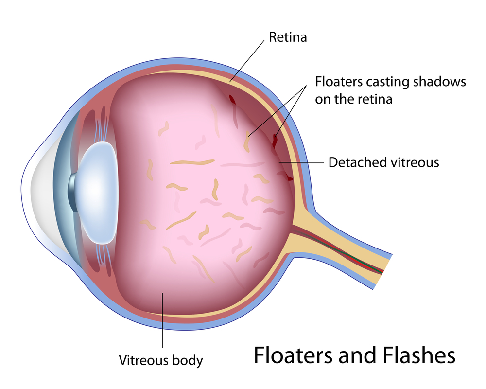
A posterior vitreous detachment (PVD) is a common cause of floaters. A PVD occurs when the vitreous separates from the retinal surface. This is a normal event and will eventually occur in everyone. Retinal tears are more likely to occur right after a PVD has started.

The vitreous is usually a clear watery gel. As we age, enough of the proteins in the vitreous liquefy and degenerate. Eventually, there is a physical separation of the vitreous from the retina due to this liquefaction.

“Floaters” can develop due to opacities/haziness due to the aging gel which is no longer absolutely clear due to changes in the optical properties of the gel or the interface between the vitreous and water inside the eye.

Tears in the Retina
Retinal tears can cause floaters by either causing a small vitreous hemorrhage, or by dislodging cells into the vitreous which are normally located underneath the retina.

Vitreous Hemorrhage
Blood in the vitreous can also cause floaters. A so-called vitreous hemorrhage may result from either a retinal tear or advanced diabetic retinopathy. Occasionally, retinal vascular occlusions can also lead to bleeding in the vitreous.

Blood in the eye often absorbs, but only your doctor can determine the exact nature of the floaters and if they are associated with a retinal tear. It’s impossible for you to tell the cause of floaters without a proper dilated eye exam.

Inflammation
Certain types of inflammation or infection can cause significant floaters. Actually, these are white cells which migrate to the retina and vitreous and can be seen a floaters.

Asteroid Hyalosis
This is a common entity where lots of fine white opacities are suspended in the vitreous. In most cases, these are not noticed by the patient. Sometimes, however, the so-called asteroid bodies are so dense they prevent good examination of the retina. Other times, especially after a PVD, the asteroid bodies are noticed and patients may complain of floaters.

New Floaters: Evaluation
All new floaters must be examined by your eye doctor within 24-72 hours after occurring. Even if the floaters disappear in that time frame, you should be examined to look for a possible tear in the retina.

Again, you, the patient, can NOT tell the cause of the floaters or if you’ve sustained a retinal tears. Retinal tears can cause a retinal detachment – a potentially blinding problem.

Treatment of Floaters
There are no medicines or eye drops to treat floaters. Most doctors advise simply putting up with the floaters.

For patients who have chronic problems with floaters, I recommend a vitrectomy. A vitrectomy is an eye operation, performed by a retinal specialist. The operation is comparable to a cataract operation in terms of safety and possible complications.

There are a handful of doctors who perform Yag laser to break up floaters.

Randall E. Wong MDRandall V. Wong, MD
Retina Specialist
Fairfax, Virginia

September is Healthy Aging Month

As your body ages, it’s common to experience changes to your eye health and vision. According to studies, over 10% of Americans over age 40 will experience permanent age-related vision loss — and almost everyone experiences normal age-related changes such as loss of close vision so that they require reading glasses.  As the U.S. population gets older, it’s important to spread the word about ways to prevent vision loss in older adults.

Genetics and the natural aging process play a role in long-term eye health. But research shows that there are controllable factors that help either lower your risk of visual impairment or slow its progress.

What Happens to Vision as You Age?

Over time your eyes will experience physical changes that will affect your vision.  On average, these changes begin around age 40 and impact the eye’s focusing ability — a natural change in vision called presbyopia. The most common effect is experiencing more difficulty in clearly seeing nearby objects, especially in dim lighting.

It’s normal for the age-related eye changes to cause:

  • Difficulty in distinguishing colors
  • A need for more light to see well and a slower adjustment between dark and bright settings
  • Headaches or tired-feeling eyes while reading or other up-close activity 

Other common causes of age-related visual decline include conditions:

Eye Floaters: Floaters are tiny spots or specks that drift throughout your field of vision. They occur when the vitreous, a jelly-like substance inside the eye, becomes more liquid as you age. This causes ocular fibers to clump, casting tiny shadows. If there is a sudden appearance of many new floaters, you should seek a dilated eye exam to rule out retinal detachment.

Dry Eye: As you age, your eye tear production decreases and this is called ‘dry eye’. Lack of enough lubrication to the surface of the eye can cause blurred vision and light sensitivity. Dry eyes can also cause uncomfortable stinging, scratching, or burning sensations.

Cataracts: As you age the clear proteins within the lens of your eye slowly become more and more cloudy interfering with the images that reach your retina. The cloudy lens causing poor vision is called a cataract. Luckily, modern eye surgery can remove the clouded lens material, which is then replaced with a man-made substitute lens that clearly focuses the image on the retina, restoring normal vision.

Glaucoma: Glaucoma causes damage to the major nerve of the eye called the optic nerve. This nerve is part of the central nervous system, that carries visual information from the eye to the brain.  In glaucoma, the eye experiences a symptom-free increase of intraocular pressure due to an imbalance of the fluid produced in the eye and the amount of fluid drained. The asymptomatic increase in pressure over time damages the optic nerve, slowly decreasing your peripheral (side) vision and can result in total vision loss. If you have glaucoma, you may experience changes to your peripheral or side vision as the eye disease progresses.  Glaucoma can be detected by a careful eye examination and the loss of vision can be stopped with medication or/and surgery.

Age-related macular degeneration (AMD): In the developed world, AMD is the most common retinal disorder and the leading cause of permanent vision loss in people over 50. AMD is due to loss of retinal cells in the macula, an area of your retina responsible for detailed vision, necessary for tasks such as driving, reading and facial recognition. In advanced cases, patients cannot recognize faces or read books without special visual aids. The good news is that there are good treatments for AMD and patients retain “getting around” peripheral vision and don’t go completely blind.


How to Care for Your Vision as You Age

Most age-related visual conditions today can be treated with medicine or outpatient surgery. A dilated eye exam performed by an eye care professional is the only way to find some common eye diseases while they’re easier to treat and before they cause permanent vision loss.

If you don’t have any symptoms or vision problems, doctors recommend getting regular eye exams based on your age:

  • Ages 20 to 39: Every 5 years.
  • Ages 40 to 54: Every 2 to 4 years.
  • Ages 55 to 64: Every 1 to 3 years.
  • Over 65: Every year

For people with diabetes, high blood pressure, or a family history of glaucoma or vision loss, the National Eye Institute recommends you get dilated eye exams yearly.

Diet and lifestyle choices can also impact your long-term eye health. To help maintain good vision as you age, you should:

  • Be alert to the early signs of vision problems.
  • Eat healthy foods – for eye healthy recipes visit: Eye Cook
  • Protect your eyes from the sun – make sure you wear UV protected sunglasses and a hat.
  • Get regular comprehensive eye exams.
  • Don’t smoke.
  • Exercise regularly to maintain healthy weight.
  • Control your blood pressure.
  • Give your eyes a break – if you work on a computer, it is recommended to take a 20-30 second break every 20 minutes. This will give your eyes a chance to relax.

Being Proactive Is the Key to Successful Aging Eye Care

There are many ways you can practice positive aging eye care. Aging may be inevitable, but exercise, a healthy diet, and positive lifestyle changes, such as stopping smoking, can prolong the health of your eyes.

May-Healthy Vision Month

When it comes to our health, we may visit our doctors and nurses regularly to make sure our bodies are healthy. But what about our eyes? They’re not always top of mind, but they’re just as important! May is Healthy Vision Month, an observance coordinated by the National Eye Institute to motivate Americans to make their eye health a priority and educate them about steps they can take to protect their vision.

Here are a few ways you can help protect your vision

  • Get an annual comprehensive dilated eye exam.
  • Know your family’s eye health history. It’s important to know if anyone has been diagnosed with an eye disease or condition, since some are hereditary.
  • Eat right to protect your sight: In particular, eat plenty of dark leafy greens such as spinach, kale, or collard greens, and fish that is high in omega-3 fatty acids such as salmon, albacore tuna, trout, and halibut. Visit Eye Cook for eye healthy recipes.
  • Maintain a healthy weight.
  • Wear protective eyewear when playing sports or doing activities around the home, such as painting, yard work, and home repairs.
  • Quit smoking or never start.
  • Wear sunglasses that block 99 -100 percent of ultraviolet A (UVA) and ultraviolet B (UVB) radiation.
  • Wash your hands before taking out your contacts and cleanse your contact lenses properly to avoid infection.
  • Practice workplace eye safety.

Taking care of your eyes also may benefit your overall health. People with vision problems are more likely than those with good vision to have diabetes, poor hearing, heart problems, high blood pressure, lower back pain and strokes, as well as have increased risk for falls, injury and depression.

In addition to your comprehensive dilated eye exams, visit an eye care professional if you have

  • Decreased vision.
  • Eye pain
  • Drainage or redness of the eye
  • Double vision
  • Diabetes
  • Floaters (tiny specks that appear to float before your eyes)
  • Circles (halos) around light sources or if you see flashes of light

For this Healthy Vision Month, take care of your eyes to make them last a lifetime.

Cancer in the EYE: Unexpected Thought

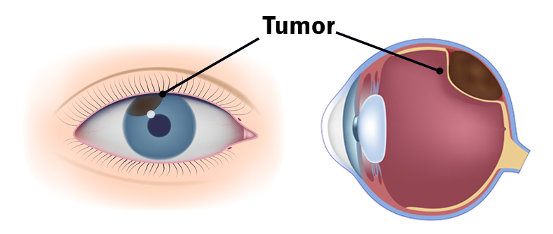
When you think of cancer, most of us do not think about the eye or vision. Though rare, cancer can start inside or outside of the eye. If cancer starts inside the eyeball it’s called intraocular and if it starts outside the eye (eyelid or in the eye socket) then it’s called extraocular tumor. It can occur in both children and adults. Most major eye centers have specialists who are trained in the diagnosis and treatment of eye cancers.

Here are a few types of cancer in eye:

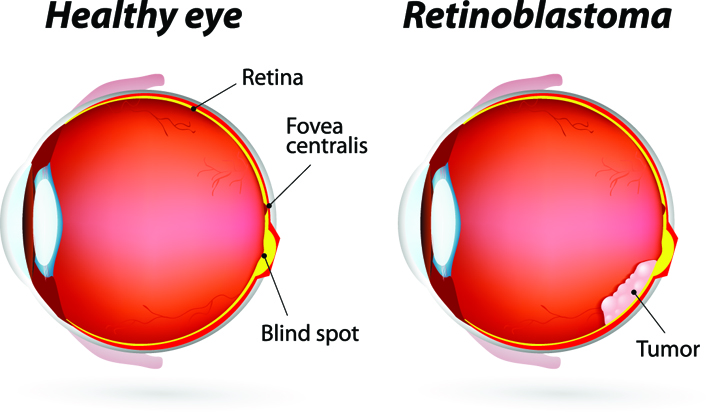
  • Eye Cancer in Children: The most common cancer seen in the eye of children is retinoblastoma. This occurs in children at very early age so these are the youngest cancer patients. This cancer starts inside the eye and affects the retinal cells. This is a cancer that presents very quietly, the child has no pain, no complaints and plays happily without any problem until one day parents notice that the pupil of the eye has some abnormal ‘White Glow’ (leukocoria) rather than the usual ‘red-eye’ reflection seen in a photo—that can be the first sign of retinoblastoma. So, it requires prompt evaluation by an ophthalmologist. Retinoblastoma is a curable cancer but if it is not treated on time, it can grow quickly and fill the eyeball. It can lead to loss of vision and life-threatening problems.

At the later stage of this cancer, the only one way to survive is to remove the eyeball (enucleation). Like many of other types of cancer, retinoblastoma has a genetic component so genetic testing needs to be done. The tumor begins with the RB1 gene mutation that stimulates retinal cells to develop into a tumor called a retinoblastoma. The RB1 mutation can be inherited from the parents, but in some cases it is sporadic and not inherited. There are various treatments such as surgery, chemotherapy, radiotherapy etc. to cure retinoblastoma cancer. Rarely it can spread beyond the eye. 

  • Eye Cancer in Adults: The development of a tumor in the back of the eye in adults can be from a metastatic cancer from elsewhere in the body or can arise in the eye itself. The most common primary eye cancer is called uveal melanoma and is a cluster of rapidly growing cells underneath the retina, which can lead to vision loss. The tumor can also appear as a dark black spot on the iris. Ideally the uveal melanoma can be treated when the tumor is still only in the eye.However, unfortunately, in approximately 50% of patients the uveal melanoma spreads (metastasis) to other part of the body, making it the most dangerous eye cancer in adults. Depending upon its location the uveal melanoma may not cause early symptoms but the patient may experience blurred vision or large numbers of floaters. The uveal melanoma can be diagnosed only when an ophthalmologist or eye care specialist examines your eye. What is the main cause of uveal melanoma? How it starts, grow and spread? These questions are still unanswered. The risk of this cancer increases in persons having fair skin (white), light eye color and inability to tan. There are certain changes in the genes linked to uveal melanoma. Currently the most common are GNAQ and BAP-1 gene mutations that are associated with greater risk of metastasis, where it spreads to other parts of the body. The standard treatment of this tumor is fine-needle aspiration biopsy (FNAB), brachytherapy (radioactive material inside a small capsule placed next to the tumor), radiation therapy and possibly enucleation. 
  • Metastasis to Eye: Finally, the other types of cancers found in the eyes are because of the spreading of a primary cancer (from a distant site such as the breast, lung or liver) to the eye.  Sometimes an eye exam can identify a metastatic tumor before the primary tumor is recognized elsewhere in the body.  Examples of cancers that spread to the eye are breast cancer in women and lung cancer in men. Less commonly, the prostate, kidney, gastrointestinal and blood cancers (leukemia and lymphomas) can spread to the eye.  Treatment depends on the type of cancer involved. 
  • Eye Damage from Chemotherapy: Eye problems can also develop through the side effects of chemotherapy or hormone therapies given for tumors outside the eye. When a person has any type of cancer, they often must undergo treatments with chemotherapeutic drugs.  A commonly used cancer drug is called cisplatin.  When treated with cisplatin, there can be damage to the retinal pigment epithelial cells in the inner part of the retina. Hemorrhages or bleeding can occur within the retina itself and vision can decrease temporarily. If a person is undergoing cancer treatments, it is a good idea to have their eyes checked, especially if they are having any vision problems. The chemotherapy does not actually cause cancers but only side effects.

Therefore, if you are having any decreased vision, it is always makes sense to have a good eye examination to identify any problems at early stage.

Myopic Degeneration

Did you ever wonder while growing up what your friends with the thick glasses meant when they said they were “nearsighted”?  What exactly does it mean to be nearsighted, and what issues related to vision arise from this?

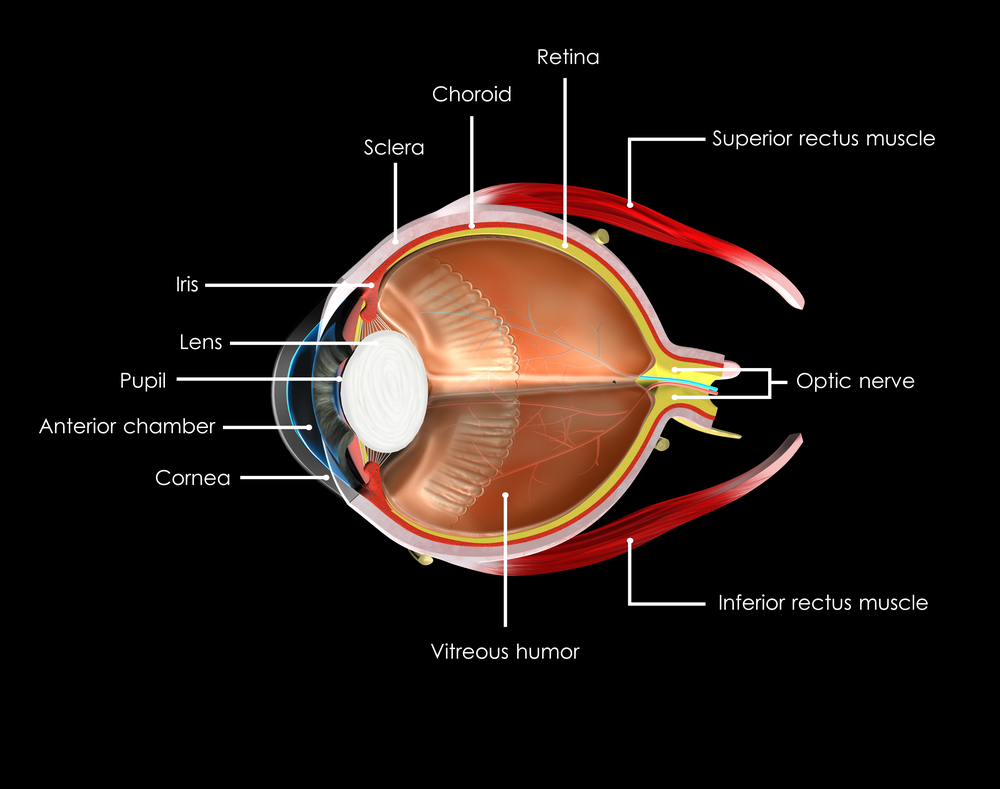
Think of your eye as a camera, which has a segment to focus the light coming into it. In the eye, this focusing segment is made up of the cornea, the clear front part of the eye, the pupil, and the natural lens inside the eye behind the pupil. Like any camera, the eye has a film to make the images, and this is the retina. The retina is a thin tissue lining the entire inside of the eye like wallpaper.

Myopic Degeneration
Figure 1. Normal retina inside the eye, the optic nerve and the blood vessels

Another term for nearsightedness is myopia. When someone is myopic, they see well up close but cannot see far away. A myopic eye is longer than average in length from the front of the eye to the back of the eye. In a sense, light and images from far away cannot reach the back of the eye where the camera film, the retina, resides. Therefore, glasses or contact lens are needed to help focus the light on to the retina to see clearly. Some people may elect to have refractive surgery to correct near sightedness. Because the eye is longer than average, the tissues inside it, like the retina, can become abnormally thinned due to stretching. It should be noted that people with myopia have an increased risk for retinal tear or detachment of the retina which can lead to rapid loss of vision. If sudden flashing lights, new floaters, or darkening of peripheral vision are seen, an eye care professional should be expeditiously consulted for an evaluation.

Myopic Degeneration
Figure 2. Myopic degeneration

Progressive thinning of the retina resulting from elongation of the eyeball is termed myopic degeneration. Individuals with more severe nearsightedness or high myopia are at greater risk for developing myopic degeneration. When thinning and atrophy occur at the part of the retina that affects central vision (macula), vision may deteriorate gradually and may not be correctable.  You can see in the Figure 2 that there are some white and black discolorations that were not present in Figure 1. These changes exemplify myopic degeneration. Glasses or contact lenses cannot correct for myopic degeneration because there is actual damage of the retina tissue when it is stretched out. There is no reversal for the actual thinning of the retina and the damage to the retina.

Myopic degeneration
Figure 3. Myopic degeneration with choroidal neovascularization

When the retina is stretched out and the eye is elongated, sometimes abnormal blood vessels can develop just below the retina. These blood vessels, termed choroidal neovascularization, can interfere with focusing of the light or bleed and can cause sudden decreased vision. The reddish discoloration in Figure 3 exemplifies bleeding from choroidal neovascularization in myopic degeneration. Fortunately there is a treatment in the form of medications that can be injected into the eye that can stop the growth of these abnormal bleeding vessels, if they are found early before permanent damage occurs to the eye.  Therefore, early diagnosis as well as treatment of choroidal neovascularization can be helpful in limiting the degree of vision loss from these bleeding blood vessels.

Not everyone with myopia develops myopic degeneration, and there is currently no algorithm to predict its development. Self-testing one eye at a time using an Amsler grid, which looks like a graph paper, to check for any distortion of straight lines on a regular basis can be a useful tool to identify any early changes. Those with myopic degeneration should have a regular dilated eye examination with an eye care professional for early detection of any treatable changes.

Image References
1.Normal Retina. Jason Calhoun MD. ASRS Image Bank.
2.Myopic degeneration. Gerrardo Garcia Aguirre MD. ASRS Image Bank.
3.Myopic degeneration and CNVM. David Callanan MD. ASRS Image Bank.

4/6/16

Judy Kim - Myopic Degeneration

Judy E. Kim, MD
Medical College of Wisconsin
NEHEP Planning Committee Member


Alessa Crossan - Myopic Degeneration

Alessa Crossan, MD
Medical College of Wisconsin

Uveitis Explained

12/18/14

Uveitis is defined as inflammation of the uveal tissue. The uvea includes the iris, ciliary body, and the choroid of the eye. The iris is located in the anterior compartment of the eye and acts like the aperture of the camera, precisely filtering the amount of light entering the eye. The ciliary body, which is attached posteriorly to the iris, is involved in both the production of the aqueous fluid in the eye as well as the accommodation of the lens apparatus. The choroid is a dense layer of blood vessels that sits underneath the retina on the back wall of the eye, helping to nourish and remove metabolic waste products from the retina. Inflammation of any of these structures will consequently cause disruption of the visual pathway and over the long term can cause permanent visual loss. In fact, uveitis is the third most common cause of preventable blindness in the developed world.
uveitis explained
Symptoms of uveitis include blurry vision, ocular pain, photophobia, redness, and floaters. These can be acute in nature, lasting a few days to weeks, and in some cases can be chronic, lasting weeks or months. Anyone with any of these symptoms should see their eye care provider as soon as possible, as faster treatment of uveitis has shown to result in better long term visual outcomes.

Uveitis can affect virtually any part of the eye, from front to back. Anterior uveitis or iridocyclitis is confined to the iris, ciliary body, anterior chamber, and cornea. Inflammation affecting the vitreous is termed intermediate uveitis, or pars planitis, and any inflammation affecting only the retina or choroid is termed posterior uveitis. The term panuveitis may be used when multiple layers of the eye are affected.

There are many possible causes of uveitis, including infection, inflammatory diseases, autoimmune diseases, and trauma. However, the majority of cases of uveitis, approximately half, are considered idiopathic, where no etiology is ever found. Trauma is the next most common cause of intraocular inflammation, accounting for approximately 20% of all cases. The remaining cases are secondary to a systemic disorder or localized ocular condition. Systemic etiologies can include inflammatory disorders such as sarcoidosis, infections such as tuberculosis and syphilis, as well as autoimmune diseases such as rheumatoid arthritis and lupus.

Treatment of uveitis is aimed at both blunting the intraocular inflammation as well as addressing any underlying systemic etiology. The most common treatment is the use of corticosteroids. These can be taken orally, or used topically as eye drops. In some cases, corticosteroids can be injected in or near the eye as well. If the uveitis is caused by an infection, such as tuberculosis or syphilis, the patient is also given antibiotics. Systemic corticosteroids can have major side effects when taken chronically, such as weight gain, hair loss, osteoporosis, hypertension, secondary diabetes, psychosis, and reduced growth in children. Because of these potential problems, the chronic use of systemic corticosteroids is not recommended. In cases of chronic uveitis that require long term treatment, immunosuppressive agents with less known side effects such as methotrexate, cyclosporine, and mycophenolate mofetil (Cellcept) are more commonly used. However, these biologic agents have their own set of potential side effects and therefore, it is recommended that a rheumatologist should also be involved in the care of the patient when using these agents. Topical and intraocular steroids localized to the eye can cause elevated intraocular pressure as well as cataracts. In most cases, elevated intraocular pressure can be controlled with topical glaucoma drops, but in some cases surgical intervention is required to prevent severe glaucomatous damage.

The most common type of uveitis is acute anterior uveitis or iridocyclitis. Many cases of anterior uveitis are idiopathic though almost half of all cases are associated with the HLA- B27 haplotype. Systemic diseases associated with HLA-B27 include psoriatic arthritis, ankylosing spondylitis, reactive arthritis, and inflammatory bowel syndrome. Signs of anterior uveitis include redness of the eye, sometimes termed ciliary flush. The conjunctiva can become extremely red, and when associated with ocular pain and photophobia, is a strong indicator of anterior uveitis. Inflammatory cells found in the anterior chamber are the hallmark of anterior uveitis, sometimes deposited on the corneal endothelium (keratic precipitates) or iris (Bussaca nodules). Patients with anterior uveitis are typically treated with topical corticosteroid and cycloplegic eye drops. A laboratory workup for systemic etiologies is usually not necessary unless the patient experiences a recurrent episode.

Inflammation affecting primarily the vitreous cavity is known as intermediate uveitis or pars planitis. Inflammatory cells in the vitreous, known as vitritis, are typically bilateral, and when severe, can be found clumped in the vitreous cavity (snowballs) or deposited on the inferior pars plana (snowbanking). Intermediate uveitis is typically idiopathic though sarcoidosis, multiple sclerosis, and Lyme disease are also possible causes. Certain malignancies such as lymphoma can also ‘masquerade’ as intermediate uveitis, and when seen in older patients, should be suspected and ruled out.

Posterior uveitis involves the retina, choroid, and/or the retinal vasculature, and usually is more difficult to treat than anterior uveitis.

Uveitis Explained
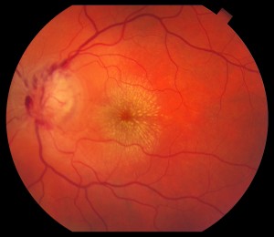
This patient with Cat-scratch disease, caused by infection with Bartonella henselae, is an example of posterior uveitis. Note the characteristic star-like pattern of exudate in the macula along with optic nerve swelling.

In many cases, patients with posterior uveitis will exhibit characteristic exam findings that help narrow the differential diagnosis. For instance, an area of active retinitis next to an old pigmented chorioretinal scar is highly suggestive of toxoplasmosis. The most common symptom in patients with posterior uveitis is blurred vision. One of the more typical findings in posterior uveitis is macular edema, which is usually treated with periocular or intraocular corticosteroids.

In summary, uveitis is a visually threatening inflammatory condition that should be diagnosed and treated immediately. It is important to determine as best as possible the etiology of the uveitis and treat appropriately. In general, most patients with uveitis have good visual recovery with the proper management. However, in some cases, severe damage can occur, either due to the inflammation itself (usually chronic) or as a side effect of therapy (corticosteroids).

RichardRoeMD-ThumbnailRichard H. Roe, MD, MHS
Retina-Vitreous Associates Medical Group

Diabetes And The Potential For Diabetic Retinopathy

11/20/14

Defining Diabetes

Diabetes mellitus is a group of metabolic disorders in which a person has high blood sugar levels. It occurs either because the pancreas does not produce enough insulin, or because the body does not respond appropriately to the insulin that is produced. Insulin is the hormone that converts sugar into energy for the body. There are several forms of diabetes.

Type 1 diabetes occurs when the pancreas does not produce adequate levels of insulin. It usually develops during childhood or adolescence; however, it can also occur in adults. Those with type 1 diabetes must take insulin injections. About 10 percent of those with diabetes have type 1.

Type 2 diabetes is the most common type of diabetes, affecting about 90 percent of those with diabetes. It is caused by the combination of the body’s resistance to insulin (not using its own insulin efficiently) and the pancreas not producing enough insulin. As a result, there is an increase in the level of sugar, or glucose, in the blood.

Gestational diabetes occurs in pregnant woman who have not previously had diabetes but whose blood sugars become elevated during pregnancy. In many patients, their blood sugars will return to normal after the pregnancy is over. Placental hormones make the mother resistant to insulin, causing a buildup of blood sugars. These women are at high risk for type 2 diabetes later on in their lives.

Diabetic Retinopathy: An Overview
diabetic retinopathy
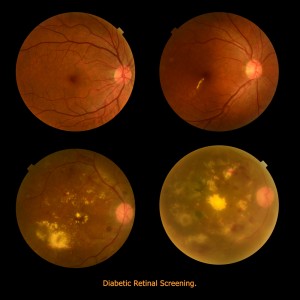
Anyone with uncontrolled diabetes is at risk for developing diabetic retinopathy. According to the National Eye Institute, between 40 to 45 percent of Americans with diabetes have some form of diabetic retinopathy, the most common eye condition associated with diabetes.

In the United States, the Centers for Disease Control and Prevention reports between 12,000 and 24,000 new cases of blindness each year due to diabetic retinopathy, making it the leading cause of vision loss among American adults, ages twenty to seventy-four. The Center also projects that by 2050, the number of Americans age forty and older with diabetic retinopathy will grow from a current 5 million individuals to about 16 million. Although these statistics are alarming, you can prevent or delay damage to your vision by controlling your diabetes, along with obtaining regular eye evaluations and treatment.

Defining Diabetic Retinopathy
Diabetic retinopathy is a disease affecting the retina, caused by elevated blood sugar levels. It usually affects both eyes and occurs when uncontrolled blood sugar levels damage the small vessels of the retina, the light-sensitive tissue in the back of your eye. The retina is responsible for processing images that make vision possible. To produce clear, distortion-free vision, the retina must lie completely flat. If the delicate retinal tissue is damaged, images that you see may be blurred or distorted.

Diabetic retinopathy is a progressive disease, and thus worsens over time. Although some effects, such as blurriness and distortions, may be mild or subtle, the long-term consequences can cause severe vision loss.

Symptoms of Diabetic Retinopathy

Because diabetic retinopathy rarely causes pain, symptoms are not always apparent in the earliest stages. In fact, damage to your retina could be occurring long before you have noticeable signs. When symptoms do occur, they’re often caused by retinopathy affecting the macula, the area at the center of the retina. Symptoms may include the following:

• blurred vision

• seeing dark spots or “floaters” (small specs in your field of vision)

• decreased night vision

• vision loss

• problems seeing colors

It’s important that you see your eye specialist immediately if you have any such symptoms. Diabetic retinopathy cannot be cured, but with careful monitoring, it can be diagnosed and treated, before your vision is impaired. The treatment is typically less invasive and more effective when diagnosed at an earlier stage, before permanent damage has occurred.

How the Retina Works

The retina is made up of specialized nerve tissue that contain microscopic receptors (called cones and rods) and other nerve cells that line the back of the eyeball.

These cells carry signals (images that we see) along the optic nerve to a special area of the brain, where they’re interpreted into what we perceive as sight. You might compare the retina to film in a camera—the film delivers the photo image that the camera captures.

There are two main areas of the retina that can be damaged by diabetic retinopathy:

• The macula, the center of the retina. The macula allows you to read and see fine details and recognize colors. At the very center of the macula is a dimple known as the fovea. It is the most sensitive portion of the macula and makes sharp vision possible.

• The peripheral retina, which is the portion of the retina that is outside the macula. It’s responsible for your side vision and also makes night vision possible.

The retina lies on a nutrient-rich flat “carpet” of vessels that nourish it with necessary oxygen and nutrients. To reach the retina, however, nutrients must pass through two buffers—a thin membrane called Bruch’s membrane and a single layer of specialized cells called the retinal pigment epithelium. Waste products are also transported away from the retina through these two membranes. Diabetic retinopathy can interfere with this constant import of necessary nutrients and export of waste products.

Risk Factors for Diabetic Retinopathy

There are many factors that can raise your risk for diabetic retinopathy. However, you’ll note that many of these risk factors can be controlled.

Duration of Diabetes and Glucose Control

The longer you’ve had poorly controlled blood glucose levels, the higher your risk for diabetic retinopathy. Most diabetic individuals develop eye problems overtime, making duration of their diabetes one of the strongest predictors that they will develop this eye disease. Research has shown that nearly all type 1 diabetics and 60 percent of type 2 diabetics develop the condition within the first two decades of their diabetes diagnosis.

The American Diabetes Association (ADA) recommends fasting glucose levels between 70 and 120 mg/dL and less than 180 mg/dL two hours after meals. They also recommend a hemoglobin A1c of 7 percent of less. Hemoglobin A1c is a protein in red blood cells that bonds with blood sugars. Since red blood cells can live from 90 to 120 days, the hemoglobin A1c stays in the blood for that length of time. Accordingly, it is effective in measuring the average blood sugars over a period of time. This test tells doctors how well your treatment plan is working. You should always know what your hemoglobin A1c values are, as they may affect the interval between your retinal examinations.

Obesity

The more fatty tissue you have, the more resistant your cells are to insulin. Obesity increases your risk for diabetes as well as other serious conditions such as heart disease. Estimates suggest that 65 percent of Americans may be overweight. Being overweight aggravates high blood pressure and cholesterol. Achieving a healthy weight is important in controlling blood sugars and diabetes related complications.

Lifestyle Choices

A sedentary lifestyle, especially if you are overweight, contributes to many diseases, including diabetes, heart disease, high blood pressure, and high cholesterol levels. On the other hand, physical exercise improves circulation, lowers blood sugars, and improves your body’s use of insulin. This results in improved blood sugar levels. This benefit of increased sensitivity to insulin continues for hours after you stop exercising.

Exercise also promotes weight loss. A sedentary lifestyle contributes to insulin resistance, and makes it more difficult to keep weight off. Even light or moderate physical activity can help lower blood sugars.

Smoking is another major risk factor for developing diabetic retinopathy. Smoking also causes diabetic retinopathy to progress faster. The nicotine in tobacco not only contributes to higher blood pressure and higher cholesterol levels, but it also impairs insulin activity. Even though quitting can be difficult, it is critical to heart health and diabetes control.
Unlike smoking, alcohol consumption doesn’t have a direct influence on diabetic retinopathy. Yet because it can affect diabetes control, drinking in excess can affect the health of your eyes. Your doctor can tell you what constitutes drinking in moderation for you.

High Cholesterol Levels

Diabetes puts you at risk for chronically high cholesterol or blood fats that promote the buildup of plaque in your arteries. Although the tiniest vessels of the retina are too small for such build-up, uncontrolled cholesterol can contribute to macular edema and the development of hard exudates, the small yellow spots or lipid deposits that may form in the macula. Both conditions are associated with a higher risk of vision loss.

Doctors advise keeping “bad” or low density cholesterol (LDLs) less than 70 mg/dL. Good cholesterol or high density lipoproteins (HDLs) should be greater than 40 mg/dL in men and 50 mg/dL in women. Both men and women should strive for triglycerides, another type of fat, at levels less than 150.

High Blood Pressure

If you have both diabetes and high blood pressure (also called hypertension), you may be at higher risk for a number of eye-related problems, including retinopathy, glaucoma and optic nerve damage. Seriously elevated blood pressure not only stresses your heart, it also raises the risk for eye problems, particularly macular edema and bleeding. Chronic hypertension combined with long-term diabetes also increases the chance that your retinopathy will be more destructive and progress more rapidly. Research has consistently shown that keeping your blood pressure below 130/80 mmHg is important in minimizing the risk of hypertension related complications.

Race/Ethnicity

Diabetic retinopathy is more common in some ethnic and racial groups than others. African Americans, Asian Americans, Hispanic/Latino Americans, American Indians and Alaskan Natives are at higher risk for type 2 diabetes than non-Hispanic whites.

African Americans and Mexicans are almost twice as likely as whites to have eye problems, according to the American Diabetes Association. Native Americans also have an increased for diabetic retinopathy. Researchers aren’t sure why some ethnic groups have higher rates of diabetes, which increase the risk for retinopathy and other problems.

Age and Gender

As mentioned earlier, the longer you have diabetes, the greater your risk for diabetic retinopathy. Not surprisingly, this complication is rare among children but common among older diabetic adults. A recent study by Prevent Blindness America and the National Eye Institute, demonstrated that older adult Americans are facing a bigger threat of all age-related eye diseases (diabetic retinopathy, age-related macular degeneration, cataracts and open angle glaucoma) today than at any other time.

Genetics

Our genetic make up has an important effect on our predisposition for many health issues such as diabetes. Scientists believe that many genes or combinations of genes either promote diabetes in certain individuals or protect them from developing it.

Scientists have yet to identify every gene involved in type 1 and type 2 diabetes, but they have shown that genetics are a factor. Research studies of identical twins, for instance, have demonstrated that if one twin has type 1 diabetes, the other twin has a 50 percent change of developing the disease. If one twin has type 2 diabetes, the other twin has a 75 percent chance of developing it.

Pregnancy

Gestational diabetes is a type of diabetes linked to pregnancy; however, diabetic retinopathy is usually not a complication in these women. However, if you’re already a diabetic and become pregnant, you are at an increased risk of developing diabetic retinopathy. This is a result of the hormonal and metabolic changes that occur during pregnancy, making the disease and its complications progress more rapidly. It is recommended that you see a retinal specialist for evaluation and monitoring.

In Summary

• Diabetic retinopathy is a serious complication of diabetes that results from high glucose levels damaging the retinal blood vessels. This can cause loss of vision.

• Between 40 and 45 percent of diabetic Americans have some form of diabetic retinopathy.

• The earliest form of the disease is called background diabetic retinopathy. With time it progresses to mild, moderate, or severe nonproliferative diabetic retinopathy.

• Without proper diagnosis and treatment of nonproliferative diabetic retinopathy, the condition can advance to proliferative diabetic retinopathy, which is a serious sight-threatening stage of the disease.

• Macular edema is due to build up of fluid and thickening of the macula and can occur with any type of diabetic retinopathy. It is the most common cause of vision loss in those with diabetes.

• The duration of your diabetes and how well blood glucose is controlled are major risk factors for the development and progression of diabetic retinopathy.

• Other risk factors that play a significant role in the development of retinopathy, include high blood pressure, high cholesterol, and smoking.

• As an individual with diabetes, you’re also at increased risk for other eye diseases, especially glaucoma, cataracts, retinal vein occlusion and optic nerve damage.

• Good blood sugar control, regular eye examination, and timely treatment are the key factors in reducing the damage to the eye and keeping your vision.

Pouya Dayani - Diabetic RetinopathyPouya N. Dayani, MD
Retina-Vitreous Associates Medical Group

Posterior Vitreous Detachment

10/30/14

Have you ever noticed floaters in your vision? Perhaps they looked like a bunch of small dots or maybe a cobweb swaying back and forth in your visual field. Were the floaters associated with flashing lights that made you think there was a lightning storm coming your way? These are typical symptoms of a posterior vitreous detachment (PVD), and if you have had these symptoms you are far from alone.
Floaters Posterior vitreous detachment
PVD is a natural process that occurs in the majority of people usually over the age of 50. The vitreous is a jelly-like substance that occupies the back portion of the eye. The vitreous is comprised primarily of water, which accounts for 99% of its volume, and the remaining 1% includes proteinaceous substances such as collagen fibers as well as hyaluronic and ascorbic acids. The collagen fibers act as a scaffold to allow the vitreous to maintain a formed shape as well as provide a means for the vitreous to attach to the retina, which is the light-sensitive tissue that lines the inner back wall of the eye and is critical for vision. As we age, changes in these fibers cause the vitreous to lose its shape and eventually pull away from the retina. When the vitreous separates from the retina, this is called a PVD.

As we age, the collagen components of the vitreous can clump together and are free to float in the eye. When the vitreous separates from the retina during the development of a PVD, the floaters may become more noticeable or numerous. It is common for patients to describe floaters of different shapes and sizes, and patients may notice just one or in some cases many. In many people, a PVD develops slowly and there may be no symptoms or just a few annoying floaters. In others, a PVD may occur abruptly and cause more dramatic symptoms that can be very anxiety provoking.

Since the normal process of PVD development involves the vitreous tugging on the retina until it can fully separate, this tugging can result in flashing lights that can commonly appear in the peripheral, or side, vision. These flashing lights are sometimes described as lightning streaks, and patients may notice them more readily in settings with low ambient light. The flashes of light typically resolve once the vitreous has fully separated from the retina and the tugging has ceased.

The good news is that PVD is usually harmless in the vast majority of cases, and the annoying floaters will become less bothersome over time. In approximately 5-10% of cases, the vitreous can tug too hard on the retina as it tries to separate and it may pull a hole or tear in the retina. Tears in the retina can predispose to retinal detachment, which is a serious condition that can lead to permanent vision loss.
It is important to recognize that the typical symptoms of a regular PVD are often similar to a PVD with an associated tear. For this reason, it is recommended that all patients with the new onset of floaters or flashes have a dilated eye exam. If a retinal tear or detachment is discovered, early treatment can help prevent loss of vision.

Treatment for PVD usually involves simple observation. With time, the flashes will go away, and the floaters will become less noticeable. More recently, few providers have claimed that floaters can be treated with a laser in order to make them less noticeable. I would caution that this is not mainstream therapy at the current time, and I do not advise my patients to pursue this option. Another treatment possibility is vitrectomy surgery, where the vitreous gel is removed as part of a surgical procedure. Due to safety advances in vitrectomy surgery, this is now a potential option for the rare patient who has floaters that are so numerous and bothersome that they are negatively impacting their activities of daily living. For the vast majority of patients this is not necessary.

When I see a patient with a PVD, I often recommend one follow-up visit in 4-6 weeks to make sure there are no retinal holes or tears that have developed in the interim. If the other eye has not had a PVD yet, I will counsel them that a PVD will most likely develop in that eye within the next few years, and when it does they need to be examined. I will also discuss the retinal detachment warning signs. Patients with retinal detachment will not only have symptoms similar to PVD, including flashes and floaters, but in addition they may also notice what looks like a black shade or curtain that starts in the peripheral vision and extends towards the central vision. My patients are taught that this symptom requires an immediate examination.

In conclusion, PVD is a natural process that the majority of people will experience in their lives. The symptoms can range from having no symptoms at all to many floaters with associated lightning flashes. In the majority of patients, there is no damage to the eye or threat to the vision. A dilated exam is recommended to look for possible holes or tears in the retina, and if these are uncovered, prompt treatment can prevent vision loss.

Dr. Esmaili posterior vitreous detachmentDaniel D. Esmaili, MD
Retina Vitreous Associates Medical Group

Our Thanks to Guest Bloggers Continues

10/7/14

More Amazing Guest Bloggers

Last week I took the opportunity to thank our very first guest bloggers for helping us launch the Discovery Eye Foundation Blog. We are pleased that so many people appreciate the wide range of eye-related information from eye care professionals, as well as the stories from people that live with eye disease on a daily basis.
Thank you part 2
Here is a round-up of guest bloggers since June 2014 that shared their time, experience and/or expertise to provide you with the best eye-related information.

Sumit “Sam“ Garg, MDwhat you should know about cataracts

Randall V. Wong, MDfloaters, causes and treatments

Roy Kennedyhis personal experiences with the miniature telescope implant

Sandra Young, ODthe importance of getting vitamins and minerals from your food and not just supplements

Jeanette Hassemanliving with keratoconus

Greg Shanetheater for the blind

Caitlin Hernandezblind actress and playwright

Jullia A. Rosdahl, MD, PhDlasers for glaucoma and genetics and glaucoma

Maureen A. Duffy, CVRTways to reduce harmful effects of sun glare

Kooshay Malekwhat is it like to lose your vision and being a blind therapist

Jeffrey J. Walline, OD PhDchildren and contact lenses

Robert Mahoneychoosing a home care agency

Robert W. Lingua, MDnystagmus in children

Buddy Russell, FCLSA, COMTcommon pediatric eye diseases, treatment options for children and pediatric contact lenses

NIH (National Institute of Health)telemedicine for ROP diagnosis

Harriet A. Hall, MDevaluating online treatment claims

Patty Gadjewskithe life-changing effects of a telescopic implant

Michael A. Ward, MMSc, FAAOproper contact lens care and wearing contacts and using cosmetics

Susan DeRemerSusan DeRemer, CFRE
Vice President of Development
Discovery Eye Foundation