Proper Contact Lens Care Provides Best Vision, Comfort and Ocular Health

9/18/14

Proper contact lens care is essential for the best contact lens wearing experience. Mr. Ward, Director of the Emory Contact Lens Service has shared some valuable information about taking care of your contact lens in the article below. On Tuesday join us for additional tips for people who wear contact lenses and wear cosmetics.contact lens case - proper contact lens care

Contact lenses provide alternatives to spectacles, and contact lens wearers report better peripheral vision, depth perception and overall vision quality. Contact lenses can correct near-sightedness, far-sightedness, astigmatism and even correct the need for reading glasses. They are also used to manage some ocular surface diseases.

Contact lenses fall into two basic material types: soft contact lenses (SCL) and rigid gas-permeable (GP) lenses. Soft lenses account for the great majority of the contact lens market. GP lenses require more precise fitting and are often used as specialty devices to correct high prescriptions and/or to manage various ocular disorders and may require longer lens-adaptation time. Regardless of lens type, careful attention to lens care instructions can provide good vision and life-long lens wearing comfort.
Proper lens care depends on the lens type, wearing schedule and other factors. Single-use or daily-disposable soft lenses are prescribed to be worn once and discarded. This is theoretically the safest lens wearing modality in that no lens cleaning, lens care or storage case is required for this modality. Other daily wear soft lenses may be replaced every 2 weeks, monthly or by other schedule. Any and all lenses that are removed each day must be cleaned and disinfected prior to their reuse. Your eye care practitioner should provide specific instructions relative to your lens wear and care needs. General lens care instructions and Dos and Don’ts are bullet-listed below.

A word of caution –
Contact lens wear is quite safe as long as proper lens and storage case care are followed. However, improper lens wear and care can put the lens wearer at risk for serious consequences. Sight-threatening microbial keratitis (corneal ulcer) is the most significant adverse event associated with contact lens wear and is largely preventable. The contact lens storage case is the most likely potential reservoir for contact lens related ocular infections. Therefore, lens storage case care should be high on the list of important lens wearing instructions. Contact lens cases are not meant to be family heirlooms; cases should be replaced regularly, at least every 1-3 months.

The Bullet List of Contact Lens Care Recommendations

  • Hand washing: Always wash your hands before handling contact lenses. Use mild, basic soap and avoid antibacterial, deodorant, fragranced or moisturizing liquid soaps (many liquid soaps have moisturizers that can contaminate your contacts from handling).
  • Cleaning, rinsing, and disinfecting: Digital cleaning (rubbing the lens with your finger in your palm) removes dirt and debris and prepares the lens surfaces for disinfection. Rub & rinse thoroughly, even if the product is labeled “No Rub”. Lens storage solutions contain chemicals that inhibit or kill potentially dangerous microorganisms while the lenses are soaked overnight.
    • Contact lenses should be cleaned when removed from the eye.
    • Do not re-use old solution or “top-off” the liquid in the lens storage case. Empty the storage case daily and always use fresh solution.
    • Do not use lens care products beyond their expiration dates. Discard opened bottles after 28 days.
    • Do not allow the tip of the solution bottle to come in contact with any surface, and keep the bottle tightly closed when not in use.
    • Do not transfer contact lens solution into smaller travel-size containers.
  • Keep your contact lens storage case clean (inside and out).
    • All lens storage cases should be emptied, rinsed, wiped, and air-dried between uses.
    • Keep the contact lens case clean and replace it regularly, every one to three months.
    • Do not use cracked or damaged lens storage cases.
    • Take care to remove residual solution from surfaces of lens case and solution bottles.

Other Dos and Don’ts

  • Do not wear your lenses during water activities (swimming, hot tubs, showering, etc).
  • Soft contact lenses should not be rinsed with or stored in water. Soft lenses will change size and shape if exposed to water.
  • Do not put your lenses in your mouth.
  • Do not use saline solution or re-wetting drops in an attempt to disinfect lenses. Neither is capable of disinfecting contact lenses.
  • Wear and replace contact lenses according to the prescribed schedule.
  • Follow the specific contact lens cleaning and storage guidelines from your eye care professional.
  • Do not change lens care products without first checking with your eye care practitioner.
  • Spare rigid (GP) lenses should be stored dry for long term storage { clean, rinse, dry}. New or dry-stored GP lenses should be re-cleaned and disinfected prior to lens wear.
  • Do not store soft lenses in the storage case for an extended period of time. “Spare” soft contact lenses should be new and stored in their original and unopened packaging.
  • Do not sleep in your contact lenses unless specifically approved to do so by your eye care practitioner.

For information from the Centers for Disease Control and Prevention, see:
www.cdc.gov/contactlenses/
www.cdc.gov/contactlenses/cdc-at-work.html

Michael Ward - proper contact lens careMichael A. Ward, MMSc, FAAO
Director, Emory Contact Lens Service
Emory University School of Medicine


The Way Eyes Work

9/16/14

Eyes are an amazing part of your body and not just because of what they do helping you see. The are also fascinating be because of the way eyes work. Here are 20 facts about how your eyes function.
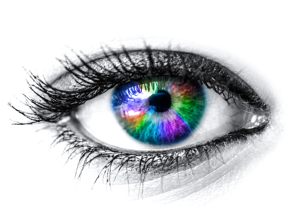
Colorful eye - the way eyes work

      1. The pupil dilates 45% when looking at something pleasant.

2. An eye’s lens is quicker than a camera’s.

3. Each eye contains 107 million cells that are light sensitive.

4. The light sensitivity of rod cells is about 1,000 times that of cone cells.

5. While it takes some time for most parts of your body to warm up their full potential, your eyes are always active.

6. Each of your eyes has a small blind spot in the back of the retina where the optic nerve attaches. You don’t notice the hole in your vision because your eyes work together to fill in each other’s blind spot.

7. The human eye can only make smooth motions if it’s actually tracking a moving object.

8. People generally read 25% slower from a computer screen compared to paper.

9. The eyes can process about 36,000 bits of information each hour.

10. Your eye will focus on about 50 things per second.

11. Eyes use about 65% or your brainpower – more than any other part of your body.

12. Images that are sent to your brain are actually backwards and upside down.

13. Your brain has to interpret the signals your eyes send in order for you to see. Optical illusions occur when your eyes and brain can’t agree.optical illusion - the way eyes work

14. Your pupils can change in diameter from 1 to 8 millimeters, about the size of a chickpea.

15. You see with your brain, not your eyes. Our eyes function like a camera, capturing light and sending data back to the brain.

16. We have two eyeballs in order to give us depth perception – comparing two images allows us to determine how far away an object is from us.

17. It is reported that men can read fine print better than women can.

18. The muscles in the eye are 100 times stronger than they need to be to perform their function.

19. Everyone has one eye that is slightly stronger than the other.

20. In the right conditions and lighting, humans can see the light of a candle from 14 miles away.

Susan DeRemerSusan DeRemer. CFRE
Vice President of Development
Discovery Eye Foundation

Rods and Cones Give Us Color, Detail and Night Vision

9/9/14

Function of Rods and Cones

Rods and cones are a vital part of the eye, helping define what we see. Here’s what you should know.
Crayons for rods and cones
1. There are three types of color-sensing cones, red, blue and green. If you are color blind one or more of these cells is missing or not working properly.

2. Men have a higher chance of being color blind than women. 1 out of 12 vs. 1 out of 255.

3. The most common type of color blindness is the disability to tell the difference between red and green.

4. The eye can distinguish between 500 shades of gray.

5. A healthy human eye can detect over 10 million different colors.

6. About 2% of women have a rare genetic mutation that gives them an extra retinal cone allowing them to see more than 100 million colors.

7. During a major depression people see less contrast, making colors appear duller.

8. All babies are color blind when they are born. Color vision begins to develop within a week after birth and by 6 months your baby can see every color you can.

9. Your eyes contain 7 million cones which help you see color and detail.

10. The stars and colors you see when you rub your eyes are call phosphenes.

11. There are 120 million light-sensing cells called rods which help you to see better in the dark.

12. Smoking reduces your night vision.

And these are just fun facts about eyes:

The Mayans believed that cross-eyes were attractive and would make efforts to ensure their children became cross-eyed.

Pirates used to wear a gold earring, believing it improved their sight. They also used eye patches to quickly adjust their eyes from above to below deck. When going below deck where it was dark, they flipped up the eye patch to see with the eye that had not be affected by light.

The phrase “it’s all fun and games until someone loses an eye” comes from Ancient Rome, as the only rule for their bloody wrestling matches was “no eye gouging.”

Susan DeRemerSusan DeRemer, CFRE
Vice President of Development
Discovery Eye Foundation

Evaluating Treatment Claims

9/03/14

The following article on evaluating treatment claims is from the Science-Based Medicine blog is being used with their permission. Since Discovery Eye Foundation provided the inspiration for Dr. Hall, we thought you might enjoy it as well.
Black board evaluating treatment options
I recently wrote about the claim that acupuncture can improve vision in patients with macular degeneration. In response, I received this e-mail:

“At Discovery Eye Foundation we have an education and outreach program for people with age-related macular degeneration, the Macular Degeneration Partnership. We are constantly getting calls from people who have heard of a new “cure” or a way to stop their vision loss. It is always hard to interject reality into the conversation and hear the hope leave their voice, replaced by frustration or despair.”

The e-mail suggested I write an article providing guidelines for consumers to help them evaluate the validity of treatment claims for themselves. On SBM we are constantly stressing the need to apply critical thinking to what you read, and the many pitfalls to be avoided. I’ll try to synthesize some of the principles into a handy list of questions.

What kind of evidence is there?

• If the claim is based on nothing but testimonials, STOP RIGHT THERE. You can forget about it, or at least file it away until there is better evidence. Anecdotes are not evidence; they only serve to suggest promising avenues of research. Science is the only reliable way to determine if a treatment is safe and effective.

• Is the claim based on a gold standard randomized, placebo-controlled, peer-reviewed study or some lesser kind of evidence like case reports? Was it published in a reputable mainstream medical journal? (If you’re not sure how reputable the journal is, you can look it up on tables of journal impact factors.”

• Keep in mind that half of all studies are wrong. There are many factors that can lead to error. Preliminary or pilot studies that are positive are frequently followed by better, larger studies that are negative. We can never rely on one study without confirmation.

• Was it a meaningful clinical study in humans?

In vitrolab studies and animal studies may not be applicable to humans; if promising, they must be confirmed in good human studies. If it was a human study, did it show meaningful outcomes that made a real difference, like a reduction in heart attacks, or did it just show an improvement in lab values or risk factors?

• Were there 10 subjects or 300? Large studies are more trustworthy than small ones. The fewer dropouts, the better.

• Did it use an appropriate placebo control that subjects really couldn’t distinguish from the active treatment?

• Have other studies found similar results? Are there any studies that show the opposite? You can search PubMed and look for them.

Who is making the claim?

  • Is it someone who is likely to be biased?
  • Is it someone who provides the treatment or sells the product?
  • Is it someone with expert medical knowledge or someone like the schoolteacher who claimed she had invented a cure for the common cold?
  • Does the person have a good reputation, or a track record of making questionable statements?
  • Is it someone who quotes or associates with unreliable sources like Mercola.com, the Weston Price Foundation, or the Health Ranger? Quackwatch has a useful list of non-recommended sources of health advice.

Where was the claim reported?

Real medical breakthroughs would be headline news. It’s not likely you would first hear about a cure for diabetes on an afternoon talk show or a Facebook page. Has your doctor heard of it? Are mainstream doctors recommending it? Is it covered on professional medical websites like the American Academy of Pediatrics or disease-focused websites like the American Diabetes Association? Is it sold only through multilevel marketing schemes?

What kind of language is being used?

Is it a sober factual report with caveats, or is it full of hype and buzzwords like “miracle,” “natural,” “known to the ancients,” “quantum,” “amazing,” “revolutionary”? If it really worked, advertising gimmicks wouldn’t be needed to sell it.

Does Gwyneth Paltrow swear it worked for her?

Testimonials can be very passionate but they are notoriously unreliable. Getting better when you use a treatment doesn’t necessarily mean you got better because of the treatment. Symptoms can fluctuate, diseases can resolve without treatment, and placebos can fool people. Perceptions can be wrong (think of optical illusions), the meaning of true perceptions can be misinterpreted, and memories can be inaccurate. Every snake oil salesman has reams of testimonials, and through the centuries there were testimonials galore for bloodletting to balance the humors. People frequently come to believe bogus remedies have worked for them. Barry Beyerstein wrote a classic article about that; it’s essential reading.

Does it make sense?

Is there a plausible mechanism of action? If someone claimed that standing on your head and whistling Dixie would cure diabetes, I think you would be skeptical. If it claims to work by a mechanism incompatible with known scientific principles, the level of evidence would have to be extraordinary for it to outweigh all the evidence those scientific principles are based on. Homeopathy’s claim that water can cure by remembering long-gone molecules, even after the water has been dripped onto a sugar pill and allowed to evaporate, would require extraordinary evidence indeed. If it’s a new antibiotic that is related to an old one, an ordinary level of evidence would suffice.

Is there a double standard?

Are they asking you to accept a “natural” or “alternative” treatment on the basis of the kind of evidence that you wouldn’t want the FDA to accept for allowing marketing of a prescription drug? There is only one science and only one standard of evidence.

Does it sound too good to be true?

Then it probably is too good to be true. Does it promise to cure a hitherto-incurable disease? Does it promise you can eat all you want and still lose weight? Does it promise there are no side effects of any kind? Does it remind you of the spiel of a used car salesman or a TV infomercial? Caveat emptor.

Who disagrees and why?

This is the most important question you can ask. It is rare for 100% of people to agree on anything. If you can find someone who disagrees, you can examine the reasons given for both opinions, and it will usually become obvious which side makes more sense. If you can’t find anyone who disagrees, it might be because it’s too new or because no scientist has taken it seriously enough to bother writing about it. In that case, withhold judgment and keep checking until someone does disagree.

Hope springs eternal, but true hope is better than false hope

If you are a desperate patient, it’s only natural to grasp at any straw of hope; but when the evidence is insufficient, the reasonable approach is to withhold judgment and wait for better evidence. You might think, “If it works, I don’t want to wait” but history teaches us that the great majority of these things don’t pan out. It might not do any harm, but then again it might; there might be adverse effects that haven’t been identified yet, it might raise false hopes only to dash them, and if nothing else it might waste time and money or interfere with getting more appropriate care. When you take an inadequately-tested medicine, you are essentially offering yourself as a guinea pig in a haphazard uncontrolled experiment that doesn’t even keep records. Of course, that’s your privilege; but I hope you would do it with your eyes open, with a realistic understanding of the state of the evidence.

Harriet Hall, MDHarriet A. Hall, MD
Retired US Air Force Physician
Editor of Science-Based Medicine Blog
Author of SkepDoc column in Skeptic Magazine

Telemedicine For Diagnosing Retinopathy Of Prematurity

8/28/14

Technology now allows patients access to highly-qualified, specialized care, no matter where they live. This potential is demonstrated in the sight-saving treatments that can be used to help children lead normal healthy lives. The following article is posted with the permission of the National Institute of Health (NIH).

Telemedicine is an effective strategy to screen for the potentially blinding disease known as retinopathy of prematurity, according to a study funded by the National Eye Institute (NEI). The investigators say that the approach, if adopted broadly, could help ease the strain on hospitals with limited access to ophthalmologists and lead to better care for infants in underserved areas of the country. NEI is a part of the National Institutes of Health.

retinopathy of prematurity
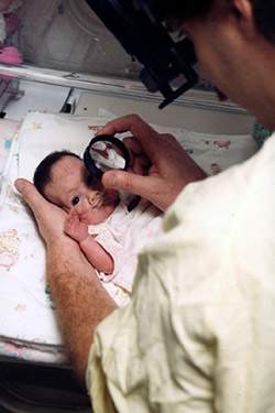
All babies born before 31 weeks of pregnancy need monitoring for retinopathy of prematurity. c. Photo credit: National Eye Institute

The telemedicine strategy consisted of electronically sending photos of babies’ eyes to a distant image reading center for evaluation. Staff at the image reading center, who were trained to recognize signs of severe ROP, identified whether infants should be referred to an ophthalmologist for evaluation and potential treatment. The study tested how accurately the telemedicine approach reproduced the conclusions of ophthalmologists who examined the babies onsite.

“This study provides validation for a telemedicine approach to ROP screening and could help save thousands of infants from going blind,” said Graham E. Quinn, MD, professor of ophthalmology at the Children’s Hospital of Philadelphia and the lead investigator for the study, which is reported today in JAMA Ophthalmology. The study was conducted by the e-ROP Cooperative Group, a collaboration that includes 12 clinics in the United States and one in Canada.

Some degree of ROP appears in more than half of all infants born at 30 weeks pregnancy or younger — a full-term pregnancy is 40 weeks — but only about 5 to 8 percent of cases become severe enough to require treatment. In ROP, blood vessels in the tissue in the back of the eye called the retina begin to grow abnormally, which can lead to scarring and detachment of the retina. Treatment involves destroying the abnormal blood vessels with lasers or freezing them using a technique called cryoablation. Early diagnosis and prompt treatment is the best prevention for vision loss from ROP, which is why the American Academy of Ophthalmology recommends routine screening for all babies who are born at gestational age 30 weeks or younger or who weigh less than 3.3 pounds at birth.

The study evaluated telemedicine for ROP screening during the usual care of 1,257 premature infants who were born, on average, 13 weeks early. About every nine days, each infant underwent screening by an ophthalmologist, who assessed whether referral for treatment was warranted. Those who were referred were designated as having referral-warranted ROP (RW-ROP). Either immediately before or after the exam, a non-physician staff member in the neonatal intensive care unit (NICU) took images of the infant’s retinas and uploaded them to a secure server at the University of Oklahoma, Oklahoma City. Trained non-physician image readers at the University of Pennsylvania, Philadelphia, then downloaded the photos, independently evaluated them following a standard protocol, and reported the presence or absence of RW-ROP.

Through the telemedicine approach, non-physician image readers correctly identified 90 percent of the infants deemed to have RW-ROP based on examination by an ophthalmologist. And they were correct 87 percent of the time when presented with images from infants who lacked RW-ROP. The examining ophthalmologists documented 244 infants with RW-ROP on exam. After referral, 162 infants were treated. Of these, non-physician image readers identified RW-ROP in all but three infants (98 percent). “This is the first large clinical investigation of telemedicine to test the ability of non-physicians to recognize ROP at high risk of causing vision loss,” said Eleanor Schron, Ph.D., group leader of NEI Clinical Applications. “The results suggest that telemedicine could improve detection and treatment of ROP for millions of at-risk babies worldwide who lack immediate in-person access to an ophthalmologist,” she said.

About 450,000 (12 percent) of the 3.9 million babies born each year in the United States are premature. The number of preterm infants who survive has surged in middle income countries in Latin America, Asia, and Eastern Europe. In these parts of the world, rates of childhood blindness from ROP are estimated at 15 to 30 percent — compared to 13 percent in the United States.

retinopathy of prematurity
NICU care providers take photos of a premature baby’s retinas in the NEI-funded e-ROP study of telemedicine for retinopathy of prematurity. Photo credit: Children’s Hospital of Philadelphia

One advantage of telemedicine ROP screening is that it can be done more frequently than screening by an ophthalmologist. “It’s much easier to examine the retina when not dealing with a wiggling baby,” said Dr. Quinn. “If a baby is too fussy or otherwise unavailable when the ophthalmologist visits the NICU, the exam may be delayed until the ophthalmologist returns — sometimes up to a week later.”

Weekly ROP screening — or even more frequently for high-risk babies — is a realistic goal for telemedicine and could help catch all cases needing treatment, according to the report. In the study, imaging was restricted to occasions when an ophthalmologist examined the baby. In practice, hospital staff could implement an imaging schedule based on the baby’s weight, age at birth, and other risk factors. “With telemedicine, NICU staff can take photos at the convenience of the baby,” said Dr. Quinn.

Telemedicine for evaluating ROP offers several other advantages.

Telemedicine may help detect RW-ROP earlier. In the study, about 43 percent of advanced ROP cases were identified by telemedicine before they were detected by an ophthalmologist — on average, about 15 days earlier.

Telemedicine could save babies and their families the hardship and hazards of being unnecessarily transferred to larger nurseries with greater resources and more on-site ophthalmologists. “Telemedicine potentially gives every hospital access to excellent ROP screening,” Dr. Quinn said.

Telemedicine might also bring down the costs of routine ROP screening by reducing the demands on ophthalmologists, whose time is better allocated to babies who need their attention and expertise. In a separate analysis, the study found that non-physicians and physicians had similar success in assessing photos for RW-ROP. Three physicians evaluated image sets from a random sample of 200 babies (100 with RW-ROP based on the eye exam findings; 100 without) using the standard grading protocol. On average, the physicians correctly identified about 86 percent of RW-ROP cases; the non-physicians were correct 91 percent of the time. The physicians correctly identified about 57 percent of babies without RW-ROP; non-physicians were correct 73 percent of the time.

The cost of establishing a telemedicine ROP screening program includes acquisition of a special camera for taking pictures of the retina, training of NICU personnel to take and transmit quality photos, and establishment and maintenance of an image reading center. “As we move along this road, advances in imaging and grading of images may streamline the process even more,” Dr. Quinn said.

The e-ROP Cooperative Group includes the following clinical sites and resource centers:

  • Children’s Hospital of Philadelphia
  • Johns Hopkins University, Baltimore
  • Boston Children’s Hospital
  • Nationwide Children’s Hospital and Ohio State University Hospital, Columbus
  • Duke University (cost-effectiveness center), Durham, North Carolina
  • University of Louisville, Kentucky
  • University of Minnesota, Minneapolis
  • University of Oklahoma (Inoveon ROP Data Center), Oklahoma City
  • University of Texas Health Science Center at San Antonio
  • University of Utah, Salt Lake City
  • Vanderbilt University, Nashville, Tennessee
  • Hospital of the Foothills Medical Center, Calgary, Alberta
  • University of Pennsylvania (data coordinating center and image reading center), Philadelphia

For more information about ROP.

Click to view a video about e-ROP.

NIH logo without bannerNational Institutes of Health
9000 Rockville Pike
Bethesda, Maryland 20892
301-496-4000,
TTY 301-402-9612

Common Pediatric Eye Diseases

8/21/14

In the third of this series, Buddy Russell, from the Emory University Eye Center, provides a great overview of common pediatric eye diseases.

Some Conditions Frequently Seen in Pediatrics

A basic understanding of some of the conditions that may be present in pediatric patients is important to not only know what they are but also understand well enough to explain to the parent or caregiver. The following is intended to be an overview of some of those conditions and not a complete explanation.Girl with eye chart-common pediatric eye diseases

  1. Nystagmus – Nystagmus is a vision condition in which the eyes make repetitive, uncontrolled movements, often resulting in reduced vision. These involuntary eye movements can occur from side to side, up and down, or in a circular pattern. As a result, both eyes are unable to hold steady on objects being viewed. Unusual head positions and head nodding in an attempt to compensate for the condition may accompany nystagmus. Most individuals with nystagmus can reduce the severity of their uncontrolled eye movements and improve vision by positioning their eyes to look to one side. This is called the “null point” where the least amount of nystagmus is evident. To accomplish this they may need to adopt a specific head posture to make the best use of their vision. The direction of nystagmus is defined by the direction of its quick phase (e.g. a right-beating nystagmus is characterized by a rightward-moving quick phase, and a left-beating nystagmus by a leftward-moving quick phase). The oscillations may occur in the vertical, horizontal or torsional planes, or in any combination. The resulting nystagmus is often named as a gross description of the movement, e.g. downbeat nystagmus, upbeat nystagmus, seesaw nystagmus, periodic alternating nystagmus. Having nystagmus affects both vision and self-concept. Most people with nystagmus have some sort of vision limitations because the eyes continually sweep over what they are viewing, making it impossible to obtain a clear image. If a refractive error is found, contact lenses may be the most effective way of obtaining best-corrected vision.
  2. Strabismus – Strabismus is any misalignment of the eyes. It is estimated that 4% of the U.S. population has strabismus. Strabismus is most commonly described by the direction of the eye misalignment. Common types of strabismus are esotropia (turn in), exotropia (turn out), hypotropia (turn down), and hypertropia (turn up). Eye misalignment can cause amblyopia in children. When the eyes are oriented in different directions, the brain receives two different visual images. The brain will ignore the image from the misaligned eye to avoid double vision, resulting in poor vision development of that eye. Also, an eye that sees poorly tends to be misaligned. The goal of strabismus treatment is to improve eye alignment, which allows for better work together (binocular vision). Treatment may involve eyeglasses, contact lenses, eye exercises, prism, and / or eye muscle surgery.
  3. Amblyopia – Amblyopia, sometimes called a “lazy eye,” occurs when one or both eyes do not develop normal vision during early childhood. Babies are not born with 20/20 vision in each eye but must develop it between birth and 6-9 years of age by using each eye regularly with an identical focused image falling on the retina of each eye. If this does not occur in one or both eyes, vision will not develop properly. Instead, vision will be reduced and the affected eye(s) are said to be amblyopic. This common condition, affecting up to 4% of all children, should be diagnosed and treated during infancy or early childhood to obtain optimum three-dimensional vision and to prevent permanent vision loss. What causes amblyopia?
      • Misaligned eyes (strabismus)
        Misaligned eyes are the most common cause of amblyopia. When both eyes are not aimed in exactly the same direction, the developing brain “turns off” the image from the misaligned eye to avoid double vision and the child uses only the better eye — the dominant eye. If this persists for a period even as short as a few weeks, the eye will not connect properly to the visual cortex of the brain and amblyopia will result.
      • Unequal refractive error (anisometropia)
        Unequal refractive error is an eye condition in which each eye has a different refractive error and therefore both eyes cannot be in focus at the same time. Amblyopia occurs when one eye (usually the eye with the greater refractive error) is out of focus because it is more nearsighted, farsighted or astigmatic than the other. Again, the brain “turns off” the image from the less focused eye and this eye will not develop normal vision. Because the eyes often look normal, this can be the most difficult type of amblyopia to detect and requires careful vision screening of acuity measurements at an early age. Treatment with glasses or contact lenses to correct the refractive error of both eyes, sometimes with part-time patching of the better seeing eye, is necessary in early childhood to correct the problem.
      • Obstruction of or cloudiness (deprivation)
        Obstruction of or cloudiness in the normally clear eye tissues may also lead to amblyopia. Any disorder that prevents a clear image from being focused can block the formation of a clear image on the retina and lead to the development of amblyopia in a child. This often results in the most severe form of amblyopia. Examples of disorders that can interfere with getting a clear image on the retina are a cataract or cloudy lens inside the eye, a cloudy and or irregular shaped cornea, or a droopy upper eyelid (ptosis) or eyelid tumor.It is not easy to recognize amblyopia. A child may not be aware of having one normal eye and one with reduced vision. Unless the child has a misaligned eye or other obvious external abnormality, there is often no way for parents to tell that something is wrong. In addition, it is difficult to measure vision in very young children at an age in which treatment is most effective.To treat amblyopia, a child and their caregiver must be encouraged to use the weaker eye. This is usually accomplished by patching the stronger eye. This covering of the stronger eye with an adhesive patch, an cclude contact lens or temporary surgery often proves to be a frustrating and difficult therapy. Patching will often continue for weeks, months, or even years in order to restore normal or near normal vision and maintain the improvement in the amblyopic eye. Occasionally, blurring the vision in the good eye with eye drops or lenses to force the child to use the amblyopic eye treats amblyopia. In some cases, cataract surgery or glaucoma surgery might be necessary to treat form deprivation amblyopia. Patching may be required after surgery to improve vision, and glasses or contact lenses may be required to restore appropriate focusing.Surprising results from a nationwide clinical trial in 2005 show that many children age seven through 17 with amblyopia may benefit from treatments that are more commonly used on younger children.
        Treatment improved the vision of many of the 507 older children with amblyopia studied at 49 eye centers. Previously, eye care professionals often thought that treating amblyopia in older children would be of little benefit. The study results, funded by the National Eye Institute (NEI), appear in the April issue of Archives of Ophthalmology.
  4. Congenital Cataract – A congenital cataract, or clouding of the crystalline lens is present in 2-3 per 10,000 live births of children. The presence of a visually significant cataract in a child is considered an urgent disorder. The resultant form deprivation of vision requires immediate surgery to remove the obstruction, prompt optical correction and amblyopia therapy in unilateral cases. Until the 1970s, it was generally believed that there was no means of restoring the vision in an eye with a unilateral congenital cataract. However, subsequent studies demonstrated that excellent visual results could be obtained with early surgical treatment coupled with optical correction with a contact lens and patching therapy of the fellow eye. However, treatment results continue to be poor in some infants with unilateral congenital cataracts due to a delay in treatment or poor compliance with contact lens wear or patching therapy of the fellow eye. The Infant Aphakia Treatment Study (IATS) was designed to compare the visual outcomes in children 1 to 6 months of age with a unilateral congenital cataract randomized to optical aphakic correction with contact lenses or an intraocular lens (IOL). Children randomized to IOL treatment had their residual refractive error corrected with spectacles. Children randomized to no IOL had their aphakia treated with a contact lens. In previous publications we have shown that the visual results are comparable for these two treatments at 1 year of age, but significantly more of the infants randomized to IOL implantation required additional intraocular surgeries.
  5. Accommodative Esotropia – Accommodative esotropia refers to a crossing of the eyes caused by farsightedness. Accommodative esotropia is a type of strabismus. Children who are farsighted easily and automatically focus on objects at distance and near through accommodation. As a result, a child who is farsighted usually does not have blurred vision. However, in some children who are farsighted, this accommodative effort is associated with a reflex crossing of the eyes. Accommodative esotropia can begin anywhere from 4 months to 6 years of age. The usual age of onset is between 2 and 3 years of age.Full-time use of the appropriate hyperopic glasses prescription or contact lenses will often control the esotropia. When wearing the correction, the child will not need to accommodate and hence the associated eye-crossing reflex will disappear. However, after removing the prescribed correction, the crossing will reappear, perhaps even more than before the child began wearing the correction. Sometimes the correction will only cause the crossing to disappear when the child views a distant object. However, when gazing at near objects, crossing may persist despite the use of the correction. In these circumstances, a bifocal lens is often prescribed to permit the child to have straight eyes at all viewing distances. One potential advantage of contact lenses compared to spectacles when correcting hyperopic powers is the decrease in accommodative demand. The increased effort to converge the eyes with spectacles requires one to over come the resultant base out prism when viewing a near object.


Buddy Russell - pediatric contact lensesBuddy Russell, FCLSA, COMT
Associate, Specialty Contact Lens Service
Emory University Eye Center

Nystagmus In Children

8/7/14

Nystagmus is a condition of uncontrolled eye movements. Patients with nystagmus are unable to maintain their eyes in a fixed position of focus. The movements can be pendular, swaying evenly side to side, or, jerk into one direction and drift toward the opposite direction. It can be present early in life or acquired as an adult. It can occur in eyes with poor vision from other anomalous development, or eyes that appear perfectly normal. In almost all patients the vision is compromised to some degree. In some patients, the eye movement is less, and the vision better, in an eccentric position that causes the patient to adopt a face turn, tilt or head posture so they can use this quieter position (“null point”) to navigate during their daily activities. To date there have been no consistently effective treatments for this condition.

Lingua and Grace - nystagmus
Dr. Lingua and Grace Nassar

Treatment efforts have been either medical (drugs to reduce the amplitude of the nytagmus movement) or surgical (to move the “null point” into straight ahead gaze to eliminate a head turn, or, directed at reducing the effective contracture of all the eye muscles to reduce the amount of movement). In general, surgical treatment of nystagmus has been disappointing.

In 2002, Dr. Robert Sinskey, noted cataract surgeon and phacoemulsification pioneer, proposed a revolutionary concept, that nystagmus could only be truly effectively controlled by removing the forward portion of the eye muscle and detach it completely from the eye. Since the twitching eye muscles were controlled by nerves sending that pulsatile information, any operation that allowed the muscles to remain attached to the eye would never quiet the movement. He performed this novel surgery in 2000 and published the results in 2002. It did not receive attention in the nystagmus surgery community, as most experts worried that the surgery would limit normal eye movements excessively. The operation does remove the forward portion of the eye muscle but, surprisingly, the eyes are still able to move to allow reading, computing, and driving.. In 2012, I had the opportunity to view a patient he operated 10 years prior and was impressed with how successful the results were even after 10 years. Coincidentaly, I was caring for a 17 year-old patient with nystagmus who had already undergone the 2 currently accepted eye muscle procedures for nystagmus without success. His movements remained uncontrolled, he could not maintain eye contact with anyone, and is his vision was less than that needed for a drivers license. In 2013, I offered him the Sinskey procedure and the results were remarkable. His nystagmus was quieted, his vision improved (20/25) enough to qualify for a drivers license and to return to school.

Since 2012, we have adapted, augmented and perfected the procedure and performed the surgery on over 12 patients with similar remarkable results. All patients experience a marked reduction in the amplitude of the nystagmus (60-100%), and all patients demonstrate improved vision (1-8 lines of the acuity chart), especially at the reading position.

Visit the YouTube posting “Meet Grace for an example of how this surgery can impact a child’s life and the hopes of their parents. Visit www.eye.uci.edu for further information, contact information and scientific data on the procedure.

Robert Lingua, MDRobert W. Lingua, MD
Director, Pediatric Ophthalmology and Strabismus
Gavin Herbert Eye Institute, UC Irvine

Learning-Related Vision Problems

7/29/14

Next month is Children’s Eye Health Month, and to kick it off a little early we are taking a look at learning-related vision problems. Next month children start heading back to school, so now is the perfect time to make sure you child has had a recent comprehensive eye exam.
child eye exam - Learning-related vision problems
Vision and learning are closely related. About 80% of what a child learns in school is presented visually, and 25% of school-aged children already have vision problems (5-10%of preschoolers do). By scheduling a comprehensive eye exam for your child before school starts, you will help your child be more successful and happier in school.

When children have difficulty in school, from being able to see what is written on the whiteboard to learning to read, it not only makes the learning difficult, but it also makes the child frustrated and can affect their love of learning as well. While most schools perform a quick eye check at some point during the school year to determine simple refractive errors such as nearsightedness, farsightedness and astigmatism, there are other visual disorders that can make learning difficult.

Vision is a complex process that involves the eyes working well with the brain. Specific learning-related vision problems can be classified as one of three types. The first two types primarily affect visual input. The third primarily affects visual processing and integration.

  1. Refractive vision problems. Your child holds a book close to their face when reading or they have difficulty seeing things that are far away. These are generally refractive issues and include nearsightedness, farsightedness and astigmatism, but could also include more subtle optical errors called higher-order aberrations.
  2. Functional vision problems. This type of problem refers to a variety of specific functions of the eye and the brain’s control of these functions, such as eye teaming (using the eyes together), fine eye movements, and focusing skills, peripheral awareness and eye-hand coordination. These issues can cause blurred or double vision, eye strain and headaches.
  3. Perceptual vision problems. Visual perception includes understanding what you see, identifying it, judging its importance and relating it to previously stored information in the brain. Examples would include recognizing words that you have seen before and being able to form a mental picture of the words you see.

Because most routine school and pediatrician eye exams evaluate only the refractive vision problems, it is wise to see optometrist who specializes in children’s vision problems to evaluate functional vision problems and perceptual vision problems that may affect learning.

Color blindness is not considered a learning-related vision problem, but it can cause problems for very young children if color-matching or identifying specific colors are part of the classroom activities. For this reason, make sure your child’s eye exam includes a color blind test prior to starting school.

Symptoms of Learning-Related Vision Problems:

  • Headaches, eye strain, excessive blinking or rubbing the eyes
  • Blurred vision or double vision
  • Crossed eyes or if the eyes move independently of each other
  • Holding a book very close to their face for reading
  • Bending way over a table to get closer to what they are drawing or writing
  • Using only one eye by tilting the head or covering the other eye
  • Losing their place while reading, or needing to use a finger to stay on line
  • Slow reading speed or poor reading comprehension
  • Omitting or repeating words, or confusing similar words
  • Persistent reversal of words or letters (after second grade)
  • Difficulty remembering, identifying or reproducing shapes
  • Poor eye-hand coordination
  • Distracted in class

If your child shows one or more of these symptoms, they could indicate a learning-related vision problem. To find out see an eye doctor who specializes in children’s vision for a comprehensive evaluation. If no vision problem is detected, your child’s problems could be caused by a non-visual issues, such as dyslexia or a learning disability, in which case you would need to contact an educational specialist for an evaluation.

Treatment of Learning-Related Vision Problems

If your child is diagnosed with a learning-related vision problem, treatment generally consists of an individualized and doctor-supervised program of vision therapy. Special eyeglasses also may be prescribed for either full-time wear or for specific tasks such as reading.

Remember that when children have a difficult time learning, especially while other classroom friends have no visual issues to impair learning, they may experience emotional problems as well, such as anxiety, depression and low self-esteem.

Reassure your child that learning-related vision problems do not relate to how smart they are and with the proper treatment and/or eyeglasses, things will become easier.

Susan DeRemerSusan DeRemer, CFRE
Vice President of Development
Discovery Eye Foundation

Ways to Reduce the Harmful Effects of Sun Glare

During the height of summer sunshine (and heat!), it’s helpful to discuss the importance of eye protection, including ways to reduce the harmful effects of sun glare.

Fundamentally, we need light to see. Approximately 80% of all information we take in is received through the sense of sight. However, too much light – and the wrong kind of light – can create glare, which can affect our ability to take in information, analyze it, and make sense of our surroundings.

Facts about Sunlight

Every type of light has advantages and disadvantages, and sunlight is no exception:

Advantages:

• Sunlight is the best, most natural light for most daily living needs.
• Sunlight is continuous and full-spectrum: the sun’s energy at all wavelengths is equal and it contains all wavelengths of light (explained below).

Disadvantages:

• It is difficult to control the brightness and intensity of sunlight.
• Sunlight can create glare, which can be problematic for many people who have low vision.
• Sunlight is not always consistent or reliable, such as on cloudy or overcast days.

Visible Light and Light Rays

An important factor to consider is the measurement of visible light and light rays, beginning with the definition of a nanometer:

• A nanometer (nm) is the measurement of a wavelength of light.
• A wavelength is the distance between two successive wave crests or troughs:

Wavelength - glare

• A nanometer = 1/1,000,000,000 of a meter, or one-billionth of a meter. It’s very small!

The human visual system is not uniformly sensitive to all light rays. Visible light rays range from 400 nm (shorter, higher-energy wavelengths) ? 700 nm (longer, lower-energy wavelengths).
Visible Light Spectrum - glare
The visible light spectrum occupies just one portion of the electromagnetic spectrum, however:

• Below blue-violet (400 nm and below), is ultraviolet (UV) light.
• Above red (700 nm and above), is infrared (IR) light.
• Neither UV nor IR light is visible to the human eye.

Ultraviolet Light and Blue Light

Ultraviolet (UV) light has several components:

• Ultraviolet A, or UVA (320 nm to 400 nm): UVA rays age us.
• Ultraviolet B, or UVB (290 nm to 320 nm): UVB rays burn us.
• Ultraviolet C, or UVC (100 nm to 290 nm): UVC rays are filtered by the atmosphere before they reach us.

Blue light rays (400 nm to 470 nm) are adjacent to the invisible band of UV light rays:

• There is increasing evidence that blue light is harmful to the eye and can amplify damage to retinal cells.
• You can read more about the effects of blue light at Artificial Lighting and the Blue Light Hazard at Prevent Blindness.

A new study from the National Eye Institute confirms that sunlight can increase the risk of cataracts and establishes a link between ultraviolet (UV) rays and oxidative stress, the harmful chemical reactions that occur when cells consume oxygen and other fuels to produce energy.

Sunlight and Glare

Glare is light that does not help to create a clear image on the retina; instead, it has an adverse effect on visual comfort and clarity. Glare is sunlight that hinders instead of helps. There are two primary types of glare.

Disability glare

• Disability (or veiling) glare is sunlight that interferes with the clarity of a visual image and reduces contrast.
• Sources of disability glare include reflective surfaces (chrome fixtures, computer monitors, highly polished floors) and windows that are not covered with curtains or shades.

Discomfort glare

• Discomfort glare is sunlight that causes headaches and eye pain. It does not interfere with the clarity of a visual image.
• Sources of disability glare include the morning and evening positions of the sun; snow and ice; and large bodies of water, (including swimming pools).

Controlling Glare

You can protect your eyes from harmful sunlight and minimize the effects of glare by using a brimmed hat or visor in combination with absorptive lenses.

• Absorptive lenses are sunglasses that filter out ultraviolet and infrared light, reduce glare, and increase contrast. They are recommended for people who have low vision and are also helpful for people with regular vision.
• Lens colors include yellow, pink, plum, amber, green, gray, and brown. Ultra-dark lenses are not the only choice for sun protection.
• Lens tints in yellow or amber are recommended for controlling blue light.
NoIR Medical Technologies: NoIR (No Infra-Red) filters absorb UVA/UVB radiation and also offer IR light protection.
Solar Shields: Solar Shields absorb UVA/UVB radiation and are available in prescription lenses.
• You can find absorptive lenses at a specialty products store, an “aids and appliances store” at an agency for the visually impaired, or a low vision practice in your area. Before you purchase, it’s always best to try on several different tints and styles to determine what works best for you.

More Recommendations

• Always wear sunglasses outside, and make sure they conform to current UVA/UVB standards.
• Be aware that UV and blue light are still present even when it is cloudy or overcast.
• Make sure that children and older family members are always protected with UVA/UVB-blocking sunglasses and brimmed hats or visors.

Maureen Duffy-editedMaureen A. Duffy, CVRT
Social Media Specialist, visionaware.org
Associate Editor, Journal of Visual Impairment & Blindness
Adjunct Faculty, Salus University/College of Education and Rehabilitation

20 Facts About Eye Color and Blinking

7/15/14

Eye color is one of the first things a person notices about another person, but blinking is so automatic we rarely think about it. Here are some intriguing facts about eye color and blinking:

1. The world’s most common eye color is brown.

2. Brown eyes are actually blue underneath.

3. Melanin affects the color of your eyes so brown eyes have more melanin than blue eyes.
Person with different colored eyes - eye color and blinking
4. Heterochromia is when you are born with two differently colored eyes.

5. Blue-eyed people share a common ancestor with every other blue-eyed person in the world.

6. We blink more when we talk.

7. It is impossible to sneeze with your eye open.

8. The average person blinks 12 times per minute or about 10,000 blinks per day.

9. The eye is the fastest muscle in the body – in the blink of an eye. They are also the most active muscles in the body.

10. A blink usually lasts 100 to 150 milliseconds making it possible to blink five times in a second.

11. You blink less when you’re reading.

12. Infants blink 10 times less than adults.

13. One blink isn’t always the same as the next.

14. Our eyes close automatically to protect us from perceived dangers.

15. The older we are the less tears we produce.

16. Tears are made of three main components – fat, mucous and water. This is so tears won’t evaporate.

17. Your nose gets runny when you cry as the tears drain into your nasal passages.

18. You blink on average 4,200,000 times a year.

19. Tears kill bacteria because they contain lysozyme, a fluid that can kill 90 to 95 percent of all bacteria.

20. A newborn baby will cry, but not produce any tears. Babies do not produce tears until they are around six weeks old.

Susan DeRemerSusan DeRemer, CFRE
Vice President of Development
Discovery Eye Foundation