
Primary Congenital Glaucoma

What is primary congenital glaucoma?

Glaucoma in children includes a variety of disorders in which drainage system of the eye does not function adequately, leading to abnormally high pressure inside of the eye (the intraocular pressure, or IOP), and resulting in damage to many different structures of the child’s eye. If not treated promptly and successfully, pediatric glaucoma can lead to severe vision loss or even blindness in one or both eyes. In primary childhood glaucoma, the drainage system usually has not formed properly (often resulting from a genetic abnormality) while in secondary childhood glaucoma, the abnormal fluid outflow problem results from other problems with the eye(s), sometimes accompanied by other medical problems outside the eyes.

Primary congenital glaucoma is the most common of the primary childhood glaucoma types, although it is still rather rare. Let’s take a moment now to review the parts of the eye, and eye’s drainage system, sometimes also called the “aqueous outflow pathway”, since it drains the fluid within the eye (the aqueous humor), which is separate from the tears that flow on the outside of the eye’s surface and then into the nose or down a child’s cheeks.

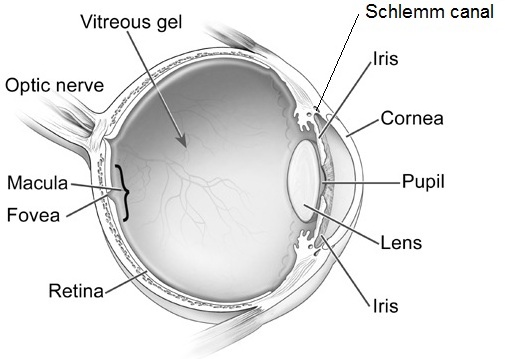
The aqueous outflow pathway of the eye (comprising both the trabecular meshwork and Schlemm canal), situated at the junction (or “angle”) between the iris (the colored portion of the eye) and the sclera (the white part of the eye), has not formed correctly (Figure 1).
primary congenital glaucoma
Figure1. Schematic eye shows different structures of the ocular globe. Note that the Schlemm canal is part of the drainage system of the eye . Modified from National Eye Institute.

The aqueous humor therefore builds up within the front portion of the eye, causing abnormal elevation of the IOP.

In contrast to the eyes of adults and older children, the entire eye in infants and young children is distensible and the high IOP in primary congenital glaucoma often causes stretching and damage to several parts of the eye; this most often results in enlargement, clouding and scaring of the cornea (the front window of the eye) as well as severe nearsightedness, damage to the optic nerve, and resulting poor vision.

Primary congenital glaucoma (also called PCG) is almost always genetic, although usually there is no one else in the family with the condition. It is not related to anything that the parents did (or did not do) during the pregnancy or afterwards, and does not have any relationship to the baby’s sex or racial background. It occurs in about 1 every 10,000 to 20,000 births in western countries, but may be more common in certain populations of the world. Most babies with this disease are otherwise normal.

How is primary congenital glaucoma diagnosed?

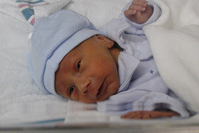
Most cases present within six months of birth, with nearly 80% presenting before one year of age. In 70- 80 % of cases both eyes are affected. Most cases present for medical attention due to the size or cloudy appearance of the cornea in one or both eyes (Figure 2).

primary congenital glaucoma
Fig 2. Left eye of child with congenital glaucoma. The eye is enlarged and the front part of the eye is cloudy (corneal edema).

In cases where only one eye is affected, a difference in size can be seen between the two eyes and this sometimes brings the baby to the ophthalmologist (Figure 3).
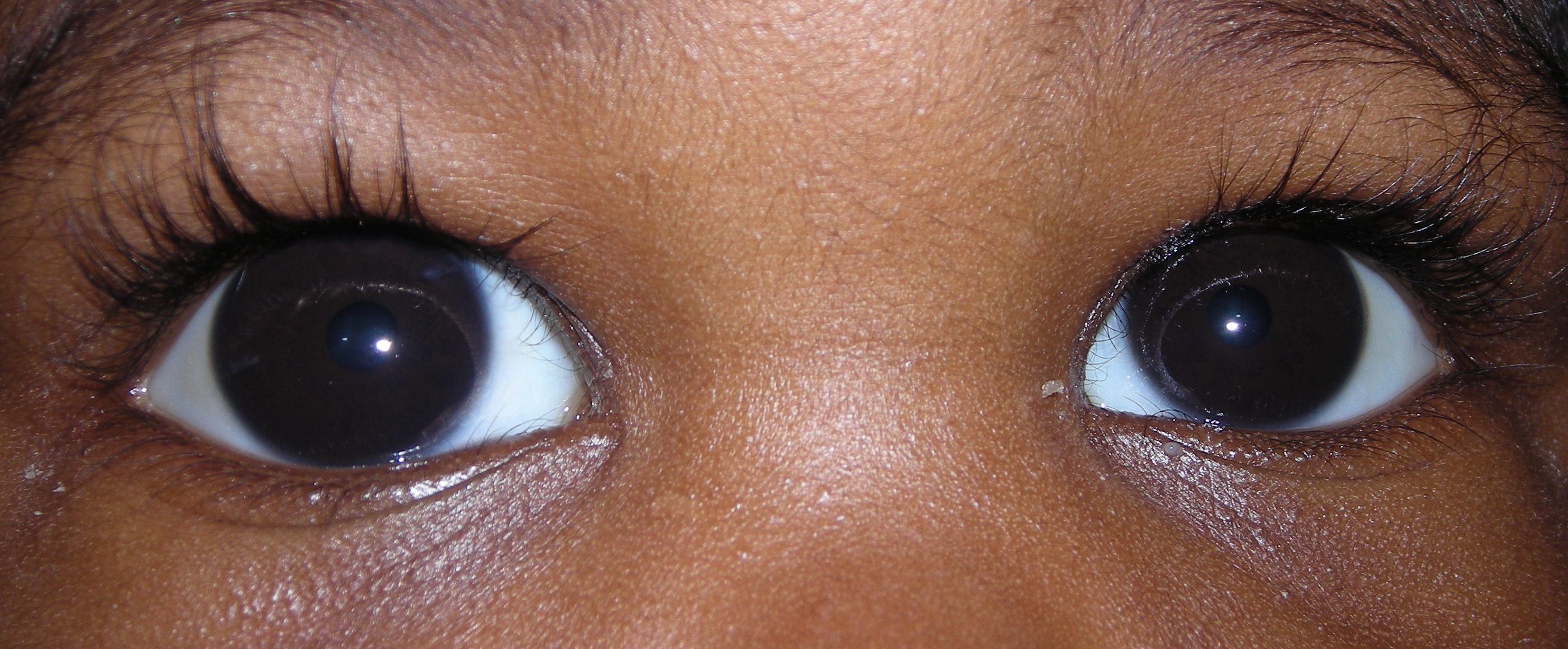
primary congenital glaucoma
Fig 3. Different size of eyes in a child with congenital glaucoma. Note the increased size of the right eye. The brown area (iris) and the transparent part in front of the color part (cornea) are significantly larger in the right eye.

The diagnosis of PCG is based on clinical findings and there are three classic signs that the child can present with:

  • abnormal sensitivity or intolerance to light (photophobia)
  • excessive blinking or squinting of the eyelids (blepharospasm)
  • excessive tearing (epiphora)

The exam in clinic can be challenging for infants and young children and most require an exam under anesthesia, to allow detailed examination of the eye(s) that would not be possible in the clinic. Often the ophthalmologist will be able to follow the examination under anesthesia with the most appropriate surgery for the glaucoma, if surgery is indeed required.

How is primary congenital glaucoma treated?

PCG is almost always treated with surgery, although medications are often needed to help in addition to the surgery. Medications are very useful before initial surgery to help reduce the IOP and decrease the clouding of the cornea. In addition, medications may be recommended to keep the IOP to a safe level after surgery has been performed. If the IOP is not controlled successfully, or if damage has been substantial prior to diagnosis and treatment, PCG causes severe vision loss and can even cause blindness. Sometimes the damage from PCG is uneven between a child’s two eyes, leading to amblyopia (“lazy eye”) in the more severely affected size.

The initial surgical procedure of choice is usually aimed at opening the trabecular meshwork and Schlemm canal (the aqueous outflow pathway) of the affected eye(s). This so-called “angle surgery” can be performed either from inside of the eye (goniotomy) or externally (trabeculotomy), and may need to be repeated more than once in some cases.

If angle surgery fails, other procedures are available to allow the aqueous humor fluid to exit the eye (glaucoma drainage device or filtration surgery), or even to decrease the amount of fluid the eye makes (cycloablation procedures). For these more difficult procedures, the child is usually referred to an ophthalmic surgeon with expertise in treating childhood glaucoma.

What is the prognosis for children with primary congenital glaucoma?

While vision loss can be severe, prompt diagnosis and effective treatment and follow-up for children with PCG usually allows affected children to have best-corrected vision of at least 20/50 vision in their better-seeing eye. Children with PCG require continued careful follow-up and treatment their lifetime, and may require more than one surgery, eye drops, and spectacles.

Successful care for children with PCG takes a dedicated team including the family, ophthalmologist, teacher and community support, and the child him/herself.

10/1/15
primary congenital glaucomaElena Bitrian, MD
Assistant Professor of Ophthalmology, Division of Glaucoma
Mayo Clinic



primary congenital glaucomaSharon F Freedman, MD
Professor of Ophthalmology and Pediatrics
Chief of Pediatric Ophthalmology
Duke Eye Center, Duke University

Cortical Visual Impairment: What Is It?

What Is It?

Cortical Visual Impairment (CVI) refers to decreased vision resulting from the visual processing parts of the brain (e.g., the posterior visual pathways and/or the occipital lobes) rather than from the eyes themselves. For example, individuals with CVI typically have normal eye exam findings. However, vision loss from CVI can range from mild to total blindness. It is also one of the more frequent causes of visual impairment in children. Regardless, children with CVI often have some level of vision that may improve over time, particularly if they receive therapy to teach them how to integrate the visual signals their brains are receiving.
cortical visual impairment
CVI may be caused by a number of different conditions that damage the visual parts of the brain. Examples include stroke, decreased blood supply to the brain, decreased oxygenation in the brain, brain malformation or infection, hydrocephalus, seizure, metabolic diseases, head trauma, and other neurologic disorders. Conditions such as these make it difficult for the brain to understand and interpret what the eyes see.

In most cases, individuals with CVI do not have other neurological problems, although epilepsy and cerebral palsy are not uncommon. The presence of CVI is not an indicator of the cognitive abilities of the individual; therefore, CVI should be distinguished from vision loss secondary to global neurological damage, where other functional deficits are also present in motor, cognitive, and physical abilities.

Indicators of Cortical Vision Loss

In children, one of the most common indicators of CVI is their poor attention to visual targets, particularly to more complex targets, such as a person’s face. Other indicators are that children with CVI often prefer to look at lights for long periods of time and that when reaching for an object, they will often look away from the object before grabbing it. This is because children with CVI have difficulty integrating visual stimuli (looking at an object) with their motor ability (grabbing the object). The diagnosis of CVI is given based on the combined results of magnetic resonance imaging (also known as an MRI) and an eye evaluation by a pediatric ophthalmologist.

Treatments

If a child is suspected of having CVI, he or she should be evaluated by a pediatric ophthalmologist as part of the initial evaluation. The pediatric ophthalmologist will assess the child’s eye health as well as the need for glasses to make sure there are no additional factors that may be limiting vision.

Often, there is concern that a child with CVI has little to no vision early in life. However, vision is a learned sense, so as the child matures, he or she may have improved visual responses. As such, early intervention is important for improved visual responses over time, as well as because the treatment period for visual development is limited to the early years of life. State and local educational agencies and early intervention programs should be contacted as soon as a visual concern is noted so that an organized plan of visual stimulation activities can be developed and implemented, based on the specific needs of each child. The professionals involved in the evaluation of a child with vision loss from CVI can include teachers of students who are blind or visually impaired, physical therapists, occupational therapists, speech therapists, and certified orientation and mobility specialists. It is important to note that although the vision of an individual with CVI may improve with intervention, rarely does the vision become totally normal.

The realization and acceptance that a child is visually impaired can be a difficult adjustment for the child’s parents. Fortunately, there are many things that can enhance the functional abilities of individuals with vision loss at any age. To learn about available resources for individuals with vision loss, visit the National Eye Health Education Program low vision program page.

9/1/15


Dr. Wilkinson - driving with vision lossMark Wilkinson, OD
University of Iowa Carver College of Medicine
Director, Vision Rehabilitation Service, UI Carver Family Center for Macular Degeneration
Medical Director, UI Optical
Chair of the National Eye Health Education Program Low Vision Subcommittee

Purpose of Eye Exams for Children

Many children who are 6 – 18 years old are now back in school or will be shortly. But have you given them everything they need to succeed in the school year ahead?

      ? New binders
      ? Notebook paper and dividers
      ? Pencil box filled with pens and pencils
      ? Calculators, protractors and rulers
      ? Backpack to carry it all

These are the tools that children and their parents focus on every year, thinking these will help their child have a fun and productive year. But the list is incomplete. For school-aged children, the AOA recommends eye exams for children every two years if no vision correction is required. Children who need eyeglasses or contact lenses should be examined annually or according to their eye doctor’s recommendations.

“But my child gets and eye screening at school every year…” While this may be true it is important to understand the difference between a screening and an eye exam.

Vision screenings are a short examination that can indicate a vision problem or a potential vision problem; however it cannot diagnose exactly what is wrong with your eyes. It can also easily miss vision issues, giving parents a false sense of security.

With an eye exam, the tests are performed by a trained professional, using specialized equipment looking for specific indicators that could affect your child’s vision. They test much more than how well your child can read letters or symbols at a distance.

Good vision is necessary for a child to succeed at school and not become frustrated or depressed. It has been estimated that as much as 80% of the learning a child does occurs through his or her eyes. Children need to read a book and see a whiteboard, write and use computers every day in the classroom and at home. When a child cannot see clearly, it becomes more difficult to learn.

It also goes beyond just seeing clearly. Your child needs their eyesight to understand and respond to what they see. This includes the ability to focus their eyes, use both eyes together, and move them effectively.

Infographic - eye exams for children

Children may not always know they have a vision problem because they think that everyone is seeing the way they do. There are some signs that may indicate a vision issue:

  • Repeated eye rubbing
  • Excessive blinking
  • Short attention span
  • Tilting the head to one side or covering one eye
  • Holding reading materials too close to the face
  • Losing their place when reading
  • Difficulty remembering what they just read
  • Trying to avoid reading or other close activities
  • Numerous headaches

So as you prepare your child to go back to school, give them the best advantage they can have – good vision. Make an appointment with your eye doctor today.

8/27/15


Susan DeRemerSusan DeRemer, CFRE
Vice President of Development
Discovery Eye Foundation

Eye Issues For Every Age Recap

Vision is something we take for granted, but when we start to have trouble seeing it is easy to panic. This blog has covered a variety of eye issues for every age, from children through older adults. Here are a few articles from leading doctors and specialists that you may have missed and might be of interest.
Artistic eye 6
Bill Takeshita, OD, FAAO – Visual Aids and Techniques When Traveling

Michelle Moore, CHHC – The Best Nutrition for Older Adults

Arthur B. Epstein, OD, FAAO – Understanding and Treating Corneal Scratches and Abrasions

The National Eye Health Education Program (NEHEP) – Low Vision Awareness
Maintaining Healthy Vision

Sandra Young, OD – GMO and the Nutritional Content of Food

S. Barry Eiden, OD, FAAO – Selecting Your Best Vision Correction Options

Suber S. Huang, MD, MBA – It’s All About ME – What to Know About Macular Edema

Jun Lin, MD, PhD and James Tsai, MD, MBA – The Optic Nerve And Its Visual Link To The Brain

Ronald N. Gaster, MD FACS – Do You Have a Pterygium?

Anthony B. Nesburn, MD, FACS – Three Generations of Saving Vision

Chantal Boisvert, OD, MD – Vision and Special Needs Children

Judith Delgado – Driving and Age-Related Macular Degeneration

David L. Kading OD, FAAO and Charissa Young – Itchy Eyes? It Must Be Allergy Season

Lauren Hauptman – Traveling With Low Or No Vision  /  Must Love Dogs, Traveling with Guide Dogs  /  Coping With Retinitis Pigmentosa

Kate Steit – Living Well With Low Vision Online Courses

Bezalel Schendowich, OD – What Are Scleral Contact Lenses?

In addition here are few other topics you might find of interest, including some infographics and delicious recipes.

Pupils Respond to More Than Light

Watery, Red, Itchy Eyes

10 Tips for Healthy Eyes (infographic)

The Need For Medical Research Funding

Protective Eyewear for Home, Garden & Sports

7 Spring Fruits and Vegetables (with some great recipes)

6 Ways Women Can Stop Vision Loss

6 Signs of Eye Disease (infographic)

Do I Need Vision Insurance?

How to Help a Blind or Visually Impaired Person with Mobility

Your Comprehensive Eye Exam (infographic)

Famous People with Vision Loss – Part I

Famous People with Vision Loss – Part II

Development of Eyeglasses Timeline (infographic)

What eye topics do you want to learn about? Please let us know in the comments section below.

7/21/15


Susan DeRemerSusan DeRemer, CFRE
Vice President of Development
Discovery Eye Foundation

Recognising Vision Problems in Children

Children typically have no idea when they have vision problems. Vision problems typically onset gradually, and children tend to believe that everyone sees similar to them, even when they have vision problems. As a result, you will rarely hear a child say any of the following: “My vision doesn’t seem to be as clear as it used to be.” “I started seeing double when reading two weeks ago.” “My eyes are feel uncomfortable when I read.”
vision problems in children
It has been estimated that 80% of what we know is learned through vision. Therefore, children with vision problems may struggle at school simply due to correctable vision issues. Because children rarely complain of vision problems, and vision problems can lead to learning difficulties, it is recommended by the American Optometric Association that children undergo annual comprehensive eye examinations if they are at risk for visual impairment. To determine if your child may be at risk, please go here.

Seeing Vision Problems in Children

Because children don’t often tell you that they have problems with their eyes, there are some signs for which you should watch. The most common vision-related problems differ by age. Beginning with the youngest children, we will provide things to consider that may portend vision issues. Infants’s eyes frequently wander because they have not yet developed the ability to control their eye movements at all times. One eye occasionally pointing in or out is common and should not be considered a problem unless the child continues to do this after eight months of age, unless it is always the same eye, or unless it is constant. A visit to the optometrist is warranted if any of those conditions exist. If the child’s eye crosses most or all of the time, then the child may have congenital esotropia, and surgery in combination with vision therapy may be necessary to correct the problem.

If the child’s eyes begin to cross frequently at around age two years, especially when the child looks at close objects, then the most likely problem is accommodative esotropia. If this is the case, wearing glasses will help to eliminate most or all of the crossed eyes.
vision problems in children
Nearsightedness (myopia) is the most common vision problem in children. It typically onsets between the ages of 8 and 10 years, although that may vary dramatically. Nearsighted children can see clearly at near, but have difficulty seeing distant objects. These children tend to squint when looking at distance objects, and may complain of headaches around their eyes, as a result of squinting. Nearsighted children are frequently detected during school vision screenings or recognized by teachers, and glasses or contact lenses can easily clear up their vision and reduce symptoms.

It is also important to remember that even children who can see details far away may have vision problems. Farsighted children may be able to read distance vision charts, but their eyes must strain to do so. Their eyes must strain even more to see up close. While a child may pass a vision screening at school by reading the bottom line on the chart across the room, he or she may struggle visually. These children often complain of headaches during the week, but less often during the weekend. They may fall asleep after a very short period of reading or struggle to maintain attention because their eyes become strained. Their headaches are typically over the eyes or on the side of the head, and they rarely wake up with a headache. They frequently try to avoid reading because it makes their eyes uncomfortable, although they rarely say that.

Another problem with similar symptoms is convergence insufficiency, which means that children have difficulty bringing their eyes toward the nose when looking at close objects. The symptoms of convergence insufficiency have frequently been misdiagnosed as attention deficit disorder because children find it difficult to attend to any single near task for even a relatively short period of time. Headaches, eye strain, double vision, and tiredness while reading are some of the main symptoms that children will exhibit. Vision therapy frequently reduces or eliminates the symptoms related to convergence insufficiency, and may improve a child’s attention during learning-related tasks.

Teachers are commonly the first people to suspect a vision problem in children because of the demanding visual environment at school. Children rarely complain at home, although homework time is an excellent time to watch your child for potential vision problems. Children with vision problems may rub their eyes frequently, fall asleep after reading for five minutes, perform better when narrating than when writing, cover an eye or close an eye when reading, or simply refuse to read. If your child exhibits these symptoms, then a visit to the eye doctor is warranted and may result in improved attitude and aptitude.

6/11/15

Jeffrey Walline - vision problems in childrenJeffrey J. Walline, OD, PhD
The Ohio State University
Associate Professor
Chair, Research and Graduate Studies

Vision and Special Needs Children

In the United States, special needs is a term used in clinical diagnostic and functional development to describe individuals who require assistance for disabilities that may be medical, mental, or psychological. Different types of special needs vary in severity. People with autism, Down syndrome, dyslexia, blindness, ADHD, or cerebral palsy, for example, may be considered to have special needs. Statistics tell us that among children ages 3 to 17, nearly 15 percent have one or more developmental disabilities. For many of these children, the kinds of disabilities they experience may require special approaches to providing care, education and/or other accommodations.
eye exam - vision and special needs children
The way a special needs child behaves or reacts can sometimes be unexpected because he/she processes sensory information differently than other children. Having the right cues in our environment can mean the difference between participation and non-participation in medical care for many of these children. Our work environment should always be arranged in a way to achieve physical and emotional comfort for the child. Patience, attentive listening, caring, and building a trusting relationship with a family and child who may be undervalued in other settings are all critical to providing good medical care.

Vision and Special Needs Children

Children with special needs are referred to pediatric ophthalmologists on a regular basis. Many of these children do not respond appropriately to standard vision screening procedures. Unidentified vision problems in this special population can further impact growth and development. The role of pediatric ophthalmologists is to ensure that the child does not have any eye conditions that could potentially interfere with his/her learning opportunities, personal development and/or overall wellbeing.

Down syndrome:
Down syndrome continues to be the most common chromosomal disorder. Each year, about 6,000 babies are born with Down syndrome, which is about 1 in every 700 babies born. It is estimated that more than 80% of these patients have some clinically significant ocular pathology. Such conditions include refractive error requiring glasses (70%), strabismus (45%), and nystagmus (35%). Other common eye problems include blepharitis, blocked tear duct, eyelid ptosis, cataracts, and keratoconus (irregularly shaped cornea). The improved quality of medical care and educational resources have allowed for a more productive life and a longer life expectancy for people with Down syndrome. Their quality of life can be further enhanced by the proper assessment and correction of eye problems.

Autism:
In 2014, the Centers for Disease Control and Prevention (CDC) released new data on the prevalence of autism in the United States. This surveillance study identified 1 in 68 children (1 in 42 boys and 1 in 189 girls) as having autism spectrum disorder (ASD). Autistic behaviors may include visual components such as lack of eye contact, starring at light or spinning objects, fleeting peripheral glances, side viewing and difficulty attending visually. An eye examination is essential in order to detect an eye condition that could potentially explain these visual behaviors. According to a study published in the June 2013 issue of Strabismus, more than 40 percent of children with autism have strabismus, or crossed eyes. This work is limited in that the researchers did not use a representative sample of children with autism. Still, the prevalence of eye problems in autism appears to be several times higher than that of the general population.
boy eye exam - vision and special needs children
Learning Disabilities:
Dyslexia is the most common neurobehavioral disorder affecting children. Visual abnormalities have not been found to affect the brain’s ability to process visual stimuli and children with learning disabilities have no increased incidence of ophthalmologic disease. However, ophthalmologic consultation should be provided to children who fail vision screening tests. This allows for diagnosis and therapy of treatable ocular conditions such as refractive errors and eye muscle imbalances.

Children with ADHD, cerebral palsy, or any other neurodevelopmental disorders should also have periodic vision screenings. Children who do not pass the vision screening should be referred to an ophthalmologist with experience in the care of children.

Physicians are not the only adults involved in the care of special needs children. At increasing rates, children with special needs are being provided with the same life experiences as their non-disabled peers. Taking part in a sport or joining a group like the Scouts, are popular activities for children. Children with disabilities are encouraged to join in such activities to help improve their health and give them opportunities to make friends. As a result, more and more adults in the community are finding themselves working with these children on a regular basis.

When considering ways to work with special needs children, we have to keep in mind that every child is different. A positive attitude and patience are probably the two most important qualities for anyone who works with these children. Parents of special needs children focus on helping their child to be “the best he/she can be”. The devotion and care these parents provide for their child have been an inspiration, and I always look forward to my visits with them.

4/2/15


Dr. Chantal BoisvertChantal Boisvert, OD, MD
Assistant Clinical Professor
Gavin Herbert Eye Institute, UC Irvine
Pediatric Ophthalmology & Strabismus
Neuro-Ophthalmology

Hadley’s Online Education for the Blind and Visually Impaired

12/4/14

The Hadley School for the Blind (www.hadley.edu) is the world’s largest educator of people who are blind or visually impaired as well as the world’s largest Braille educator. The school’s curriculum has always been delivered completely through online education, meaning students can study from anywhere at any time. Hadley serves nearly 10,000 students in all 50 states and approximately 100 countries. Hadley serves adult students, age 14 and over. Our mission is to promote independent living through lifelong, distance education programs for individuals who are blind or visually impaired, their families and blindness service providers.
Hadley School
Hadley courses are tuition-free for people who are blind or visually impaired and their family members; there is modest tuition for professionals. Students study at their own pace and receive personalized, one-on-one instruction from their instructor by phone, email, virtual “office hours” or mail.

Students can take courses in four formats: large print, Braille, audio or online. Since we began offering courses in an online format in 2002 as part of our eHadley initiative, more than 13,000 students have completed one or more online courses. 89 of our approximately 100 courses are offered in an online format, and 20 new online courses were introduced in 2014. Most of our online courses also are now accessible on mobile devices, so that students can literally study on the go if they choose.

If you’re not familiar with blindness, you may be asking yourself how a blind person can take courses online. The answer is through adaptive technology, such as screen magnifiers or screen readers. A screen reader is a software application that converts text on a screen to speech. One of the most popular is called JAWS. If a student doesn’t know how to use a screen reader, Hadley has a series of courses to teach them how to use this technology!

While many students come to us to learn to read and write Braille, we offer much more than just Braille education. In addition to Braille, some of our most popular areas of study include access technology, independent living skills, employment skills and recreation. These courses fall within Hadley’s Adult Continuing Education Program, which represents the largest number of students at Hadley. Some examples of ACE courses include:

Independent Living Series: This series of two-lesson courses focuses on a variety of topics central to the tasks of daily living. When individuals lose their sight, it can be overwhelming. They must relearn key daily living skills, such as cooking, dressing themselves and getting around. These courses give our students the tools they need to retain their independence and lead productive lives. The individual course topics include: orientation and mobility basics; clothing care and dressing confidently; socializing and dining; and cooking.

Self-Esteem and Adjusting with Blindness: Whether a student was born visually impaired or lost vision later in life, this course will help them understand the adjustment to life’s demands. Maintaining self-esteem with the changes and adaptations that come with blindness are discussed in-depth and illustrated with quotes from many blind persons.

Human Eye 1 and 2: These courses explain the parts of the human eye, how it works and how corrective lenses work. They enable the student to describe the basic anatomy, routine examination procedures and some common conditions of the human eye. Hadley also offers courses on some of the specific conditions that can lead to vision impairment, including: macular degeneration, diabetes and glaucoma. These courses are designed to help students living with these conditions to better understand them and how to manage them.

Developing Your Technology Toolkit: Technology plays a significant role in how people interact with each other, and most will likely encounter some aspect of technology in their everyday life. This course presents a variety of prominent desktop and mobile solutions. It also discusses how adaptive technology solutions provide access to mainstream hardware and software. In addition, it focuses on proprietary, adaptive technology products designed to be used specifically by persons who are visually impaired.

Enjoying Bird Songs: Listening to birdsongs helps people reduce stress, improve cognition and memory, interact with nature and even have spiritual experiences. This course guides students through the many bird songs presented in Hadley student John Neville’s audio CD set Beginner’s Guide to Bird Songs of North America. This course helps students become able to appreciate nature and birdsongs, as well as reflect on their experiences with birdsong.

Also part of the ACE program is the Forsythe Center for Employment and Entrepreneurship (FCE), designed to address the 70 – 80% un- and underemployment rate among people who are blind or visually impaired. The FCE is not an academic, college-level business program, but instead was designed to provide the requisite computer training; relevant social security, tax, accounting, legal, marketing, management and communications information; and content specific to the needs and concerns of visually impaired individuals who want to launch and grown their own businesses. All FCE courses are online, and many are just one-lesson modules, which makes learning quick and easy. Some examples of modules include: The Marketing Plan, The Business Plan, Forms of Ownership and Networking Skills.

Since the 1930s, The Hadley School for the Blind also has offered a nationally-recognized, accredited High School Program, available to students with visual impairments age 14 and up who live in the United States. Students may transfer credits from courses taken at Hadley to their high school to graduate locally. Students may also earn their high school diploma directly through Hadley. Frustration with local schools not offering quality vision services or accessible formats, difficulties passing the state-required exit exam for graduation or inability to travel long distances have prevented many of our students from earning their diploma in the past, so the Hadley High School Program is a much-needed “second chance.”

Courses are supplemented by Seminars@Hadley, free 60 to 90-minute webinars that are available to the public. These seminars are designed to bring together our blind and visually impaired students from around the world to discuss various topics in a virtual conference room with a panel, moderator and guest speakers. Participants listen to the seminar from their computer and post questions electronically. They also ask questions and/or make comments using a microphone. Seminar topics are timely, practical and determined by what our students tell us they need. Some of our most popular seminars include: Learning to Put the “You” in YouTube, Simplifying Internet Searching and Crafting with Vision Loss. Some seminars are approved for Continuing Education Credits (CEUs).

Hadley also offer a series of instructional videos on YouTube called iFocus (www.youtube.com/hadleyschool). These videos explain how to use the vision accessibility features on iDevices (Apple products such as the iPhone and iPad). Each video focuses on a specific task on iDevices, such as sending a text message or creating a calendar event . There are now 25 videos in the series, and they have been viewed nearly 10,000 times!

For more information about Hadley or to enroll, visit our website at www.hadley.edu, call our Student Services Department at 800-526-9909 or send an email to student_services@hadley.edu.

Kate Streit Hadley SchoolKate Streit
Media and Marketing Specialist
The Hadley School for the Blind

Our Thanks to Guest Bloggers Continues

10/7/14

More Amazing Guest Bloggers

Last week I took the opportunity to thank our very first guest bloggers for helping us launch the Discovery Eye Foundation Blog. We are pleased that so many people appreciate the wide range of eye-related information from eye care professionals, as well as the stories from people that live with eye disease on a daily basis.
Thank you part 2
Here is a round-up of guest bloggers since June 2014 that shared their time, experience and/or expertise to provide you with the best eye-related information.

Sumit “Sam“ Garg, MD – what you should know about cataracts

Randall V. Wong, MD – floaters, causes and treatments

Roy Kennedy – his personal experiences with the miniature telescope implant

Sandra Young, OD – the importance of getting vitamins and minerals from your food and not just supplements

Jeanette Hasseman – living with keratoconus

Greg Shane – theater for the blind

Caitlin Hernandez – blind actress and playwright

Jullia A. Rosdahl, MD, PhD – lasers for glaucoma and genetics and glaucoma

Maureen A. Duffy, CVRT – ways to reduce harmful effects of sun glare

Kooshay Malek – what is it like to lose your vision and being a blind therapist

Jeffrey J. Walline, OD PhD – children and contact lenses

Robert Mahoney – choosing a home care agency

Robert W. Lingua, MD – nystagmus in children

Buddy Russell, FCLSA, COMT – common pediatric eye diseases, treatment options for children and pediatric contact lenses

NIH (National Institute of Health) – telemedicine for ROP diagnosis

Harriet A. Hall, MD – evaluating online treatment claims

Patty Gadjewski – the life-changing effects of a telescopic implant

Michael A. Ward, MMSc, FAAO – proper contact lens care and wearing contacts and using cosmetics

Susan DeRemerSusan DeRemer, CFRE
Vice President of Development
Discovery Eye Foundation

Our First Three Months Of Eye Care

9/30/14

Discovery Eye Foundation Blog’s First Three Months

It is hard to believe, but this blog has been providing information and insights into eye disease, treatment options, personal experiences of living with vision loss, and other eye-related information for seven months.

All of this would not have been possible without the expertise of remarkable eye care professionals who took time out of their busy schedules to share information to help you cope with vision loss through a better understanding of your eye condition and practical tips. Since so much information was shared in the seven months, here is a look at the first three months, with the additional four months to be reviewed next Tuesday.
Thank You - first three months
I am very thankful to these caring eye professionals and those with vision loss who were willing to share their stories:

Marjan Farid, MD – corneal transplants and new hope for corneal scarring

Bill Takeshita, OD, FAAO, FCOVD – proper lighting to get the most out of your vision and reduce eyestrain

Maureen A. Duffy, CVRT – low vision resources

M. Cristina Kenney, MD, PhD – the differences in the immune system of a person with age-related macular degeneration

Bezalel Schendowich, OD – blinking and dealing with eyestrain

Jason Marsack, PhD – using wavefront technology with custom contact lenses

S. Barry Eiden, OD, FAAO – contact lens fitting for keratoconus

Arthur B. Epstein, OD, FAAO – dry eye and tear dysfunction

Jeffrey Sonsino, OD, FAAO – using OCT to evaluate contact lenses

Lylas G. Mogk, MD – Charles Bonnet Syndrome

Dean Lloyd, Esq – living with the Argus II

Gil Johnson – employment for seniors with aging eyes

We would like to extend our thanks to these eye care professionals, and to you, the reader, for helping to make this blog a success. Please subscribe to the blog and share it with your family, friends and doctors.

Susan DeRemerSusan DeRemer, CFRE
Vice President of Development
Discovery Eye Foundation

Telemedicine For Diagnosing Retinopathy Of Prematurity

8/28/14

Technology now allows patients access to highly-qualified, specialized care, no matter where they live. This potential is demonstrated in the sight-saving treatments that can be used to help children lead normal healthy lives. The following article is posted with the permission of the National Institute of Health (NIH).

Telemedicine is an effective strategy to screen for the potentially blinding disease known as retinopathy of prematurity, according to a study funded by the National Eye Institute (NEI). The investigators say that the approach, if adopted broadly, could help ease the strain on hospitals with limited access to ophthalmologists and lead to better care for infants in underserved areas of the country. NEI is a part of the National Institutes of Health.

retinopathy of prematurity
All babies born before 31 weeks of pregnancy need monitoring for retinopathy of prematurity. c. Photo credit: National Eye Institute

The telemedicine strategy consisted of electronically sending photos of babies’ eyes to a distant image reading center for evaluation. Staff at the image reading center, who were trained to recognize signs of severe ROP, identified whether infants should be referred to an ophthalmologist for evaluation and potential treatment. The study tested how accurately the telemedicine approach reproduced the conclusions of ophthalmologists who examined the babies onsite.

“This study provides validation for a telemedicine approach to ROP screening and could help save thousands of infants from going blind,” said Graham E. Quinn, MD, professor of ophthalmology at the Children’s Hospital of Philadelphia and the lead investigator for the study, which is reported today in JAMA Ophthalmology. The study was conducted by the e-ROP Cooperative Group, a collaboration that includes 12 clinics in the United States and one in Canada.

Some degree of ROP appears in more than half of all infants born at 30 weeks pregnancy or younger — a full-term pregnancy is 40 weeks — but only about 5 to 8 percent of cases become severe enough to require treatment. In ROP, blood vessels in the tissue in the back of the eye called the retina begin to grow abnormally, which can lead to scarring and detachment of the retina. Treatment involves destroying the abnormal blood vessels with lasers or freezing them using a technique called cryoablation. Early diagnosis and prompt treatment is the best prevention for vision loss from ROP, which is why the American Academy of Ophthalmology recommends routine screening for all babies who are born at gestational age 30 weeks or younger or who weigh less than 3.3 pounds at birth.

The study evaluated telemedicine for ROP screening during the usual care of 1,257 premature infants who were born, on average, 13 weeks early. About every nine days, each infant underwent screening by an ophthalmologist, who assessed whether referral for treatment was warranted. Those who were referred were designated as having referral-warranted ROP (RW-ROP). Either immediately before or after the exam, a non-physician staff member in the neonatal intensive care unit (NICU) took images of the infant’s retinas and uploaded them to a secure server at the University of Oklahoma, Oklahoma City. Trained non-physician image readers at the University of Pennsylvania, Philadelphia, then downloaded the photos, independently evaluated them following a standard protocol, and reported the presence or absence of RW-ROP.

Through the telemedicine approach, non-physician image readers correctly identified 90 percent of the infants deemed to have RW-ROP based on examination by an ophthalmologist. And they were correct 87 percent of the time when presented with images from infants who lacked RW-ROP. The examining ophthalmologists documented 244 infants with RW-ROP on exam. After referral, 162 infants were treated. Of these, non-physician image readers identified RW-ROP in all but three infants (98 percent). “This is the first large clinical investigation of telemedicine to test the ability of non-physicians to recognize ROP at high risk of causing vision loss,” said Eleanor Schron, Ph.D., group leader of NEI Clinical Applications. “The results suggest that telemedicine could improve detection and treatment of ROP for millions of at-risk babies worldwide who lack immediate in-person access to an ophthalmologist,” she said.

About 450,000 (12 percent) of the 3.9 million babies born each year in the United States are premature. The number of preterm infants who survive has surged in middle income countries in Latin America, Asia, and Eastern Europe. In these parts of the world, rates of childhood blindness from ROP are estimated at 15 to 30 percent — compared to 13 percent in the United States.

retinopathy of prematurity
NICU care providers take photos of a premature baby’s retinas in the NEI-funded e-ROP study of telemedicine for retinopathy of prematurity. Photo credit: Children’s Hospital of Philadelphia

One advantage of telemedicine ROP screening is that it can be done more frequently than screening by an ophthalmologist. “It’s much easier to examine the retina when not dealing with a wiggling baby,” said Dr. Quinn. “If a baby is too fussy or otherwise unavailable when the ophthalmologist visits the NICU, the exam may be delayed until the ophthalmologist returns — sometimes up to a week later.”

Weekly ROP screening — or even more frequently for high-risk babies — is a realistic goal for telemedicine and could help catch all cases needing treatment, according to the report. In the study, imaging was restricted to occasions when an ophthalmologist examined the baby. In practice, hospital staff could implement an imaging schedule based on the baby’s weight, age at birth, and other risk factors. “With telemedicine, NICU staff can take photos at the convenience of the baby,” said Dr. Quinn.

Telemedicine for evaluating ROP offers several other advantages.

Telemedicine may help detect RW-ROP earlier. In the study, about 43 percent of advanced ROP cases were identified by telemedicine before they were detected by an ophthalmologist — on average, about 15 days earlier.

Telemedicine could save babies and their families the hardship and hazards of being unnecessarily transferred to larger nurseries with greater resources and more on-site ophthalmologists. “Telemedicine potentially gives every hospital access to excellent ROP screening,” Dr. Quinn said.

Telemedicine might also bring down the costs of routine ROP screening by reducing the demands on ophthalmologists, whose time is better allocated to babies who need their attention and expertise. In a separate analysis, the study found that non-physicians and physicians had similar success in assessing photos for RW-ROP. Three physicians evaluated image sets from a random sample of 200 babies (100 with RW-ROP based on the eye exam findings; 100 without) using the standard grading protocol. On average, the physicians correctly identified about 86 percent of RW-ROP cases; the non-physicians were correct 91 percent of the time. The physicians correctly identified about 57 percent of babies without RW-ROP; non-physicians were correct 73 percent of the time.

The cost of establishing a telemedicine ROP screening program includes acquisition of a special camera for taking pictures of the retina, training of NICU personnel to take and transmit quality photos, and establishment and maintenance of an image reading center. “As we move along this road, advances in imaging and grading of images may streamline the process even more,” Dr. Quinn said.

The e-ROP Cooperative Group includes the following clinical sites and resource centers:

  • Children’s Hospital of Philadelphia
  • Johns Hopkins University, Baltimore
  • Boston Children’s Hospital
  • Nationwide Children’s Hospital and Ohio State University Hospital, Columbus
  • Duke University (cost-effectiveness center), Durham, North Carolina
  • University of Louisville, Kentucky
  • University of Minnesota, Minneapolis
  • University of Oklahoma (Inoveon ROP Data Center), Oklahoma City
  • University of Texas Health Science Center at San Antonio
  • University of Utah, Salt Lake City
  • Vanderbilt University, Nashville, Tennessee
  • Hospital of the Foothills Medical Center, Calgary, Alberta
  • University of Pennsylvania (data coordinating center and image reading center), Philadelphia

For more information about ROP.

Click to view a video about e-ROP.

NIH logo without bannerNational Institutes of Health
9000 Rockville Pike
Bethesda, Maryland 20892
301-496-4000,
TTY 301-402-9612