Diabetes Awareness Month & Diabetic Eye Disease

11/11/14

Even though people with diabetes are at a greater risk of developing blinding eye diseases, a recent study of Medicare beneficiaries show that very few of the people at risk have a preventative yearly eye exam.

Facts About Diabetes

In the US there are 29.1 million that have diabetes, 21 million have been diagnosed and 8.1 million are undiagnosed. Unfortunately, if left undiagnosed or untreated, diabetes can lead to serious health problems such as high blood pressure, increased LDL cholesterol, heart disease, stroke, kidney disease and blindness.

Although African-Americans and Hispanics are more likely to have diabetes, less than a third are aware of diabetic eye disease. And for those that have been diagnosed with diabetes, where a yearly eye exam is considered essential, ¾ of them have not had an eye exam in five years.

Diabetic Eye Disease – 3 Ways Diabetes Affects Vision

diabetic eye disease
Courtesy of National Eye Institute, National Institutes of Health
Diabetic retinopathy affects 28.5% of people 40 and older that have been diagnosed with diabetes. It happens when the blood vessels in the retina are damaged by leaking or blocking blood flow to the retina (the source of your central vision for reading, driving, recognizing faces, etc.) and if untreated, it can lead to complete blindness. In the very early stages there are no symptoms. And while there are some treatments that may help slow the progression of the macular edema, there is no way to regain sight that is lost.

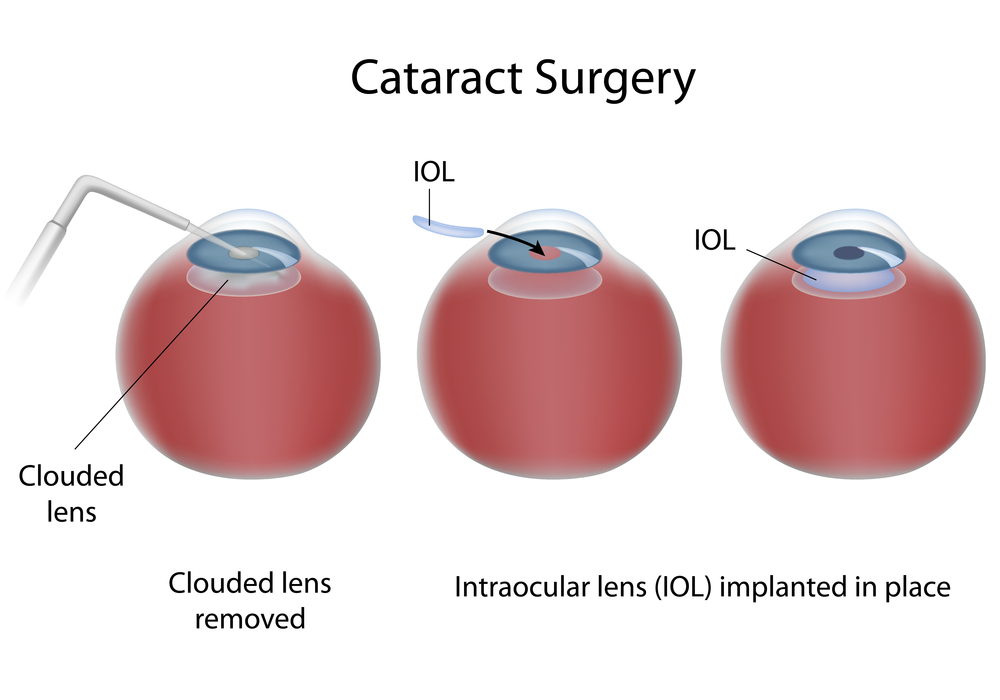
Cataracts occur when there is a clouding of the eye’s lens, making your vision blurry or cloudy. While it is a normal for this to happen as a person ages, someone with diabetes is more likely to develop them at an earlier age. While beginning cataracts can be treated with glasses, when they become more advanced, cataract surgery will be needed to replace the cloudy lens with an artificial lens.

glauccoma diabetic eye disease
Courtesy of National Eye Institute, National Institutes of Health
A person with diabetes is nearly twice as likely to get glaucoma as other adults. Glaucoma is an eye disease that damages the optic nerve. The damage occurs when the pressure in the eye increases, squeezing the optic nerve and restricting its visual transmissions to the brain. Like diabetic retinopathy, you rarely notice any changes in your vision in the early stages, but as it progresses you begin to lose your peripheral vision. It can be treated with eye drops or surgery, but left untreated it can result in blindness.

Controlling Diabetes

Diabetes can be controlled, and some cases prevented, with careful attention to diet, watching your weight and exercise. Also learn your family medical history. You are at a higher risk of diabetes if a mother, father, brother or sister has the disease. If you are diagnosed with diabetes, make sure you have a yearly comprehensive eye exam to avoid vision loss. To learn more about diabetes go to www.ndep.nih.gov.

Susan DeRemerSusan DeRemer, CFRE
Vice Pesident of Development
Discovery Eye Foundation

Night Blindness

10/28/14

As the number of daylight hours decrease and daylight savings time is about to end, many of us feel that the days are getting much shorter. If you suffer from night blindness, your days are shorter, because getting around or driving at night, are sometimes impossible.

Night blindness is a condition that makes it difficult for a person to see in low-light situations or at night. Some types are treatable, while others are not. You will need to consult your eye doctor to determine the underlying cause of your night blindness to determine what can or cannot be done.

night blindness
Courtesy of wikipedia
There are several things that could cause night blindness:
•Cataracts
•Genetic eye disease
•Vitamin A deficiency
•Diabetes
•Aging eye
•Sunlight exposure

Here is a brief look at each.
Cataracts – This is when the lens of the eye becomes gradually becomes clouded, reducing vision. Besides reducing vision at night you may also experience halos around lights. This is a treatable condition requiring cataract surgery and replacing your clouded lens with a clear artificial lens. Your vision should improve considerably.

Genetic Eye Disease – Both retinitis pigmentosa or Usher syndrome are progressive genetic eye diseases where the rods that regulate light, and cones that control color perception and detail die. Progressive night blindness is one of the first visual symptoms of these two diseases. Currently there is no treatment for them as there is no way to treat or replace the dying rods.

Vitamin A Deficiency – While rare in the US, it can be a result of other diseases or conditions such as Crohn’s disease, celiac disease, cystic fibrosis or problems with the pancreas. Options to help with the deficiency include vitamin supplements suggested by your doctor, or increasing your intake of orange, yellow or green leafy vegetables.

Diabetes – People with diabetes are at higher risk for night vision problems because of the damage to the blood vessels and nerves in the resulting in diabetic retinopathy. Not only can it cause poor night vision, it may also take longer to see normally after coming indoors from bright light outside. There is no cure, but controlling blood sugar levels with medicine and diet can help prevent developing retinopathy or help slow the progression.

Aging Eye – As we age several things happen to our eyes. Our iris, which regulates the amount of light going into the eye, gets weaker and less responsive. This can make adapting from light to dark more difficult and slower. Our pupils shrink slightly allowing less light into the eye. The lens of the eye becomes cloudier, as explained above in cataracts, limiting the amount of light into the eye. We also have fewer rods for light perception. Aside from cataract surgery there is no treatment for age-related night blindness. However, eating a diet rich in fruits and vegetables and low in saturated fat is the best way to slow the progression. Here is more information on how the aging eye is affected.

Sunlight Exposure – If your night vision seems temporarily worse after a trip to the beach or a day on the ski slopes, it probably is. Sustained bright sunlight can impair your vision, especially if you fail to wear sunglasses or goggles.

Night blindness due to genetic conditions or aging cannot be prevented. However if you protect your eyes from extreme sunlight, eat a healthy diet, and monitor blood sugar levels if needed, you can reduce your chances for night blindness.

As we head into the holiday season, you should know that some great sources of vitamin A include sweet potatoes, butternut squash and … pumpkins!

Susan DeRemerSusan DeRemer, CFRE
Vice President of Development

20 Tips For Cooking With Low Vision

10/21/14

People cook for different reasons. Some just so they can eat, others enjoy the creativity and find it relaxing and for some it is a activity they have fun doing with others. But cooking can become difficult and dangerous as you lose your vision. Here are some useful tips to help keep you safe and give you confidence to enjoy cooking with low vision.

Cooking with low vision
1. To avoid burns, place a pot or pan on the burner before turning it on and, turn the burner off before removing the pot or pan.

2. Turn pan handles inward from the stove or counter to avoid spills and burns.

3. Mark common or often used settings on appliances with bright nail polish, bright colored tape, or raised dots.

4. Purchase a microwave with a sensor reheat feature to automatically reheat food to the proper temperature.

5. Use long oven mitts to protect your hands and arms from hot surfaces.

6. Turn on the oven light while using it to remind you the oven is on.

7. Use a double spatula to help avoid spills when turning food.

8. Use a special measuring cup that enhances contrast.

9. Use a knife with an adjustable slicing guide.

10. Have light and dark cutting boards to be able to provide the most contrast depending on what you are slicing or chopping – such as black for slicing onions and white for chopping kale.

11. Use a large print, low-vision kitchen timer.

12. Use an automated coffee maker that makes a single cup of coffee or tea to eliminate the need to pour boiling liquids and risk burns.

13. Attach low-glare fluorescent lighting to the underside of cabinets to make it easier to see.

14. Outline the end of countertops with colored duct tape, or paint in a color that contrasts with the work surface. Also choose kitchenware that contrast with the countertop.

15. Organize shelves in a logical way, possibly alphabetically, with most use items easily accessible. Be sure to return items to where they were originally.

16. Use a color for the interior of cupboards that contrasts with your dishes to make them easier to see. Do not use clear glasses or dishes as they appear invisible.

17. If you have glaucoma and experience tunnel vision, remove cabinet doors or replace with sliding doors.

18. Place rubber bands around the milk carton to distinguish it from the orange juice carton. This also works well with distinguishing a can of chicken noodle soup from tomato, etc.

19. Avoid overflows by hooking a “liquid Level” indicator to your glass or cup that will play a tune when you are ¾” from the top.

20. For cold drinks you can also use your finger by placing the tip of it over the edge of the glass and stop pouring when you feel the liquid.

Once you have prepared your food, eating can also be a challenge, so here are five bonus tips.

1. Use dinner plates with a raised lip to avoid spills or pushing the food off while eating.

2. Avoid patterns on dinnerware.

3. Make sure your dishes contrast with the table or table linens.

4. Have a system for putting food on your plate such as meat at 6:00, vegetables at 3:00 and starches at 9:00.

5. Use a gooseneck lamp directly over you plate.

Susan DeRemerSusan DeRemer, CFRE
Vice President of Development

15 Things Doctors Might Like Us To Know

10/16/14

The Doctor Patient Relationship

Doctors are human. Professional decorum may not let them speak their minds and for that reason, I have gathered comments heard from eye doctors with whom I have been privileged to associate as a so-called “expert patient”. These may not speak for all, but I do think they represent the majority. Here are 15 of the most important, which we might benefit from hearing.15 - doctor patient relationship

1. Your eyes are growing older, and I can’t turn back the years in an aging retina. I can, however, help you try to maintain your current vision for as long as possible.

2. Your appreciation means a lot. Don’t hesitate to compliment me if I have earned it.

3. I have quite a few patients who need my help and don’t want to be kept waiting. So I don’t have to keep repeating myself, please take notes, or bring someone with you who can refresh your memory later.

4. It is important that you comply with my instructions and show up for appointments if you want the best results.

5. My specialty is eye care. I will, however, try to provide you with resources for other ailments you may have.

6. Please be concise with your questions and stay on topic, so I’ll have time to treat other deserving patients.

7. Don’t believe everything you see in the media unless the source is reliable. If the information isn’t based on good evidence, my response to you may be disappointingly negative.

8. My services may seem expensive, but I have so many expenses and debts resulting from my profession that my bottom line may not be much different than yours.

9. An ethical physician will not choose a particular drug or treatment by how much of a profit he can make prescribing it.

10. I might not agree with everything you say, but rest assured that I will never let it affect the level of care I will provide.

11. Don’t expect me to keep all eye research in my head. I do know where to look it up, so allow me some time to get back to you on some questions.

12. Conferences I attend are sometimes in exotic places, but most of the time is spent going from one event to another, so it is really not a vacation.

13. Even if there is no current effective treatment for your eye disease, I still need to see you regularly to monitor your condition. Regular check-ups are important to your health and preventative care and treatment options are changing all the time.

14. I may not remember you personally, especially in the beginning or if I see you only see you once or twice a year, but your records contain everything I need to know to care for your vision.

15. I may not agree with your decision to try an alternative treatment, but I respect your right to do so. All I ask is that you seriously consider my opinion, and if you decide to go ahead with the treatment on your own, please keep me in the loop so I can monitor your condition.

We should try to understand that doctors are human beings who truly care about our welfare. Doing so could turn a top-down doctor-patient relationship into one of mutual respect and understanding. We might even get a few extra minutes of precious time in the chair, just because we’re a pleasure to have around.

dan robertsDan Roberts
“Expert Patient”

Our Thanks to Guest Bloggers Continues

10/7/14

More Amazing Guest Bloggers

Last week I took the opportunity to thank our very first guest bloggers for helping us launch the Discovery Eye Foundation Blog. We are pleased that so many people appreciate the wide range of eye-related information from eye care professionals, as well as the stories from people that live with eye disease on a daily basis.
Thank you part 2
Here is a round-up of guest bloggers since June 2014 that shared their time, experience and/or expertise to provide you with the best eye-related information.

Sumit “Sam“ Garg, MDwhat you should know about cataracts

Randall V. Wong, MDfloaters, causes and treatments

Roy Kennedyhis personal experiences with the miniature telescope implant

Sandra Young, ODthe importance of getting vitamins and minerals from your food and not just supplements

Jeanette Hassemanliving with keratoconus

Greg Shanetheater for the blind

Caitlin Hernandezblind actress and playwright

Jullia A. Rosdahl, MD, PhDlasers for glaucoma and genetics and glaucoma

Maureen A. Duffy, CVRTways to reduce harmful effects of sun glare

Kooshay Malekwhat is it like to lose your vision and being a blind therapist

Jeffrey J. Walline, OD PhDchildren and contact lenses

Robert Mahoneychoosing a home care agency

Robert W. Lingua, MDnystagmus in children

Buddy Russell, FCLSA, COMTcommon pediatric eye diseases, treatment options for children and pediatric contact lenses

NIH (National Institute of Health)telemedicine for ROP diagnosis

Harriet A. Hall, MDevaluating online treatment claims

Patty Gadjewskithe life-changing effects of a telescopic implant

Michael A. Ward, MMSc, FAAOproper contact lens care and wearing contacts and using cosmetics

Susan DeRemerSusan DeRemer, CFRE
Vice President of Development
Discovery Eye Foundation

Our First Three Months Of Eye Care

9/30/14

Discovery Eye Foundation Blog’s First Three Months

It is hard to believe, but this blog has been providing information and insights into eye disease, treatment options, personal experiences of living with vision loss, and other eye-related information for seven months.

All of this would not have been possible without the expertise of remarkable eye care professionals who took time out of their busy schedules to share information to help you cope with vision loss through a better understanding of your eye condition and practical tips. Since so much information was shared in the seven months, here is a look at the first three months, with the additional four months to be reviewed next Tuesday.
Thank You - first three months
I am very thankful to these caring eye professionals and those with vision loss who were willing to share their stories:

Marjan Farid, MDcorneal transplants and new hope for corneal scarring

Bill Takeshita, OD, FAAO, FCOVDproper lighting to get the most out of your vision and reduce eyestrain

Maureen A. Duffy, CVRTlow vision resources

M. Cristina Kenney, MD, PhDthe differences in the immune system of a person with age-related macular degeneration

Bezalel Schendowich, ODblinking and dealing with eyestrain

Jason Marsack, PhDusing wavefront technology with custom contact lenses

S. Barry Eiden, OD, FAAOcontact lens fitting for keratoconus

Arthur B. Epstein, OD, FAAOdry eye and tear dysfunction

Jeffrey Sonsino, OD, FAAOusing OCT to evaluate contact lenses

Lylas G. Mogk, MDCharles Bonnet Syndrome

Dean Lloyd, Esqliving with the Argus II

Gil Johnsonemployment for seniors with aging eyes

We would like to extend our thanks to these eye care professionals, and to you, the reader, for helping to make this blog a success. Please subscribe to the blog and share it with your family, friends and doctors.

Susan DeRemerSusan DeRemer, CFRE
Vice President of Development
Discovery Eye Foundation

Common Pediatric Eye Diseases

8/21/14

In the third of this series, Buddy Russell, from the Emory University Eye Center, provides a great overview of common pediatric eye diseases.

Some Conditions Frequently Seen in Pediatrics

A basic understanding of some of the conditions that may be present in pediatric patients is important to not only know what they are but also understand well enough to explain to the parent or caregiver. The following is intended to be an overview of some of those conditions and not a complete explanation.Girl with eye chart-common pediatric eye diseases

  1. Nystagmus – Nystagmus is a vision condition in which the eyes make repetitive, uncontrolled movements, often resulting in reduced vision. These involuntary eye movements can occur from side to side, up and down, or in a circular pattern. As a result, both eyes are unable to hold steady on objects being viewed. Unusual head positions and head nodding in an attempt to compensate for the condition may accompany nystagmus. Most individuals with nystagmus can reduce the severity of their uncontrolled eye movements and improve vision by positioning their eyes to look to one side. This is called the “null point” where the least amount of nystagmus is evident. To accomplish this they may need to adopt a specific head posture to make the best use of their vision. The direction of nystagmus is defined by the direction of its quick phase (e.g. a right-beating nystagmus is characterized by a rightward-moving quick phase, and a left-beating nystagmus by a leftward-moving quick phase). The oscillations may occur in the vertical, horizontal or torsional planes, or in any combination. The resulting nystagmus is often named as a gross description of the movement, e.g. downbeat nystagmus, upbeat nystagmus, seesaw nystagmus, periodic alternating nystagmus. Having nystagmus affects both vision and self-concept. Most people with nystagmus have some sort of vision limitations because the eyes continually sweep over what they are viewing, making it impossible to obtain a clear image. If a refractive error is found, contact lenses may be the most effective way of obtaining best-corrected vision.
  2. Strabismus – Strabismus is any misalignment of the eyes. It is estimated that 4% of the U.S. population has strabismus. Strabismus is most commonly described by the direction of the eye misalignment. Common types of strabismus are esotropia (turn in), exotropia (turn out), hypotropia (turn down), and hypertropia (turn up). Eye misalignment can cause amblyopia in children. When the eyes are oriented in different directions, the brain receives two different visual images. The brain will ignore the image from the misaligned eye to avoid double vision, resulting in poor vision development of that eye. Also, an eye that sees poorly tends to be misaligned. The goal of strabismus treatment is to improve eye alignment, which allows for better work together (binocular vision). Treatment may involve eyeglasses, contact lenses, eye exercises, prism, and / or eye muscle surgery.
  3. Amblyopia – Amblyopia, sometimes called a “lazy eye,” occurs when one or both eyes do not develop normal vision during early childhood. Babies are not born with 20/20 vision in each eye but must develop it between birth and 6-9 years of age by using each eye regularly with an identical focused image falling on the retina of each eye. If this does not occur in one or both eyes, vision will not develop properly. Instead, vision will be reduced and the affected eye(s) are said to be amblyopic. This common condition, affecting up to 4% of all children, should be diagnosed and treated during infancy or early childhood to obtain optimum three-dimensional vision and to prevent permanent vision loss. What causes amblyopia?
      • Misaligned eyes (strabismus)
        Misaligned eyes are the most common cause of amblyopia. When both eyes are not aimed in exactly the same direction, the developing brain “turns off” the image from the misaligned eye to avoid double vision and the child uses only the better eye — the dominant eye. If this persists for a period even as short as a few weeks, the eye will not connect properly to the visual cortex of the brain and amblyopia will result.
      • Unequal refractive error (anisometropia)
        Unequal refractive error is an eye condition in which each eye has a different refractive error and therefore both eyes cannot be in focus at the same time. Amblyopia occurs when one eye (usually the eye with the greater refractive error) is out of focus because it is more nearsighted, farsighted or astigmatic than the other. Again, the brain “turns off” the image from the less focused eye and this eye will not develop normal vision. Because the eyes often look normal, this can be the most difficult type of amblyopia to detect and requires careful vision screening of acuity measurements at an early age. Treatment with glasses or contact lenses to correct the refractive error of both eyes, sometimes with part-time patching of the better seeing eye, is necessary in early childhood to correct the problem.
      • Obstruction of or cloudiness (deprivation)
        Obstruction of or cloudiness in the normally clear eye tissues may also lead to amblyopia. Any disorder that prevents a clear image from being focused can block the formation of a clear image on the retina and lead to the development of amblyopia in a child. This often results in the most severe form of amblyopia. Examples of disorders that can interfere with getting a clear image on the retina are a cataract or cloudy lens inside the eye, a cloudy and or irregular shaped cornea, or a droopy upper eyelid (ptosis) or eyelid tumor.It is not easy to recognize amblyopia. A child may not be aware of having one normal eye and one with reduced vision. Unless the child has a misaligned eye or other obvious external abnormality, there is often no way for parents to tell that something is wrong. In addition, it is difficult to measure vision in very young children at an age in which treatment is most effective.To treat amblyopia, a child and their caregiver must be encouraged to use the weaker eye. This is usually accomplished by patching the stronger eye. This covering of the stronger eye with an adhesive patch, an cclude contact lens or temporary surgery often proves to be a frustrating and difficult therapy. Patching will often continue for weeks, months, or even years in order to restore normal or near normal vision and maintain the improvement in the amblyopic eye. Occasionally, blurring the vision in the good eye with eye drops or lenses to force the child to use the amblyopic eye treats amblyopia. In some cases, cataract surgery or glaucoma surgery might be necessary to treat form deprivation amblyopia. Patching may be required after surgery to improve vision, and glasses or contact lenses may be required to restore appropriate focusing.Surprising results from a nationwide clinical trial in 2005 show that many children age seven through 17 with amblyopia may benefit from treatments that are more commonly used on younger children.
        Treatment improved the vision of many of the 507 older children with amblyopia studied at 49 eye centers. Previously, eye care professionals often thought that treating amblyopia in older children would be of little benefit. The study results, funded by the National Eye Institute (NEI), appear in the April issue of Archives of Ophthalmology.
  4. Congenital Cataract – A congenital cataract, or clouding of the crystalline lens is present in 2-3 per 10,000 live births of children. The presence of a visually significant cataract in a child is considered an urgent disorder. The resultant form deprivation of vision requires immediate surgery to remove the obstruction, prompt optical correction and amblyopia therapy in unilateral cases. Until the 1970s, it was generally believed that there was no means of restoring the vision in an eye with a unilateral congenital cataract. However, subsequent studies demonstrated that excellent visual results could be obtained with early surgical treatment coupled with optical correction with a contact lens and patching therapy of the fellow eye. However, treatment results continue to be poor in some infants with unilateral congenital cataracts due to a delay in treatment or poor compliance with contact lens wear or patching therapy of the fellow eye. The Infant Aphakia Treatment Study (IATS) was designed to compare the visual outcomes in children 1 to 6 months of age with a unilateral congenital cataract randomized to optical aphakic correction with contact lenses or an intraocular lens (IOL). Children randomized to IOL treatment had their residual refractive error corrected with spectacles. Children randomized to no IOL had their aphakia treated with a contact lens. In previous publications we have shown that the visual results are comparable for these two treatments at 1 year of age, but significantly more of the infants randomized to IOL implantation required additional intraocular surgeries.
  5. Accommodative Esotropia – Accommodative esotropia refers to a crossing of the eyes caused by farsightedness. Accommodative esotropia is a type of strabismus. Children who are farsighted easily and automatically focus on objects at distance and near through accommodation. As a result, a child who is farsighted usually does not have blurred vision. However, in some children who are farsighted, this accommodative effort is associated with a reflex crossing of the eyes. Accommodative esotropia can begin anywhere from 4 months to 6 years of age. The usual age of onset is between 2 and 3 years of age.Full-time use of the appropriate hyperopic glasses prescription or contact lenses will often control the esotropia. When wearing the correction, the child will not need to accommodate and hence the associated eye-crossing reflex will disappear. However, after removing the prescribed correction, the crossing will reappear, perhaps even more than before the child began wearing the correction. Sometimes the correction will only cause the crossing to disappear when the child views a distant object. However, when gazing at near objects, crossing may persist despite the use of the correction. In these circumstances, a bifocal lens is often prescribed to permit the child to have straight eyes at all viewing distances. One potential advantage of contact lenses compared to spectacles when correcting hyperopic powers is the decrease in accommodative demand. The increased effort to converge the eyes with spectacles requires one to over come the resultant base out prism when viewing a near object.


Buddy Russell - pediatric contact lensesBuddy Russell, FCLSA, COMT
Associate, Specialty Contact Lens Service
Emory University Eye Center

Ways to Reduce the Harmful Effects of Sun Glare

During the height of summer sunshine (and heat!), it’s helpful to discuss the importance of eye protection, including ways to reduce the harmful effects of sun glare.

Fundamentally, we need light to see. Approximately 80% of all information we take in is received through the sense of sight. However, too much light – and the wrong kind of light – can create glare, which can affect our ability to take in information, analyze it, and make sense of our surroundings.

Facts about Sunlight

Every type of light has advantages and disadvantages, and sunlight is no exception:

Advantages:

• Sunlight is the best, most natural light for most daily living needs.
• Sunlight is continuous and full-spectrum: the sun’s energy at all wavelengths is equal and it contains all wavelengths of light (explained below).

Disadvantages:

• It is difficult to control the brightness and intensity of sunlight.
• Sunlight can create glare, which can be problematic for many people who have low vision.
• Sunlight is not always consistent or reliable, such as on cloudy or overcast days.

Visible Light and Light Rays

An important factor to consider is the measurement of visible light and light rays, beginning with the definition of a nanometer:

• A nanometer (nm) is the measurement of a wavelength of light.
• A wavelength is the distance between two successive wave crests or troughs:

Wavelength - glare

• A nanometer = 1/1,000,000,000 of a meter, or one-billionth of a meter. It’s very small!

The human visual system is not uniformly sensitive to all light rays. Visible light rays range from 400 nm (shorter, higher-energy wavelengths) ? 700 nm (longer, lower-energy wavelengths).
Visible Light Spectrum - glare
The visible light spectrum occupies just one portion of the electromagnetic spectrum, however:

• Below blue-violet (400 nm and below), is ultraviolet (UV) light.
• Above red (700 nm and above), is infrared (IR) light.
• Neither UV nor IR light is visible to the human eye.

Ultraviolet Light and Blue Light

Ultraviolet (UV) light has several components:

• Ultraviolet A, or UVA (320 nm to 400 nm): UVA rays age us.
• Ultraviolet B, or UVB (290 nm to 320 nm): UVB rays burn us.
• Ultraviolet C, or UVC (100 nm to 290 nm): UVC rays are filtered by the atmosphere before they reach us.

Blue light rays (400 nm to 470 nm) are adjacent to the invisible band of UV light rays:

• There is increasing evidence that blue light is harmful to the eye and can amplify damage to retinal cells.
• You can read more about the effects of blue light at Artificial Lighting and the Blue Light Hazard at Prevent Blindness.

A new study from the National Eye Institute confirms that sunlight can increase the risk of cataracts and establishes a link between ultraviolet (UV) rays and oxidative stress, the harmful chemical reactions that occur when cells consume oxygen and other fuels to produce energy.

Sunlight and Glare

Glare is light that does not help to create a clear image on the retina; instead, it has an adverse effect on visual comfort and clarity. Glare is sunlight that hinders instead of helps. There are two primary types of glare.

Disability glare

• Disability (or veiling) glare is sunlight that interferes with the clarity of a visual image and reduces contrast.
• Sources of disability glare include reflective surfaces (chrome fixtures, computer monitors, highly polished floors) and windows that are not covered with curtains or shades.

Discomfort glare

• Discomfort glare is sunlight that causes headaches and eye pain. It does not interfere with the clarity of a visual image.
• Sources of disability glare include the morning and evening positions of the sun; snow and ice; and large bodies of water, (including swimming pools).

Controlling Glare

You can protect your eyes from harmful sunlight and minimize the effects of glare by using a brimmed hat or visor in combination with absorptive lenses.

• Absorptive lenses are sunglasses that filter out ultraviolet and infrared light, reduce glare, and increase contrast. They are recommended for people who have low vision and are also helpful for people with regular vision.
• Lens colors include yellow, pink, plum, amber, green, gray, and brown. Ultra-dark lenses are not the only choice for sun protection.
• Lens tints in yellow or amber are recommended for controlling blue light.
NoIR Medical Technologies: NoIR (No Infra-Red) filters absorb UVA/UVB radiation and also offer IR light protection.
Solar Shields: Solar Shields absorb UVA/UVB radiation and are available in prescription lenses.
• You can find absorptive lenses at a specialty products store, an “aids and appliances store” at an agency for the visually impaired, or a low vision practice in your area. Before you purchase, it’s always best to try on several different tints and styles to determine what works best for you.

More Recommendations

• Always wear sunglasses outside, and make sure they conform to current UVA/UVB standards.
• Be aware that UV and blue light are still present even when it is cloudy or overcast.
• Make sure that children and older family members are always protected with UVA/UVB-blocking sunglasses and brimmed hats or visors.

Maureen Duffy-editedMaureen A. Duffy, CVRT
Social Media Specialist, visionaware.org
Associate Editor, Journal of Visual Impairment & Blindness
Adjunct Faculty, Salus University/College of Education and Rehabilitation

Corneal Donor’s Age Not Critical for Transplant Success

6/17/14

In the United States, more than 40,000 corneal transplants are performed each year with a high success rate in comparison to other types of organ transplants. According to the Eye Bank Association of America (EBAA), keratoconus was the leading cause of anterior lamellar keratoplasty (DALK/ALK partial thickness transplant) and was the fourth most common indication for penetrating keratoplasty surgery in 2012 (their last reporting period).
corneal transplant-original size
Advances in technology have led to increasingly successful outcomes for all who need corneal transplants. New long-term research of corneal transplant patients have shown that the age of corneal donors is no longer as important as once thought by eye health providers. According to a study funded by the National Institutes of Health, ten years after a transplant, a cornea from a 71-year-old donor is likely to remain as healthy as a cornea from a donor half that age.

The Cornea Donor Study (see www.ClinicalTrials.gov), funded by NIH’s National Eye Institute (NEI), was designed to compare graft survival rates for corneas from two donor age groups, aged 12-65 and aged 66-75. It was coordinated by the Jaeb Center for Health Research in Tampa, Fla., and involved 80 clinical sites across the United States. The study enrolled 1,090 people eligible for transplants, ages 40-80. Donor corneas were provided by 43 eye banks, and met the quality standards of the Eye Bank Association of America. The study found that 10-year success rates remained steady at 75 percent for corneal transplants from donors 34-71 years old. In the United States, three-fourths of cornea donors are within this age range, and one-third of donors are at the upper end of the range, from 61-70 years old.

Prior to this study, many surgeons would not accept corneas from donors over 65. Since the supply of young donor corneas is limited, these study results are encouraging for those who face a corneal transplant . The high level of success rates using corneas from older donors (over age 60) greatly increases the pool of donated corneas and corneal tissue available for transplant. In 2012, corneal donors under age 31 comprised less than 10 percent of the U.S. donor pool. “Our study supports continued expansion of the corneal donor pool beyond age 65,” said study co-chair Edward J. Holland, M.D., professor of ophthalmology at the University of Cincinnati and director of the Cornea Service at the Cincinnati Eye Institute. “We found that transplant success rates were similar across a broad range of donor ages.”

“Overall, the findings clearly demonstrate that most corneal transplants have remarkable longevity regardless of donor age,” said Mark Mannis, M.D., chair of ophthalmology at the University of California, Davis, and co-chair of the study. “The majority of patients continued to do well after 10 years, even those who received corneas from the oldest donors.”

SOURCE: National Eye Institute Press Releases

For information about Eye Bank Association of America

CathyW headshotCathy Warren, RN
Executive Director
National Keratoconus Foundation

What You Need to Know About Cataracts

6/5/14

Do you feel like your vision is getting worse? Do you feel like colors are not as vibrant as they used to be? Are you having more trouble with glare? If you have any of these symptoms, you may be experiencing the effects of cataracts.

Cataracts are a normal aging process of the crystalline lens in the eye. Well you may ask — what is the crystalline lens? It is easiest to think of the eye as a camera. The eye has a lens (actually two) – the cornea (the front window of the eye) and the crystalline lens (inside the eye). It also has an aperture (the colored iris), and film (the retina). All these structures work together to focus light and allow us to see – just like a camera. When we are young (less than 40), the crystalline lens is flexible. This is why we are able to see distance and then near without the need for reading glasses. The crystalline lens is able to change its shape depending on where one is looking.

Figure 1 – Slit-lamp photo of a visually significant cataract.

As we age, the crystalline lens becomes less flexible, thereby causing one’s near vision to be more blurry. This necessitates the need for reading glasses. As the crystalline lens become less flexible with age, the lens also starts to become more yellow and can also become cloudy. When the yellowing and/or clouding become visually significant, we refer to this as a cataract (figure 1).

Are cataracts dangerous? The simple answer is no. In the vast majority of cases, a cataract can be monitored until it becomes visually significant (drop in vision, glare, decreased contrast, vision related difficulties with day to day activities, etc). However, there are a few instances in which cataract removal is a medical necessity. Routine examinations by your eyecare provider can help you determine if you are at risk for these less common instances.

Figure 2 – Intraoperative photo during cataract surgery (prior to cataract removal).

What can I expect during cataract surgery? Do you have to replace the crystalline lens with anything? Cataract surgery involves removing the clouded crystalline lens (figure 2) and replacing it with an artificial lens known as an intraocular lens (IOL) (figure 3). Surgery generally takes 10-15 minutes under a mild sedative, and you don’t have to stop any of your current medications. Anesthesia is achieved with drops and you will only feel mild pressure during the surgery. IOLs come in different styles – Standard IOLs grossly correct your vision and you can fine tune your vision (distance and near) with glasses post-operatively;

Figure 3 – Intraoperative photo during cataract surgery (after implantation of an IOL).

Toric (astigmatism correcting) IOLs allow for increased spectacle independence, but glasses will still be needed for distance or near; Accommodating IOLs “flex” within the eye to decrease your dependence on distance and near glasses; Multifocal IOLs allow spectacle independence for distance and near. I always counsel patients that there is no perfect IOL and you have to determine which IOL is best for your particular situation. Your doctor can help you decide which IOL is best for you. Generally, cataract surgery is extremely safe. Your doctor will discuss particular risks specific to your eye.

How do I know if cataract surgery is right for me? The best way to know if you have a cataract and if it time to consider surgery is to consult with your local ophthalmologist. If you have experienced a recent drop in vision, that is not correctable with glasses, cataract surgery may be able to restore your vision!

Garg feb 2014 thumbSumit “Sam“ Garg, MD
Medical Director
Vice Chair of Clinical Ophthalmology
Assistant Professor of Ophthalmology
Gavin Herbert Eye Institute – UC, Irvine