4 Super Greens for Better Sight

A healthy diet not only helps your heart, but also your eyes. Your diet should include lots of fruits and vegetables to provide you with a natural source of nutrients to help protect your sight. As wonderful as supplements are, eating the actual foods is always better. Some of the best vegetables for you are the dark, leafy greens that are rich in valuable vitamins and nutrients. These are the super greens for better sight.

With the US experiencing bitter, freezing temperatures on the East Coast, while the West Coast is having summer in February, with record-breaking hot temperatures, I thought it would be interesting to see how you could enjoy those super greens, no matter what the temperature is outside. Below is a quick look at four dark leafy greens that are a great addition to a healthy diet, watercress, arugula, spinach and kale. For each vegetable I have included a recipe that is served hot, along with one that is served cold.

Here is what you need to know about super greens for better sight.
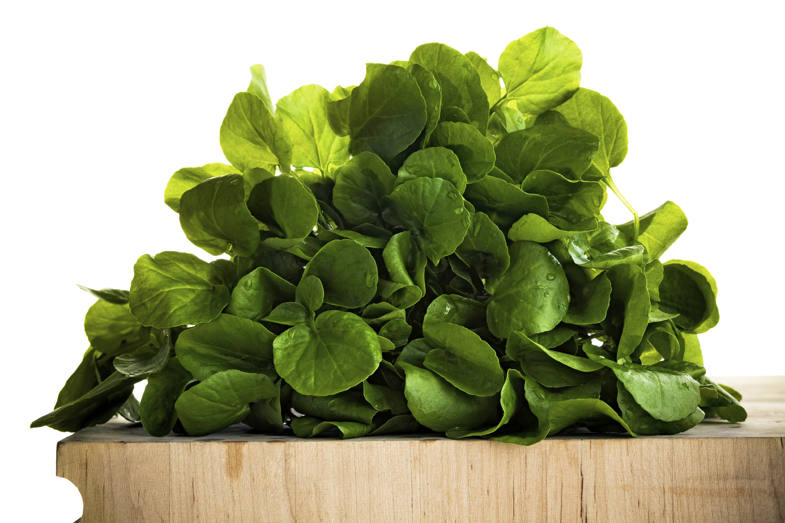
super greens
WATERCRESS

Watercress is a cruciferous plant and part of the brassica family, like arugula and kale. It contains vitamins A, B6, B12, C, K, iron, magnesium, calcium, phosphorus, lutein and zeaxanthin. In fact, weight for weight, watercress contains more vitamin C than an orange, more calcium than milk, more iron than spinach and more folate than bananas. Watercress has the highest amount of nutrients for the smallest amount of calories.

The health benefits of watercress include boosting immunity, cancer & diabetes prevention, reducing cognitive decline, thyroid support, better cardiovascular health and stronger bones. As for your eyesight, it can help prevent or slow the onset of age-related macular degeneration and possibly cataracts.

Watercress is most commonly enjoyed fresh in salads, but can also be use in pastas, casseroles, soups and sauces. Choose watercress with deep green, crisp leaves, with no signs of wilting. Trim the stems, rinse the greens in cold water and dry. It is best if used immediately, but can be store for up to four days in the refrigerator.

Watercress Soup by William Anatooskin

Watercress and Grapefruit Salad by Martha Stewart
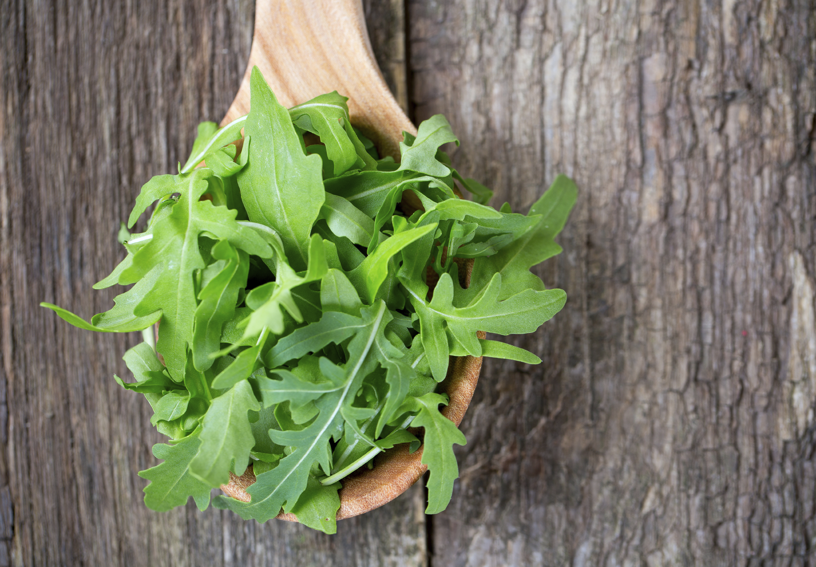
super greens
ARUGULA

Arugula is also known as a salad or garden rocket. It is a small low growing herb that is packed with vitamins, minerals and antioxidants. It is rich in folates, vitamin A, B-complex, C and K and has copper, iron, calcium, potassium, manganese and phosphorus.

The health benefits of arugula include a lowered risk of cancer, healthy bones, strengthened brain function, improved mineral absorption and it boosts the immune system. Because of being a source of carotenoids, it also helps to slow the progression of age-related macular degeneration.

Arugula is popular in salads, used with milder greens to add a peppery flavor. But it can also be used in pastas, casseroles, pizzas, soups and sauces. Choose arugula that is crisp with green young leaves. Avoid the flowered harvest as those leaves are tough and have a bitter taste. Wash leaves in a bowl of water, swishing thoroughly to get rid of all sand and soil. Drain and pat dry before storing in the vegetable bin of your refrigerator for no more than a few days.

Grilled Stuffed Swordfish by Stormy Scarlett

Pasta Salad with Goat Cheese and Arugula by Martha Stewart
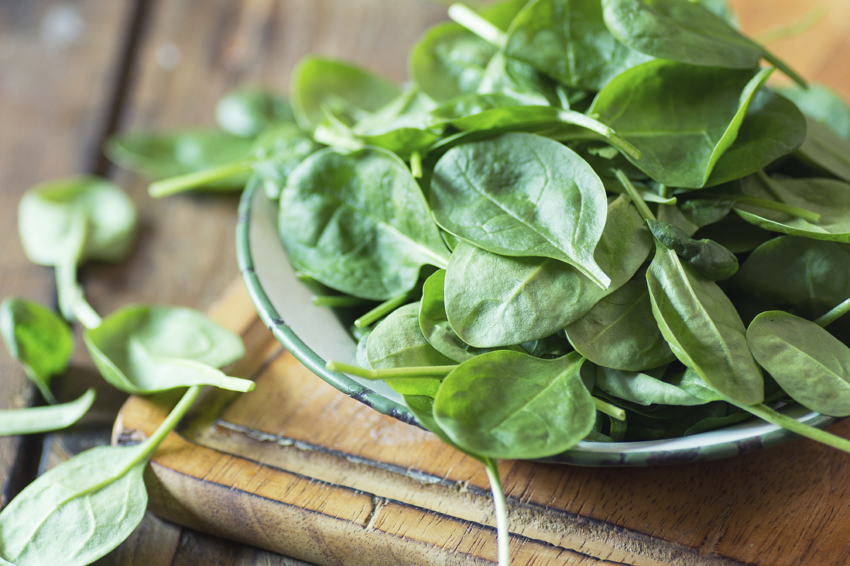
super greens
SPINACH

Spinach is a very popular leafy green vegetable, with two common varieties cultivated for food; the savory-type with dark green crinkled leaves and the flat-leaf type with smooth surfaced leaves. Spinach contains vitamin A, B-complex, C and K, along with lutein, zeaxanthin beta-carotene, potassium manganese, magnesium, copper and zinc.

The nutrients in spinach help improve blood glucose control in diabetes, lower the risk of certain cancers, reduce blood pressure, increase bone health and help iron deficiency. The lutein, zeaxanthin and beta-carotene help to potentially prevent and slow the progression of age-related macular degeneration. Lutein also works to protect the eye from free radical damage by helping filter out damaging blue and ultraviolet light.

Spinach is a universally popular vegetable, used around the world in a variety of ways, including salads, soups, noodles, pies, casseroles, dips, sauces, etc. Look for leaves that are dark green in color, crisp and not dull or yellow and spotted. Wash thoroughly to remove sand and soil, dry, trim away tough stems and store in the refrigerator for up to a week.

Turkey-Spinach Meatballs from Bon Appètit

Spinach Salad with Dates from Bon Appètit
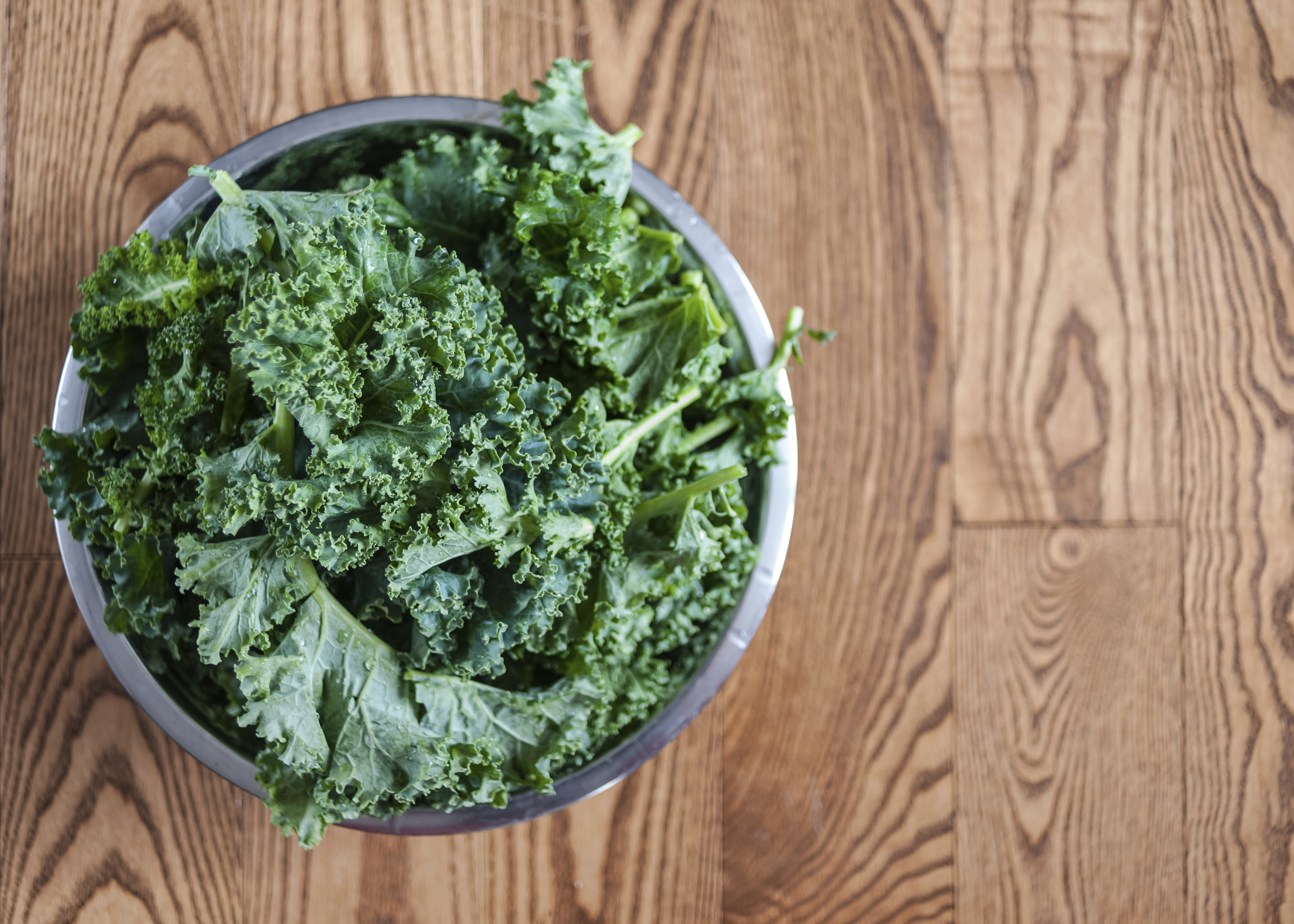
super greens
KALE

Kale is a member of the mustard and cabbage families and has more nutrients than spinach. Less than ½ cup has 333% of the recommended daily allowance (RDA) of vitamin A, 587% RDA of vitamin K and 200% RDA of vitamin C. This frilly-leafed vegetable also has vitamin B-complex, lutein, zeaxanthin, beta-carotene, copper, calcium, sodium, potassium, iron, manganese, and phosphorus.

The health benefits of kale include healthy muscles and skin, improved blood glucose control, lower colon and prostate cancer risk, better cardiovascular health, stronger bone health, reduced neuronal brain damage and support for red blood cell formation. The advantage for your eyes comes from the lutein, zeaxanthin, beta-carotene and vitamin A, all which work to support a healthy retina. They help protect against blue and ultraviolet light as well as the early onset and progression of age-related macular degeneration. Because of the positive impact on diabetes it also reduces the onset and progression of diabetic retinopathy.

Kale is very versatile and can be served in a variety of ways including salads, soups and casseroles. It can also be braised, broiled, sautéed and even made into kale chips by tossing them in extra-virgin olive oil and sprinkled with your choice of cumin, curry powder, chili powder, red pepper flakes or garlic powder and baking at 275 degrees for 15-30 minutes depending on how crisp you want them to be. When shopping for kale look for leaves that are crispy and crunchy with a brilliant dark blue-green color. Wash thoroughly to remove soil and sand, dry well, and remove all tough stems. It is extremely perishable, so use it as quickly as possible.

Kale and Chicken Casserole by Martha Stewart

Kale with Pomegranate Dressing and Ricotta Salata from Bon Appètit

All of these dark green leafy vegetables are not only healthy for you, but can be used in many ways to make it easy to incorporate them into your diet. Here are a few ideas:

  • Throw a small handful into your blender when making your favorite smoothie
  • Add them to your next omelet or egg scramble
  • Use them for making pesto or adding to pasta sauce
  • Sauté with a small amount of extra-virgin olive and season with freshly ground black pepper and freshly grated Parmesan cheese to serve at a topping for your baked potato
  • Add it to your wrap, sandwich or flatbread

2/17/16

Susan DeRemerSusan DeRemer, CFRE
Vice President of Development
Discovery Eye Foundation

Layers of the Retina

The retina at the back of the eye is essential for all vision. Each layer of cells in this tissue serves a specific purpose. As we prepare for Age-Related Macular Degeneration Awareness Month in February, a closer look at the layers of the retina and their function.

layers of the retina

Layers of the Retina

Choroid – This is made up of a layer of blood vessels that supply oxygen and nutrients to the retina. Defect in the CHM gene can cause choroideremia, leaky blood vessels can expand in the retina causing wet age-related macular degeneration (AMD) and diabetic retinopathy.

Retinal pigment epithelium – This is a single layer of cells that provide essential nutrition and waste removal for the photoreceptor cells. Accumulation of waste can lead to AMD and Stargardt disease.

Photorecptors – This is where the rods and cones are located that convert light into electrical signals. Rods help you with night and peripheral vision. Cones are more concentrated in the macula (the central part of the retina) and proved central and color vision. Death of the rods can cause vision loss called retinitis pigmentosa, while AMD is the loss of central vision.

Horizontal cells – These cells are connect to the photoreceptors that surround the bipolar connected photoreceptor cells and help the help integrate and regulate the input from multiple photoreceptor cells, increasing your visual acuity.

Bipolar cells – The dependence of each layer of the retina on each other is exemplified here. These cells take the electrical information from the photoreceptor cells and pass it along to other retinal cells.

Ganglion cells – These cells extend to form an optic nerve that conveys information to the brain and take the electrical information from the bipolar cells and process it to determine shapes, contrast and color. Glaucoma vision loss results from high intraocular pressure that affects the optic nerve, interrupting the signals to the brain.


5 Elements of Accessible Web Design

A common misconception is that all you need to have a successful blog or website is good content. While content people want to read is important, if you can’t see it, or it is difficult to read, very few people are going to take the time to try. There is lots of good content to choose from.

Making your content easy to scan and read and using great graphics that can tell a story are just as important. Especially if you want to reach the millions of potential readers that are blind or visually impaired.

While a person with low vision (due to age-related macular degeneration, cataracts,glaucoma or other eye diseases that are related to aging) can increase font size or graphics by enlarging them with a pinch of the fingers or scroll of the mouse, the result is often blurry and still difficult to see.

If a person is blind and using a screen reader, what they hear may not match what is written or displayed. A picture without underlying descriptive text is worthless. And when a blind person is using a screen reader to read a web site, they will often tab from link to link to scan your article, skipping over your text, to get a sense of what the options are. “Click here” says doesn’t tell the reader anything.

Here are 5 essential elements of accessible web design.

5 elements of accessible web design

12/15/15

Susan DeRemerSusan DeRenmer, CFRE
Vice President of Development
Discovery Eye Foundation

Low Vision Aging Adults at Higher Risk for Falls

Low vision in aging adults varies as do the occupational therapy techniques that might help older adults in becoming more independent. Mr. P has glaucoma resulting in a narrowed field of view. He is light sensitive, and keeps his blinds closed, darkening the house. He is responsible for doing his laundry, yet his washer and dryer are in the basement, causing a safety concern. Mrs. K has macular degeneration with 20/400 visual acuity. She has severely reduced contrast sensitivity, and can no longer drive or recognize faces easily. She no longer gets to the senior center for regular exercise sessions, which is concerning to her and her OT. Ms. T was diagnosed with diabetic retinopathy. She has struggled for years to accurately see her glucometer and insulin. She has recently developed peripheral neuropathy, leading to wheelchair dependency. Each of these patients is likely to leave their home less often because of their low vision, eventually leading to a decline in lower extremity weakness, balance, endurance and confidence. The fear of falling leads to more isolation, which can cause even more lower extremity weakness, problems with balance, decreased endurance, and even depression. The irony is that this cycle, which began with caution and a fear of falling, lends itself to exactly that. . .a fall.

Why are low-vision aging adults at higher risk for falls?

According to the CDC, “Each year, millions of older people-those 65 and older-fall. In fact, one out of three older people fall each year, but less than half tell their doctor. Falling once doubles your chances of falling again.” The typical aging adult with low vision faces challenges that others do not. Difficulty seeing details combined with reduced contrast sensitivity leads to a decline in mobility and socialization.
risk for falls
risk for falls
So how do we help older adults with low vision lower their risk for falling? When considering safety within the home, persons with low vision must make it a priority to add lighting & contrast whenever possible. Consider all rooms of the house, including entrances, hallways and stairways.

  • Placement of night lights in hallways, stairway, etc.
  •  

  • Keep flashlights in accessible places throughout the home where night lights are not possible.
  •  

  • Instead of closing blinds, keep them open & wear appropriate colored filters to manage glare/light sensitivity.
  •  

  • Small battery operated stick-on lights or rope lighting is inexpensive, and easy to place on steps to light up a stairwell.
  •  

  • Line edge of steps, or stairway railing with brightly colored duct tape to increase visibility.
  •  

  • Make sure grab bars, tub benches, shower chair are high contrast, to be most visible.
  •  

  • Remove throw rugs, with the exception of those providing function, such as the one at the entrance and bathtub. Their purpose is for providing dry shoes and feet, but they should have a non-skid back and a high contrast color to “stand out.”
  •  

  • Reduce clutter in rooms to increase safety by removing items from floors, walkways and stairwells.
  •  

  • Consider investing in a Medical Alert System to provide added piece of mind, confidence, especially when living alone.

Persons with low vision can decrease their frequency of falls by staying social and walking in the community. Unfortunately, many low vision adults become more house bound when they can no longer drive. Locating sources of alternative transportation may be helpful. Seeing faces is difficult, making socializing a challenge, which can lead to depression. The following recommendations take into consideration the challenges of not seeing details or across the visual field normally, while improving lower body strength, endurance, coordination, confidence, and hopefully reducing the risk of a fall.

  • Encourage regular trips to the grocery stores and the mall to keep physically fit. Malls are safe environments to walk around because of wide, straight hallways. Use magnifiers to see price tag/label details, or take advantage of personal shoppers who assist with locating items.
  •  

  • Participate in regularly scheduled exercise sessions (videos, groups, etc). Sit/stand in the front row, ask instructor to provide clear verbal instruction, instead of only demonstration. Consider hiring a personal trainer in order to get 1:1 instructions for how to use exercise equipment. If watching a video, move closer to your largest TV screen. Home exercise equipment can be labeled with high contrast markings to increase visibility of its details.
  •  

  • Consider joining a senior center in the community. Some have low vision support groups.
  •  

  • Access driving alternatives, such as the Smart Bus, Senior Centers, Local Volunteer or Church groups. Consider using money previously spent for a car and its expenses to hire a private driver.
  •  

  • Use appropriate colored filters in bright outside conditions or darker inside environments (i.e. Amber outside in the sun; Yellow inside a dark restaurant or outside with overcast weather).
  •  

  • Consider using a walker or support cane when walking longer distances in the community or neighborhood. Many individuals decline using an assistive device, not realizing how active and fit it can make them. Rolling walkers are even available with seats, allowing for rest breaks wherever necessary.

While the fear of falling is great amongst all seniors, those with low vision need to consider adding strategies that specifically benefit them. Making changes within the home may be as simple as improving lighting and contrast. Remaining physically fit outside of the home may be done with shopping or exercise equipment, but either way staying active will improve confidence, the fear of falling, and hopefully decrease the chance of a fall. If you are unsure of what approach is best for you, consult with your ophthalmologist about scheduling a low vision eye exam and occupational therapy.

Reference:
Centers for Disease Control and Prevention, National Center for Injury Prevention and Control-CDC 24/7: Saving Lives, Protecting People

12/3/15

Annette Babinski's headshot thumbnailAnnette M. Babinski, OTR/L CLVT
Henry Ford Health System
Vision Rehabilitation Center


Michelle Buck's headshot thumbnailMichelle Buck, MS, OTR/L CLVT
Henry Ford Health System
Vision Rehabilitation Center

#GivingTuesday – Giving For Eye Research

Medical research funding from the US government, the medical device and pharmaceutical industries, biotech and foundations has dropped 0.8% per year, every year from 2007 to 2012. Even with the economic recovery, the funding of eye research from the NIH in 2014 decreased by 17% from 2012.
giving for eye research
What makes this particularly disturbing is that the need for eye research is more important now than ever, as demonstrated by the infographic below.

Giving to eye research
This infographic by the National Institute of Health (NIH) is used with their permission.

Giving For Eye Research

The eye diseases in the above graphic are related to aging. The number of people being affected is more than doubling for each eye diseases, except for diabetic retinopathy which will increase by 47%, by 2050.

While we are more health conscious than in previous years, knowing the importance of exercise, healthy eating and not smoking, the US population is aging.

Aging baby boomers, the largest population group in US history, are creating a dramatic shift in the age composition of the U.S. population. It is projected that the entire senior population, including the pre-boomer silent generation, will reach 71.4 million people by 2029. This means that those people 65 and older will make up about 20%of the US population by 2029, up from almost 14% in 2012.

We need your help to find treatments and cures for these sight-threatening eye diseases.

December 1st will be the fourth year of #GivingTuesday. It is a global day of giving using combined power of social media and collaboration. Always the Tuesday following Thanksgiving, it has become as widely recognized as Black Friday and Cyber Monday and kicks off the philanthropic season, when many focus on their holiday and end-of-year giving.

We are asking that you support eye research through the Discovery Eye Foundation this philanthropic season with a gift on #GivingTuesday. It is easy to do; you can do it from the comfort of your home – just click here.

Also share this post with family and friends so they can also give the gift of sight this holiday season.

Thank you for reading our blog and for your support.

11/24/15

Susan DeRemerSusan DeRemer, CFRE
Vice President of Development
Discovery Eye Foundation

Smoking and Your Eyes

Smoking is the single largest preventable cause of eye disease.
smoking and your eyes
On the third Thursday of November each year, smokers across the nation take part in the Great American Smokeout, sponsored by the American Cancer Society. This might be the ideal time for you to stop smoking and ACS has information and resources you may find helpful.

Here are some things you should know about smoking and your eyes.

  • Smoking at any age, even in your teens or twenties, increases your future risk for vision loss.
  •  

  • The more you smoke, the higher your risk for eye disease.
  •  

  • If you quit smoking, your risk for these eye diseases decreases considerably.
  •  

  • Smoking increases your risk for cardiovascular diseases that indirectly influence your eyes’ health.
  •  

  • Women who smoke during pregnancy increase their chance for a premature birth and a potentially blinding eye disease called retinopathy of prematurity (ROP).
  •  

  • A smoker is two times more likely to develop macular degeneration compared with a nonsmoker.
  •  

  • Smoking double your chance of forming cataracts and the risk continues to increase the more you smoke.
  •  

  • Smoking doubles your diabetes risk which can lead to the blinding eye disease, diabetic retinopathy.
  •  

  • Smokers are more than twice as likely to be affected by dry eye syndrome as a non-smoker.
  •  

  • Second-hand smoke also makes dry eye worse, especially for contact lens wearers and post-menopausal women.
  •  

  • If you smoke you can have a three-fold increase in the risk of developing AMD compared with people who have never smoked.
  •  

  • Smoking appears linked to the development of uveitis with smokers having more than twice the risk of non-smokers.

If you are looking to stop smoking you may also want to check out Smokefree.gov which provides free, accurate, evidence-based information and professional assistance to help support the immediate and long-term needs of people trying to quit smoking.

11/19/15

Susan DeRemerSusan DeRemer, CFRE
Vice President of Development
Discovery Eye Foundation

Creating Cybrids to Study Age-Related Diseases

DEF Research Director Dr. M. Cristina Kenney’s research has shown that the mitochondrial DNA from different ethnic/racial populations may play a key role in determining that population’s resistance or susceptibility to disease (see previous article on 11/12/15 – Mitochondria and Age-Related Macular Degeneration). In order to study these effects, Kenney has developed the cybrid model using mitochondria from subjects of different ethnic/racial groups (Figure 1). The comparison of an individual’s mitochondria with that from other ethnic/racial groups (African, European, Asian or Ashkenazi Jewish) allows us to determine if their mitochondria determine that population’s susceptibility or resistance to disease and to response to drugs.

cybrids to study age-related disease
Figure 1 – Cybrids are cell lines with identical nuclei but the mitochondrial DNA from individuals of different ethnic/racial groups.

Personalized cybrids
Kenney’s cybrids are made with mitochondria from the blood taken from individual living donors. Looked at individually they are all really “personalized cybrids” because each cybrid test system has the mitochondria from the original donor and reflects the responses of that donor.

Using Cybrids to Study Age-Related Diseases

How is Kenney using these personalized cybrids?
Kenney is partnering with Dr. Pinchas Cohen, dean of the University of Southern California, Leonard Davis School of Gerontology, to explore how novel, small proteins produced from mitochondria might be used to treat a variety of age-related diseases such as age-related macular degeneration, Alzheimer’s, Parkinson’s, stroke and cholesterol. Cohen’s laboratory has discovered and characterized many of these new, small proteins called “mitochondrial derived peptides” (MDPs). His work has shown that these MDPs can protect brain cells from damage and early death, such as occurs in Alzheimer’s disease. Cohen and Kenney are now testing these MDPs in the K and H cybrids to assess their protective effects to stop retinal cell death, such as seen in AMD.

Kenney explains her approach:
“Our cybrid system represents a very powerful technique. We are now using the Ashkenazi Jewish population as an excellent model to learn how the mitochondria, with their unique mtDNA, influence the risk factors for AMD. We plan to extend the study to investigate Ashkenazi Jewish people’s susceptibility to Alzheimer’s disease, heart disease and stroke. Eventually, we believe the findings for the K haplogroup mitochondrial DNA will be applicable to other groups, as well.”

11/17/15


Anthony B. Nesburn, MD FACSAnthony B. Nesburn, MD, FASC
President/Medical Director
Discovery Eye Foundation

Mitochondria and Age-Related Macular Degeneration

Research on mitochondrial DNA shows promise for treating AMD

For the past few years, DEF Research Director Dr. M. Cristina Kenney has been researching the relationship of mitochondria and age-related macular degeneration (AMD). She found that damaged mitochondria from people with AMD send signals that can cause retinal cells to die at an increased rate, compared with people who had healthy mitochondria and no AMD. That research led to the exploration of stimulating mitochondria to support retinal cell health in an effort to retain or restore vision for people with AMD.

Mitochondria in Cells
Cells are the basic building blocks of all living things. The human body is composed of trillions of cells. They provide structure for the body, take in nutrients from food, convert those nutrients into energy and carry out specialized functions. Cells also contain the body’s hereditary material (DNA) and so they can make copies of themselves.

Mitochondria are tiny structures inside cells whose function is to produce energy, like a battery in a flashlight, to keep cells alive. Each cell contains hundreds to thousands of mitochondria, which are located in the fluid that surrounds the nucleus. Although most DNA is packaged in chromosomes within the nucleus of a cell (nuclear DNA), mitochondria also have a small amount of their own DNA, known as mitochondrial DNA or mtDNA.

Because only egg cells contribute mitochondria to a developing embryo, only females can pass on the mitochondrial DNA to their children.

Mitochondrial Haplogroups
The mtDNA can be classified into categories called haplogroups, which represent different ancient, geographically separated groups of people. For example, African-Americans and people of ancient African lineage have inherited L haplogroup mitochondria from their mothers no matter where they currently live.
mitochondria and age-related macular degeneration
Similarly, most Ashkenazi Jewish populations (primarily those Jews whose families originated in Eastern or Central Europe) possess mitochondria of the K haplogroup. People with this haplogroup of mtDNA seem to be susceptible to a variety of age-related diseases, including age-related macular degeneration (AMD).

The incidence of AMD varies a lot among different ethnic/racial populations. For example, in the United States, the likelihood of losing vision from AMD is very low for a person with an African maternal background but it is much higher in people of European descent. Similarly, in an Israeli eye clinic, of the people who had AMD, 96% were Jewish while only 4% were of Arab descent. This suggests that European mtDNA in retinal cells of Caucasians may be the reason they are more susceptible to AMD.

Mitochondria and Age-Related Macular Degeneration
Figure 1

It has been recognized that AMD is a very complex disease with many factors involved (Figure 1). There are more than 30 genes associated with AMD, representing many different biological pathways. In addition, mitochondrial damage and specific mtDNA haplogroups have been associated with AMD. Finally, it is recognized that environmental factors, such as smoking and obesity, increase the risk to develop AMD.

Although millions of dollars and thousands of man-hours have been invested in finding the causes and treatments for AMD, we still do not understand how to prevent the most common form of AMD. One major difficulty has been that when we study a diverse group of individuals, each with hundreds of different nuclear and mitochondrial genes, it is very difficult to identify the causes and pathways involved with developing AMD and determining effective treatments. One drug may not help everyone and different people develop different types and severities of AMD.

Mitochondria and Age-Related Macular Degeneration
Figure 2

Kenney’s approach to this dilemma has been to SIMPLIFY THE TESTING SYSTEM (Figure 2). In her research with different ethnic/racial groups, Kenney has found that the Ashkenazi Jewish population (K haplogroup) is an excellent group in which to study age-related diseases. This group has very well characterized nuclear and mitochondrial genes, the population tends to relatively homogenous and to marry within their community. Finally, the Ashkenazi Jewish population has longevity, which increases the likelihood that they will develop aging diseases, such as AMD.

Kenney’s laboratory has created a “cybrid” test system, which are cell lines with identical nuclei and nuclear DNA, but different mitochondrial DNA so that the differences in the cell behavior can be attributed to the different mitochondrial DNA (see the following cybrid story on 11/17/15). Using the cybrid system, Kenney has compared cell behavior of mitochondria from subjects with the K (Ashkenazi Jewish) haplogroups and the mitochondria from people of the H haplogroup (Figure 3), the most common European haplogroup.

Mitochondria and Age-Related Macular Degeneration
Figure 3

There are:

  • Major differences in production of cholesterol and lipid molecules
  • Altered levels of inflammation
  • Differences in their responses to toxic effects of amyloid-? (a toxic protein associated with AMD and Alzheimer’s disease)

These differences are important contributors to AMD and other age-related diseases.

Significance of the Findings
Maternally inherited mitochondrial DNA can influence how a person’s cells respond to stress and this can contribute to age-related diseases. This is a completely new way of thinking about common aging diseases and offers new approaches to treatment and prevention of those diseases.

Future Studies
Kenney’s laboratory will continue to use the K haplogroup cybrid model to study the mitochondrial DNA, with the goal of blocking the harmful events that cause early retinal cell death, such as that seen in AMD. An additional advantage of cybrids is that they are unique to the donor whose blood was used to make them. Therefore with these “personalized cybrids,” Kenney can test the responses of the personalized cybrids to drugs that are currently being used for AMD (Lucentis™, Avastin™ and Eylea™). They can also be used to identify novel, new drugs that can protect the cells from early cellular death, a major event in the dry form of AMD. This research shows great promise in developing personalized treatments for AMD and other age-related diseases.

11/12/15


Anthony B. Nesburn, MD  FACSAnthony B. Nesburn, MD FACS
President/Medical Director
Discovery Eye Foundation

Technology for Vision

In just over 10 years, the technology that has been developed to help people see has been amazing. While medical research continues to move forward to find treatments and potential cures of the future, those with the technological know-how have created ways for people to see NOW. Here is a very brief look at some of these technologies.

Technology for Vision

Second Sight and the Argus II

One of the pioneers in the field of vision technology, Second Sight started in 1998, and they are now currently producing the second version of their device. It is made up of two parts:

The Implant: Requiring a 4-hour surgery, a device is surgically implanted in the eye on top of the retina, and along the outside of the eye. It includes an antenna, an electronics case, and an electrode array.
technology for vision
The External Equipment: It includes glasses, a video processing unit (VPU) and a cable.
technology for vision
In a healthy eye, photoreceptors (rods and cones) in the retina convert light into tiny electrochemical impulses that are sent to the brain, where they are translated into images. If the photoreceptors don’t function correctly your brain can’t produce images. The Argus II Retinal Prosthesis System (“Argus II”) is designed to bypass the damaged photoreceptors.

A miniature video camera housed in the patient’s glasses captures an object. The video is sent to the small VPU that the patient wears. It is then processed and transmitted back to the glasses via a cable. This information is sent wirelessly to the antenna in the implant and signals are sent to the electrode array, which emits small pulses of electricity. These pulses bypass the damaged photoreceptors and stimulate the retina’s remaining cells. The visual information is then sent to the brain to create the perception of patterns of light which patients can learn to interpret as objects. Significant training is required to use the system.

The implant is designed to give you a visual field of about 3.5 inches by 6.5 inches at arm’s length; however, the actual size of light you see may be larger or smaller. Since it is strictly based on light, there is no color perception.

eSight

Another system that relies upon the user wearing glasses is eSight which started in 2012. While the Argus II is for people that have very little or no vision, the eSight is for people with low vision or that are legally blind. eSight glasses require the individual to have a certain degree of sight remaining to be successful. If you can only see shadows you probably don’t have enough remaining sight for the glasses to work for them.

This system is also composed of two parts, but it does not require any surgery:

The Headset: It contains a high-definition camera, OLED screens, and the ability to capture and display a real-time video feed. The headset is mounted on carrier frames, which enables eSight’s “bioptic tilt” feature so the user can shift between viewing modes and engage their peripheral vision.
technology for vision
The Controller: A small, lightweight processing unit that adjusts every pixel of the video in real time. It also houses the battery, which powers eSight.
technology for vision
Because most legally blind individuals retain limited sight concentrated in their peripheral vision, their eyes do not receive an adequate signal for the brain to recognize what is being seen. This can create blind spots, blurriness, inability to detect contrast, and other symptoms that reduce vision. eSight is able to significantly corrects these issues by using a high-speed camera, video processing software, a computer processor and the high quality video OLED screens to project a real-time image on the inside of the glasses, allowing people to see.

eSight requires considerably less training than the Argus II. It is intuitive, but as with learning anything new, the more you use it the easier it is to use. It is best to practice on a daily basis.

CentraSight from VisionCare Ophthalmic Technologies

The CentraSight uses a tiny telescope that is implanted inside the eye. The telescope implant was created to improve for people with end-stage age-related macular degeneration (AMD). The tiny telescope – about the size of a pea – is implanted inside one eye, behind the iris and is barely noticeable in your eye.
technology for vision
In end-stage AMD, the macula, where central vision occurs, is degenerated in both eyes without any healthy macular areas left for detailed central vision. There is no way for the individual to see around the central blind spot in their vision. It does not affect peripheral vision, which is blurry so you can’t use it to read, but you can use it to detect objects and movement.
technology for vision
Once implanted inside the eye, the tiny telescope works like a telephoto lens of a camera. It magnifies images onto the healthy areas of the retina to help improve central vision. Because the image is enlarged it reduces the effect the blind spot has on central vision. The surgical procedure is only performed on one eye because the peripheral (side) vision will be restricted in the eye with the telescope implant. This means the peripheral vision in the untreated eye will need to work in conjunction with the implanted eye. “A person uses the eye with the telescope implant for detailed central vision (such as reading “WALK” signs at a crosswalk). The other eye is used for peripheral vision (such as checking to see if cars are coming from the side).

Training with a CentraSight low vision specialist will be needed to develop the skills you need to use your new vision, such as how to switch your viewing back and forth between the eye with the telescope implant and the eye without the implant. You will still need to wear eye glasses and may need to use a hand-held magnifier with the telescope-implanted eye to read or see fine details clearly. However, in general, less magnification will be needed.

Ocumetics Bionic Lens

After eight years of research, a Canadian optometrist, Dr. Gareth Webb, has invented a tiny bionic lens that is able to enhance eyesight so that an individual can see three times better than the sharpness of 20/20 vision. The Ocumetics Bionic Lens is a button-shaped lens that can be injected into the eye in eight minutes in a procedure identical to cataract surgery.

As people get older, the lens inside the human eye becomes cloudy over time, causing blurred vision, known as cataracts. The Bionic Lens would be inserted, replacing the person’s clouded lens, similar to the intraocular lenses currently used in cataract surgery.

The Bionic Lens features a patented Ocumetics camera optics system, which is a tiny bio-mechanical camera that is able to shift focus from a close range object to optical infinity – as far as the eye can see – much faster than the human brain.

This device is still not available to the public, but Webb is hopeful that clinical trials will start soon. Depending on regulatory processes in each country, Webb hopes the Bionic Lens will be commercially available by 2017.

11/10/15


Susan DeRemerSusan DeRemer, CFRE
Vice President of Devlopment
Discovery Eye Foundation

The Importance of An Eye Exam

Why You Need An Eye Exam

The end of the year is fast approaching – when was the last time you had an eye exam? Was it a comprehensive eye exam?
eye exam
To keep your eyes healthy and maintain your vision, the American Optometric Association (AOA) recommends a comprehensive eye exam every two years for adults ages 18 to 60, and annual exams for people age 61 and older. However, if you have a family history of eye disease (glaucoma, macular degeneration, etc.), diabetes or high blood pressure, or have had an eye injury or surgery, you should have a comprehensive exam every year, unless otherwise indicated by your doctor.
Also, adults who wear contact lenses should have annual eye exams.

An important part of the comprehensive eye exam is the dilated eye exam to look inside your eye. Drops are placed in each eye to widen the pupil and allow more light to enter the eye. This gives your doctor a clear view of important tissues at the back of the eye, including the retina, the macula, and the optic nerve. This allows for early diagnosis of sight-threatening eye diseases like age-related macular degeneration, diabetic retinopathy, glaucoma, etc.

To better understand the importance of the dilated eye exam, here is a video from the National Eye Institute (NE) that explains what to expect.

At the end of your comprehensive eye exam your doctor should raise any concerns he has with you. But it is up to you to be prepared to react and ask questions for peace of mind and to help save your vision.

Questions To Ask After Your Eye Exam

It is always important to know if anything about your eyes have changed since your last visit. If the doctor says no, then the only thing you need to know is when they want to see you again.

If the doctor says the have been some minor changes, you need to know what questions to ask, such as:

  • Is my condition stable, or can I lose more sight?
  • What new symptoms should I watch out for?
  • Is there anything I can do to improve or help my vision?
  • When is the next time you want to see me?

If the doctor sees a marked change in your vision or give you a diagnosis of eye disease, you would want to ask:

  • Are there treatments for my eye disease?
  • When should I start treatment and how long will it last?
  • What are the benefits of this treatment and how successful is it?
  • What are the risks and possible side effects associated with this treatment?
  • Are there any foods, medications, or activities I should avoid while I am undergoing this treatment?
  • If I need to take medication, what should I do if I miss a dose or have a reaction?
  • Are there any other treatments available?
  • Will I need more tests necessary later?
  • How often should I schedule follow-up visits? Should I be monitored on a regular basis?
  • Am I still safe to drive?

Your vision is a terrible thing to lose, but with proper diet, exercise and no smoking, along with regularly scheduled eye exams, you improve your chances of maintaining your sight.

11/5/15


Susan DeRemerSusan DeRemer, CFRE