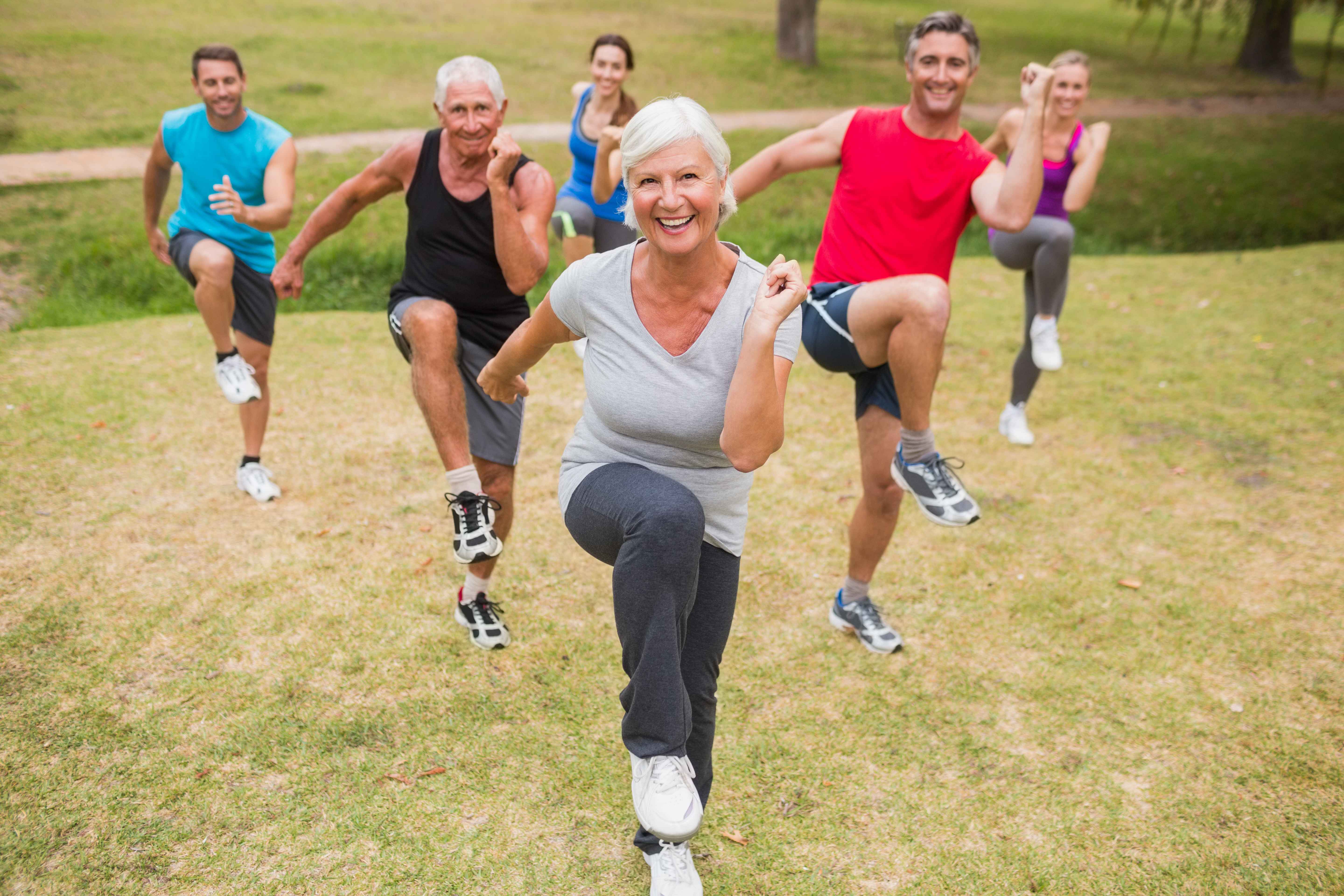
It’s no secret that exercise is the best way to get in shape and to prevent serious health conditions. But did you know that you can actually exercise your way to healthy eyes too!?
Like your heart, brain, and lungs, your eyes are impacted by how you care for your body. By eating a healthy diet and exercising regularly, you can help prevent eye conditions linked to obesity and being out of shape.
Studies have found connections between regular exercise and reducing risks for several common eye ailments such as cataracts, age-related macular degeneration and glaucoma. Vision problems and eye disease also stem from high blood pressure and high cholesterol. A healthy diet and regular exercise are two of the most important steps you can take to lower both.
What you should know about the common eye diseases and exercise
Cataracts – Studies have found that a lack of physical activity may be associated with an increased risk of getting cataracts. Whether it’s a brisk walk around your block or a run through the park, both activities may be associated with decreased risk of age-related cataract.
Age-related Macular Degeneration (AMD)– Studies found that that those who were active and exercised three or more times a week were less likely to develop AMD.
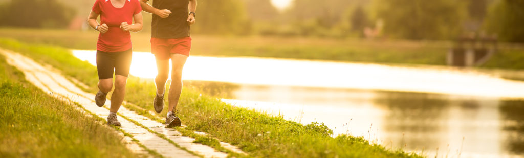
Glaucoma – Doctors treat glaucoma by lowering high intraocular (eye) pressure. Research that focused on young adults found that moderate intensity, low-impact exercise led to significant reduction in eye pressure. Regular, ongoing exercise, such as walking or jogging, will help reduce eye pressure.
How to kick off your exercise routine
- Start simple and set realistic goals. Keep a log of your goals and daily exercise routine, this will help you stick to your plan and track your progress.
- Keep it fun.
- Mix up your fitness routine with combinations of strength and cardio.
- Not feeling all that motivated? Try jump-starting your fitness routine with a little Zumba or ballroom dancing; these are super fun ways to get in shape.
- Head outside and roller skate, bike, or go on a hike—all are great calorie burners too.
- If it’s too cold to go outdoors, pick up the latest yoga fitness DVD and create a calming atmosphere in your living room. Or you could try a circuit training fitness DVD for a more upbeat cardio workout.
- Don’t get stuck in a rut: change up your routine frequently to get the biggest bang out of your exercise time!
No time for exercise? You can squeeze it in between everyday tasks such as:
- Taking the stairs at work instead of the elevator.
- Walking to your co-worker’s desk instead of sending an e-mail.
- Doing lunges or squats while brushing your teeth.
- Contracting and holding your abs while working on your computer – try for 10 contractions every hour.
- Taking the dog for a long walk or jog—try doing a few lunges on the trail.
Stay active, get plenty of exercise and keep a healthy diet. When you’re tempted to slack off, or quit exercising all together, just remember how terrific you’ll look and feel when you keep moving. You’ll probably agree it’s worth working out a few minutes each day to reap the rewards of good health and great vision!
Click here for Eye Healthy Recipes.
 The Difference Between an
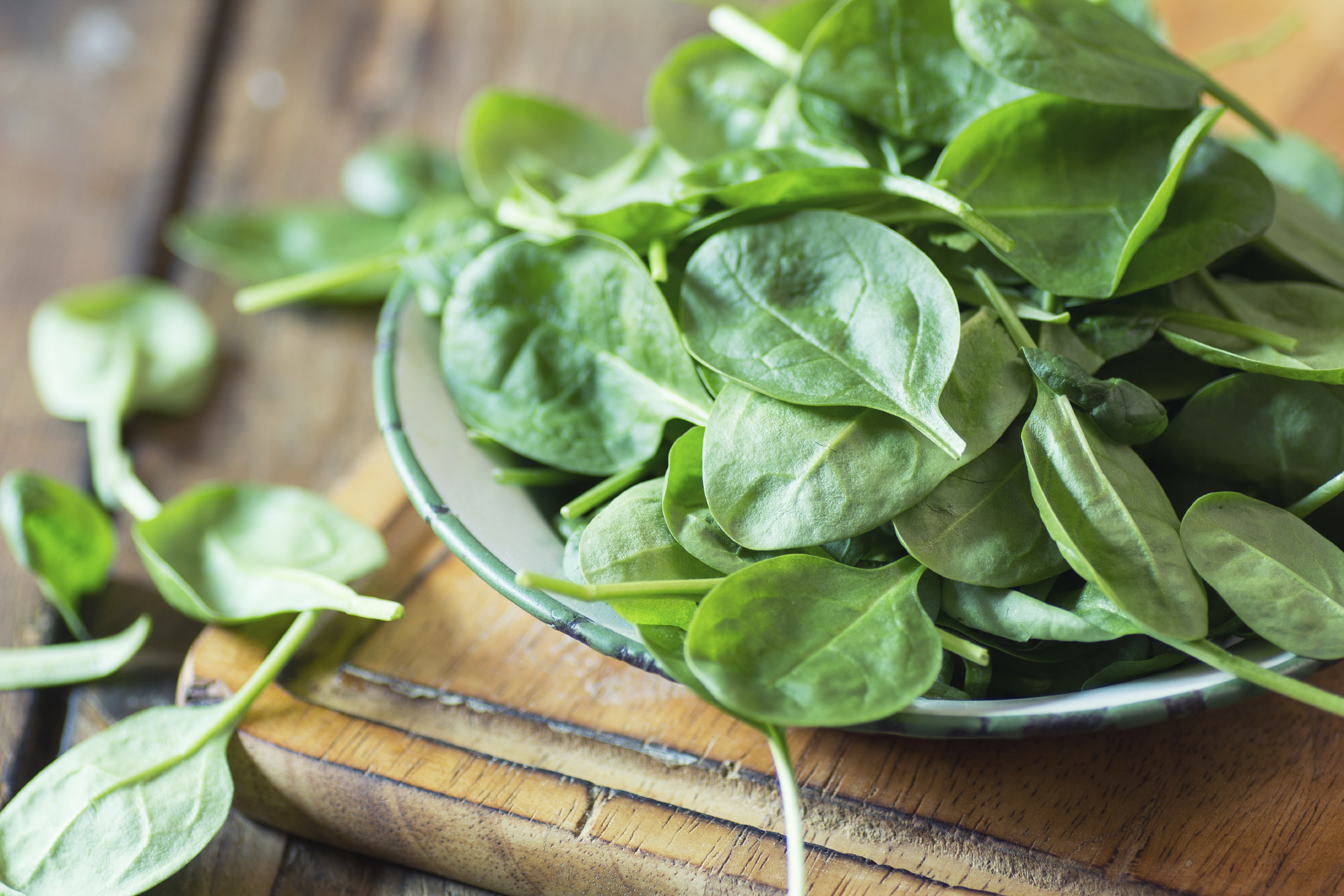
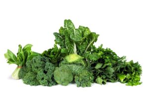 Be sure to start your holiday meal with a salad, it’s an excellent way to ensure that you and your guests get plenty of zeaxanthin and lutein, two nutrients that help protect your central vision. Adding kale, spinach, or romaine lettuce to salads helps your eyes absorb damaging blue light, combats the effects of cigarette smoke and pollution, and also decreases your risk of developing
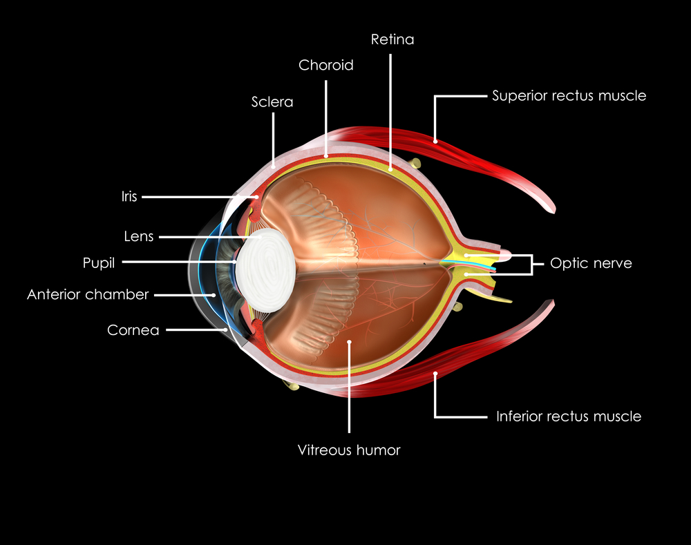
  Turkey and lean beef, two of the main ingredients in many holiday meals, keep your eyes strong and healthy. Both foods are high in zinc, a nutrient important to the retina and the choroid layer under the retina.
 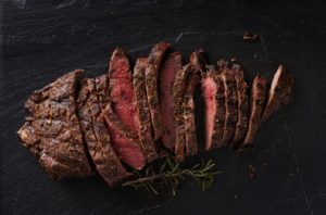 Zinc is essential for good night vision. Eating foods that are high in the nutrient can also reduce your risk of cataracts and AMD. Other foods that contain zinc include pork, dairy products, chick peas, black-eyed peas, crab, oysters, beans, spinach, mushrooms, cashews, and almonds.
 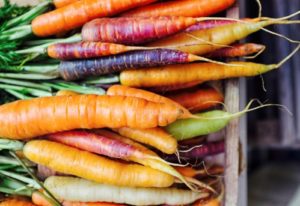 It wasn’t an old wives tale, it is true Carrots are good for your eyes! They contain beta carotene, a substance that turns into vitamin A when eaten. Eating carrots can benefit your night vision and could possibly reduce your risk of cataracts, AMD, and dry eyes. Other foods that contain beta carotene include pumpkin, sweet potatoes, and butternut squash. All great ingredients to include into your holiday feast.
  Fish contain omega-3 fatty acids, which can reduce your risk of developing AMD, dry eye, and glaucoma. Salmon, mackerel, flounder, tuna, halibut, herring, and sardines would be a great addition to your holiday meals.
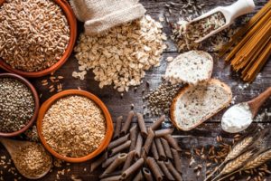 Whole grains reduce your risk of heart disease, obesity, and type 2 diabetes and can also decrease your risk of AMD. Substituting whole grain flour for white flour in holiday breads and muffins is a simple way to boost your whole grain intake. Other good whole grain sources include wild rice, brown rice, popcorn, oatmeal, bulgur, barley, buckwheat, and couscous.
 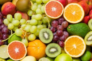 Fruits high in vitamin C, such as strawberries and oranges, also offer important vision benefits. Vitamin C is an antioxidant, a substance that can prevent cell damage caused by free radicals. Vitamin C-rich foods help keep the collagen in your cornea healthy and reduce the risk of cataracts and AMD. You can also find vitamin C in grapefruit, kiwi, blueberries, peas, broccoli, and tomatoes.
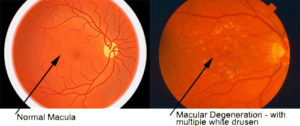
 Many of us just use the basics on our smart phone and never personalize them for our own needs. It is worth taking the time to adjust our phones to take advantage of the special services that may be available and unused. Making a phone call or sending a text message with a smart phone can be challenging, however, with simple modifications, keeping in touch with the world can become a snap. Getting comfortable with your smart phone will make staying in touch with your loved ones very easy.
 In a presentation to the American Society of Retina Specialists, Dr. Ajay E. Kuriyan of the University of Rochester reported that three patients who underwent bilateral intravitreal injection of stem-cells for age-related macular degeneration (AMD) suffered bilateral vision loss. The clinic at which all three procedures were performed did not have a licensed ophthalmologist on-site, and the stem-cell injections were administered by a nurse practitioner, Ocular Surgery News reported. Each patient paid $5,000 for the procedure.

 The National Eye Institute has recommended that people who are high-risk for developing AMD eat diets rich in green leafy vegetables, whole fruits, any type of nuts and omega 3 fatty acids. Many of these foods have anti-oxidant properties that help to “turn off” genes involved with inflammation, an important factor of retinal diseases. Salmon, mackerel and sardines have the highest levels of omega-3 fatty acids. An analysis that combined the data from 9 different studies showed that fish intake at least twice a week was associated with reduced risk of early and late AMD. Other studies show that Omega-3 fatty acids improve mitochondrial function, decreases production of reactive oxygen species (free radicals that damage cells) and leads to less fat accumulation in the body. The green leafy vegetables contain important protective macular pigments (carotenoids) called lutein and zeaxanthin that reduce the risk of AMD by 43%. High levels of lipid or fat deposits in the body (obesity) can “soak-up” the lutein and zeaxanthin so that they are not available to protect the retina.