
As I write this blog, I’m thinking about tomorrow – one of the landmark days of my life, my 50th Wedding Anniversary. Fifty years married to Patty, my wife, my best friend, my lover, the mother of our children, and my eyes on the world. It’s Patty’s eyes that have navigated our live’s journey. From describing a beautiful California sunset to deciding what I should wear to that very important meeting, Patty’s eyes have effortlessly and lovingly turned the darkness of my blindness into a life full of light, love, and joy.
Tomorrow evening we will dress for each other and as always I will feel a twinge of sadness because although in every way I appreciate Patty’s beauty and believe I have a special ability to see her inside-out, I’m sure that she wishes that I could take in the complete picture of who she is without limitation. It’s said that the eyes are the windows to the soul. If that’s really true, it’s Patty’s eyes that bond us together heart to heart and soul to soul.
 So make sure that you take a look – a real look at the people you love and realize the importance of what it means to see. At Discovery Eye, we are committed with your help to finding the answers that will allow people to enjoy the vision that makes every day and every anniversary worth living.
 Tom Sullivan
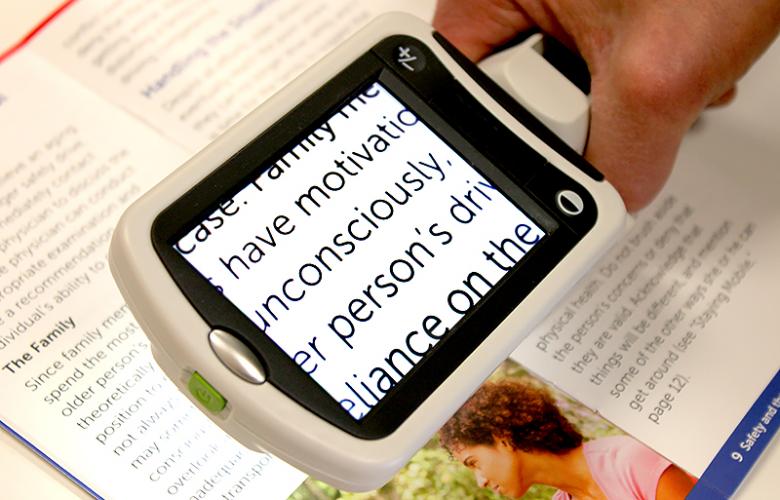 Portable magnifiers and lighted magnifiers- offer magnified reading on the go. Perfect for menus, shopping lists, label reading, and more, portable magnifiers can fit in your pocket, purse, or be worn on the belt for quick, easy use.
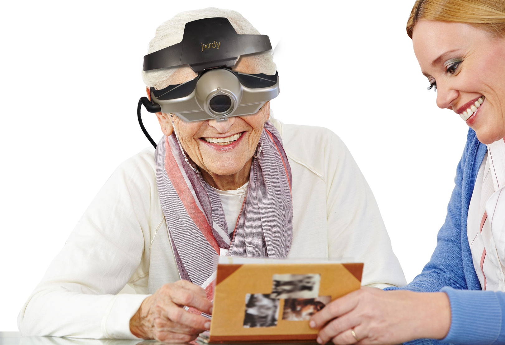 Wearable magnifiers – wearable technology is the future for those with low vision who live an active lifestyle. Wearable options make it possible to see and take part in everyday tasks, such as reading and recognizing faces.
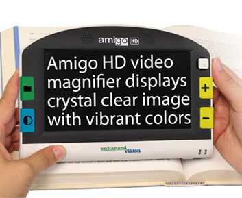 Transportable magnification screens– are perfect for close up viewing as well as distance viewing. These great viewers offer great flexibility, from watching TV to using the mirror image feature for self-viewing. There are APPS for smart phones that can be used to magnify reading material.
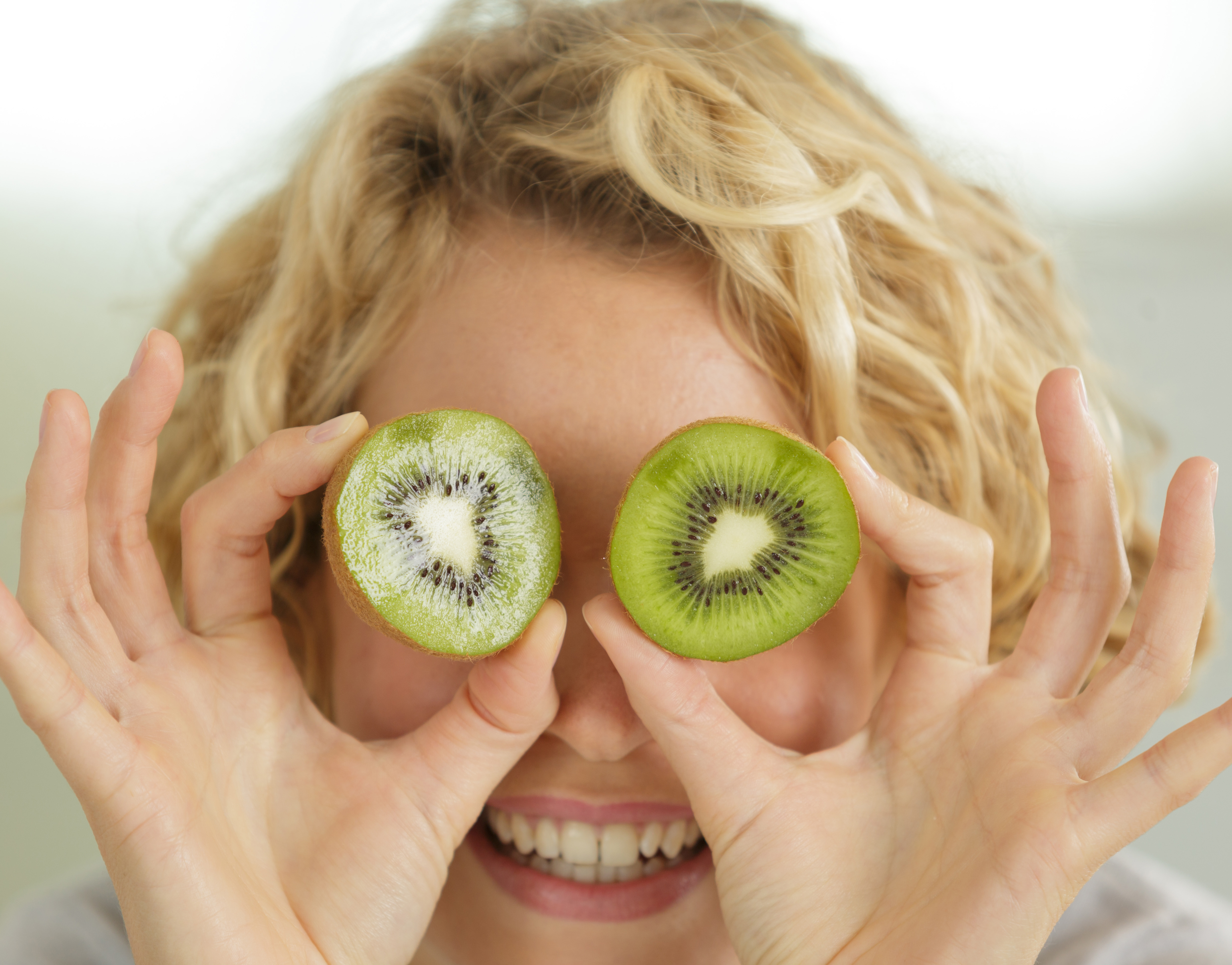 Good nutrition is important to keep your eyes healthy. Researchers have linked two very important eye nutrients that play a key role in healthy vision. Lutein (LOO-teen) and Zeaxanthin (zee-ah-ZAN-thin), both are potent antioxidants and are best known for protecting your eyes and may reduce your risk for
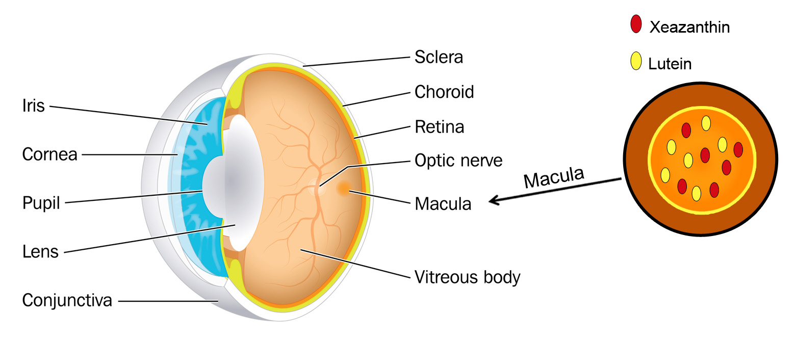
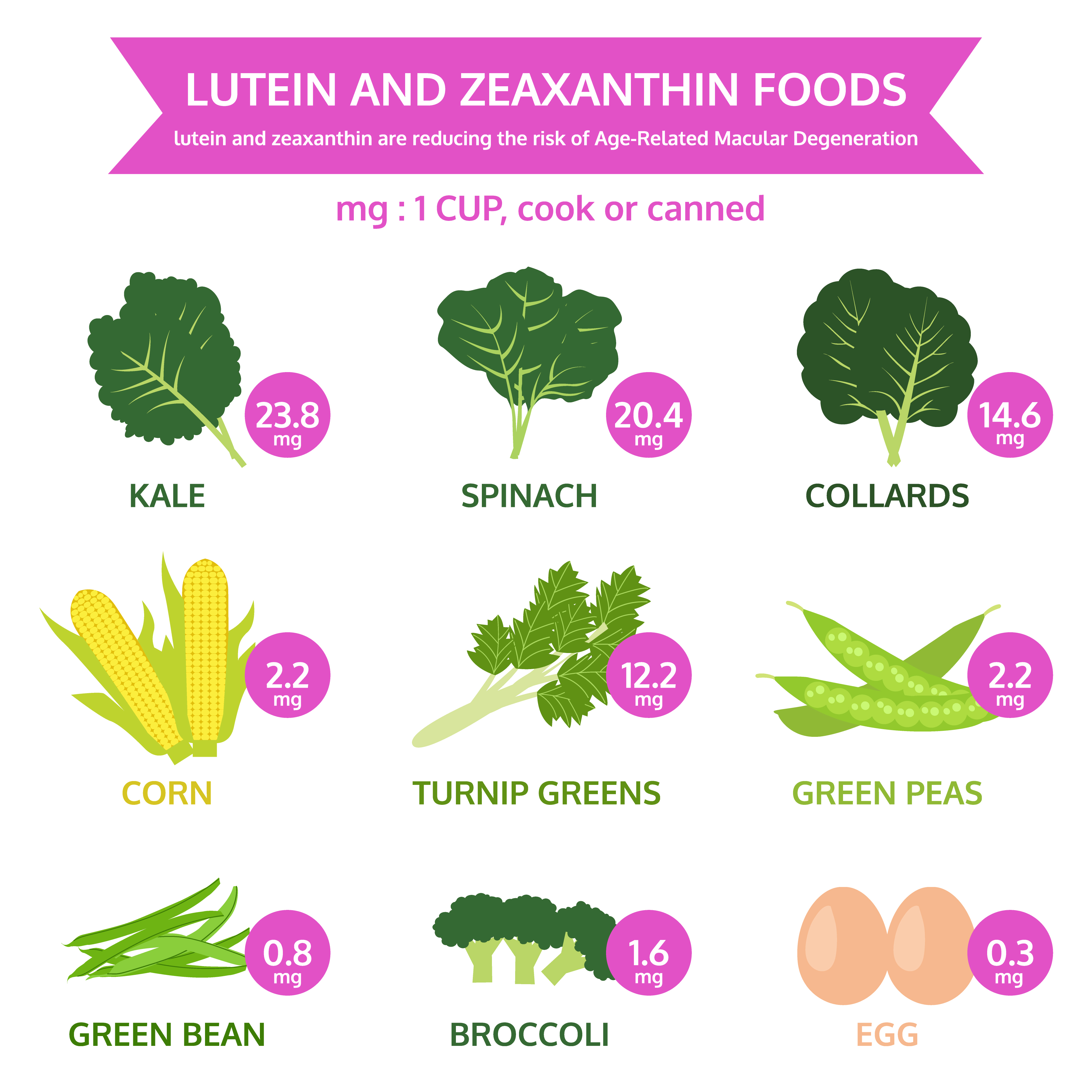 Diets rich in these two nutrients may help hold off age-related eye diseases. The best natural food sources of lutein and zeaxanthin are green leafy vegetables and other green or yellow vegetables. Among these, cooked kale and cooked spinach top the list.
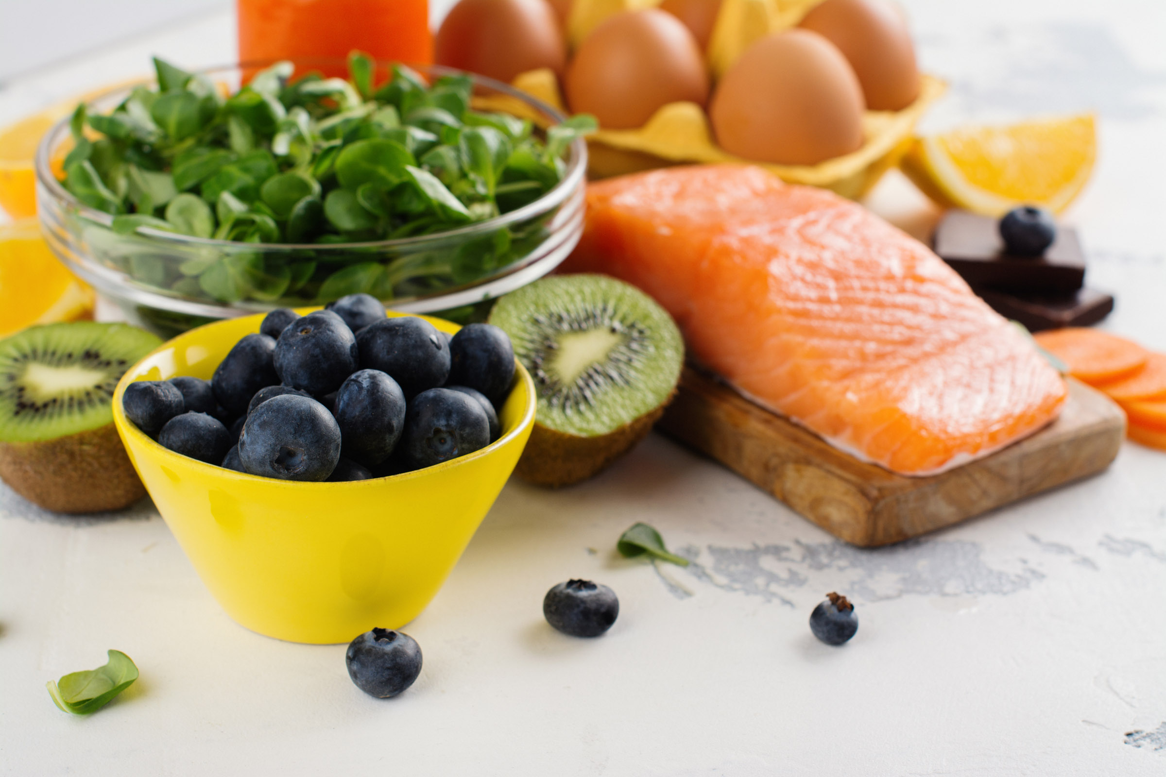

 Lauren Hauptman
 The other day my daughter Blythe asked me which Christmas I consider to be my favorite. I had to think a minute, because as a family, the Sullivan’s have had some great ones. I was about to say the first time you and your brother Tom were old enough to really get into Santa, being absolutely sure that the fat man brought your presents right down the chimney. I was about to say that, and then I remembered.
  Colorado, when our children were teenagers and our friend, the marvelous Betty White, joined us for a Christmas Eve sleigh ride none of us will ever forget. The night was perfect. It had snowed earlier that day, and the air had a feeling of Christmas that you could almost taste. Oh, sure, it was cold, but we were bundled up under tons of blankets as two beautiful Clydesdale horses with bells jingling took us through the woods to a magical barn where dinner would be served and carols sung.
  Harsh weather conditions can reduce the natural moisture in your eyes and the irritation usually results in a burning or itching sensation that often leads to rubbing or scratching your eyes which can worsen the symptoms. Sometimes it feels like there is a foreign object in your eye and for some, dry eyes can even cause excessive tearing, as your eyes try to overcompensate for their lack of protective tears. Prolonged, untreated dry eyes can lead to blurred vision as well. Between the harsh winter winds outside and the dry heat radiating inside, our eyes are very quickly irritated and dried in the winter months. The result is itchy, dry eyes that may cause pain, blurred vision, a burning sensation, or even watery vision as our eyes try to compensate for the dryness.
 I could hear Charlie rubbing his wife’s shoulders and telling her that everything would be alright. But, Rose kept saying “I know we’ll have to sell the house and move into something smaller, and I am going to be blind Charlie. Blind.”
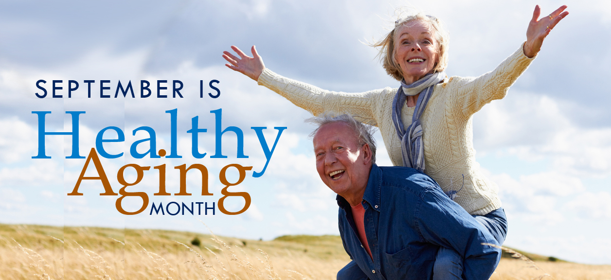 Today, people are living longer than ever before so it’s important to be proactive and take responsibility for your health as you age.
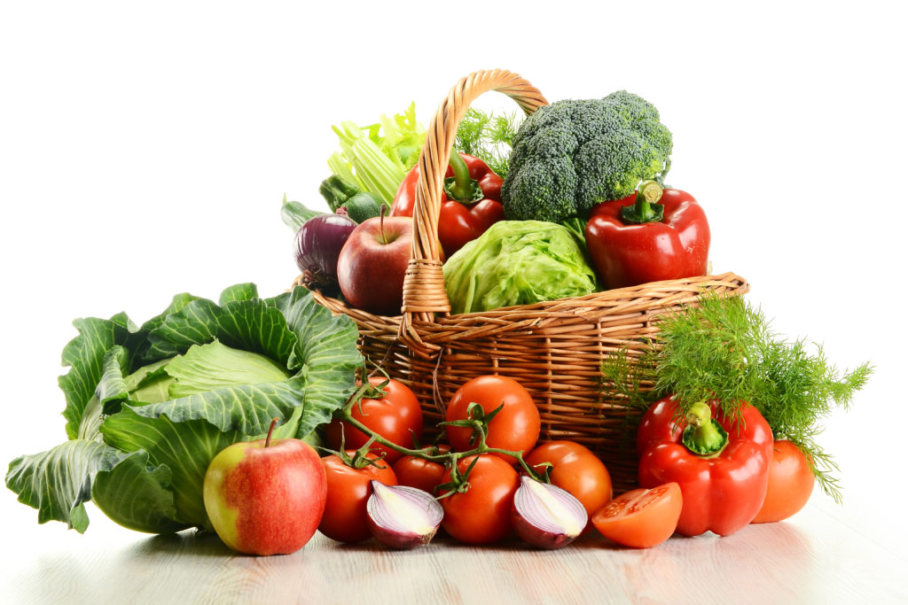
 Maintain a healthy weight. Being overweight increases your risk for diabetes. By exercising regularly, you can help keep your body healthy and prevent vision loss.
  Don’t smoke. Smoking increases your risk for age-related macular degeneration, cataract, and other eye diseases and conditions that can damage the optic nerve.
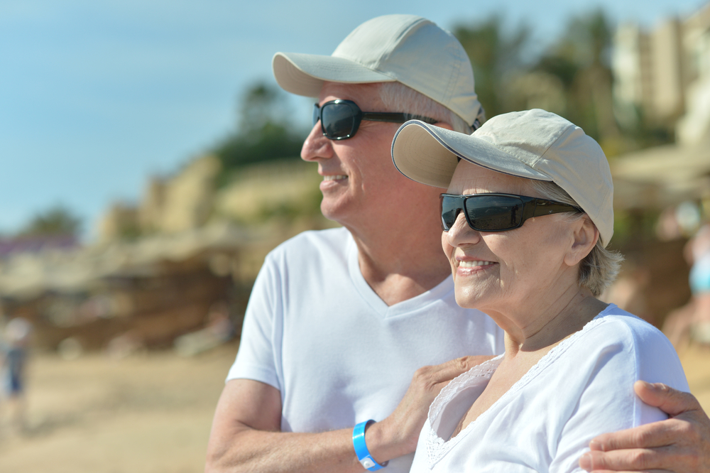 Wear protective eyewear when outdoors. Protecting your eyes from the sun’s ultraviolet rays when you are outdoors is vital for your eye health. Wearing sunglasses that block 99 to 100 percent of both UV-A and UV-B radiation.
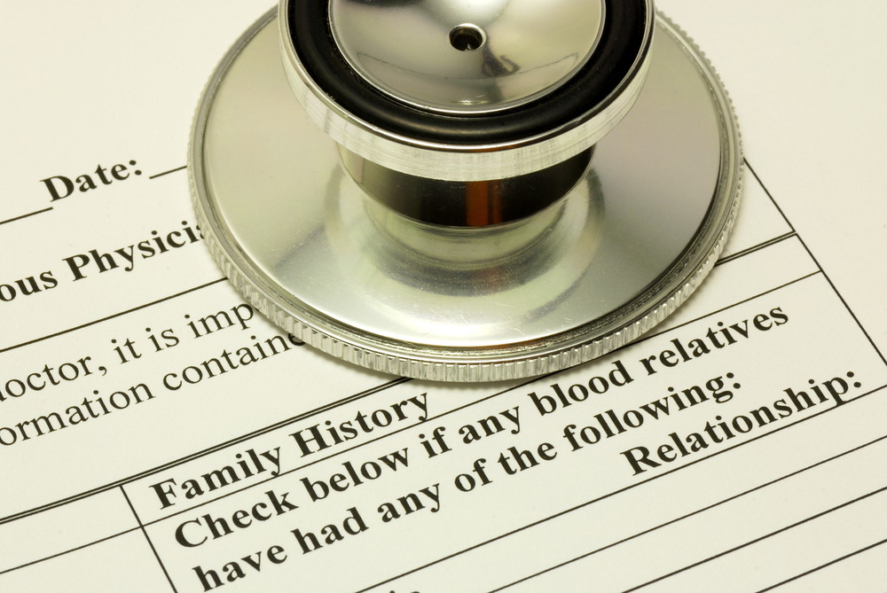 Know your family history. Talk to your family members about their eye health history. It’s important to know if anyone has been diagnosed with a disease or condition since many are hereditary, such as glaucoma, macular degeneration, and diabetes . This will help determine if you are at higher risk for developing an eye disease or condition.
 Consider a multivitamin. Vitamins C, E and the mineral zinc have been shown to promote eye health. Vitamins with Lutein and Zeaxanthin have been known to help patients with moderate to severe age-related macular degeneration.
 Give your eyes a rest. If you spend a lot of time at the computer or focusing at any one distance, you sometimes forget to blink, resulting in dryness and eye fatigue. Every 20 minutes, look away about 20 feet in front of you for 20 seconds. This can help reduce eyestrain. Consider using a lubricant eye drop during long periods of intense eye use and rest your eyes for 5 minutes.
 Tom Sullivan