Posterior Vitreous Detachment

10/30/14

Have you ever noticed floaters in your vision? Perhaps they looked like a bunch of small dots or maybe a cobweb swaying back and forth in your visual field. Were the floaters associated with flashing lights that made you think there was a lightning storm coming your way? These are typical symptoms of a posterior vitreous detachment (PVD), and if you have had these symptoms you are far from alone.
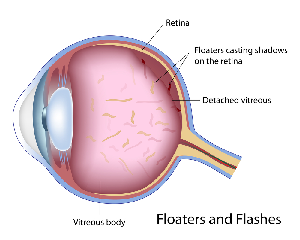
Floaters Posterior vitreous detachment
PVD is a natural process that occurs in the majority of people usually over the age of 50. The vitreous is a jelly-like substance that occupies the back portion of the eye. The vitreous is comprised primarily of water, which accounts for 99% of its volume, and the remaining 1% includes proteinaceous substances such as collagen fibers as well as hyaluronic and ascorbic acids. The collagen fibers act as a scaffold to allow the vitreous to maintain a formed shape as well as provide a means for the vitreous to attach to the retina, which is the light-sensitive tissue that lines the inner back wall of the eye and is critical for vision. As we age, changes in these fibers cause the vitreous to lose its shape and eventually pull away from the retina. When the vitreous separates from the retina, this is called a PVD.

As we age, the collagen components of the vitreous can clump together and are free to float in the eye. When the vitreous separates from the retina during the development of a PVD, the floaters may become more noticeable or numerous. It is common for patients to describe floaters of different shapes and sizes, and patients may notice just one or in some cases many. In many people, a PVD develops slowly and there may be no symptoms or just a few annoying floaters. In others, a PVD may occur abruptly and cause more dramatic symptoms that can be very anxiety provoking.

Since the normal process of PVD development involves the vitreous tugging on the retina until it can fully separate, this tugging can result in flashing lights that can commonly appear in the peripheral, or side, vision. These flashing lights are sometimes described as lightning streaks, and patients may notice them more readily in settings with low ambient light. The flashes of light typically resolve once the vitreous has fully separated from the retina and the tugging has ceased.

The good news is that PVD is usually harmless in the vast majority of cases, and the annoying floaters will become less bothersome over time. In approximately 5-10% of cases, the vitreous can tug too hard on the retina as it tries to separate and it may pull a hole or tear in the retina. Tears in the retina can predispose to retinal detachment, which is a serious condition that can lead to permanent vision loss.
It is important to recognize that the typical symptoms of a regular PVD are often similar to a PVD with an associated tear. For this reason, it is recommended that all patients with the new onset of floaters or flashes have a dilated eye exam. If a retinal tear or detachment is discovered, early treatment can help prevent loss of vision.

Treatment for PVD usually involves simple observation. With time, the flashes will go away, and the floaters will become less noticeable. More recently, few providers have claimed that floaters can be treated with a laser in order to make them less noticeable. I would caution that this is not mainstream therapy at the current time, and I do not advise my patients to pursue this option. Another treatment possibility is vitrectomy surgery, where the vitreous gel is removed as part of a surgical procedure. Due to safety advances in vitrectomy surgery, this is now a potential option for the rare patient who has floaters that are so numerous and bothersome that they are negatively impacting their activities of daily living. For the vast majority of patients this is not necessary.

When I see a patient with a PVD, I often recommend one follow-up visit in 4-6 weeks to make sure there are no retinal holes or tears that have developed in the interim. If the other eye has not had a PVD yet, I will counsel them that a PVD will most likely develop in that eye within the next few years, and when it does they need to be examined. I will also discuss the retinal detachment warning signs. Patients with retinal detachment will not only have symptoms similar to PVD, including flashes and floaters, but in addition they may also notice what looks like a black shade or curtain that starts in the peripheral vision and extends towards the central vision. My patients are taught that this symptom requires an immediate examination.

In conclusion, PVD is a natural process that the majority of people will experience in their lives. The symptoms can range from having no symptoms at all to many floaters with associated lightning flashes. In the majority of patients, there is no damage to the eye or threat to the vision. A dilated exam is recommended to look for possible holes or tears in the retina, and if these are uncovered, prompt treatment can prevent vision loss.

Dr. Esmaili posterior vitreous detachmentDaniel D. Esmaili, MD
Retina Vitreous Associates Medical Group

What Are A Macular Pucker and Macular Hole?

10/9/14

What is the macula?
The eye is very much like a camera, taking light from the outside world and converting it into picture information that our brains perceive as vision. The retina is the light sensitive layer in the back of the eye that is very much like the film in that camera. The central retina, also known as the macula, is essential for crisp, high definition vision. Conditions that damage or distort the macula can therefore result in blurred or distorted vision. Two common conditions that affect the macula are macular puckers and macular holes.

What is a macular pucker or macular hole?
A macular pucker is a thin layer of scar tissue that forms on top of the retina. The amount of scar tissue can range from mild to severe. Mild macular puckers may be barely noticeable during an eye exam and resemble a fine layer of cellophane resting on the macula. More severe macular puckers can cause wrinkling or distortion of the macula. In contrast to a macular pucker, a macular hole is a small gap that extends through the entire thickness of the macula.

What are the symptoms of a macular pucker or a macular hole?
At first, a macular pucker may lead to mild blurring of the central vision. Because the problem involves the back of the eye, glasses will not completely restore vision. More severe macular puckers may result in wavy or distorted vision. For instance, objects that normally appear straight, such as venetian blinds or a printed line of text, might appear to have a dip or bend in the center. Small macular holes can cause similar symptoms of blurring or distortion. Larger macular holes often result in a central blind spot. This can also result in straight lines appearing broken or having a piece missing in the middle. Patients with a macular pucker or hole do not normally experience difficulty with peripheral vision.

What can cause a macular pucker or macular hole?
Recall that a macular pucker is a scar tissue. Anything that causes scar tissue, such as trauma or inflammation in the eye, can result in scar tissue and hence a macular pucker. Certain diseases that affect the retinal blood vessels such as diabetes can also cause a macular pucker to form. However, one of the most common causes of macular pucker is simple aging of structures within the eye. As the eye ages, the clear jelly that fills it, called the vitreous gel, shrinks. When enough shrinkage occurs, the vitreous gel detaches from its normal position adjacent to the retina. This process of vitreous detachment can cause microscopic damage or inflammation leading to macular pucker formation. In some cases, the vitreous gel does not detach cleanly from the retina. Instead it can put traction on the macula, pulling its delicate structures apart in the center, resulting in a macular hole.

How are macular puckers and macular holes diagnosed?
A simple examination from an ophthalmologist or retina specialist is often enough to diagnose a macular pucker or hole. However, additional testing is often useful in diagnosing subtle cases or monitoring eyes for changes. An optical coherence tomography (OCT) scan is a specialized photograph that allows your physician to look for microscopic changes in the contour of the macula. The following figures show an OCT of a normal macula, a macular hole, and a macular pucker. Note that the normal macula has a central dip known as the fovea, shown in Figure 1. In Figure 2, the dip is replaced by a gap which is a macular hole. Finally, Figure 3 shows a macular pucker where the dip is no longer visible. This is because the macular pucker, seen as a thin white line is distorting the normal shape of the macula.

Normal - Macular Pucker and Macular Hole
Figure 1: Normal Macula
Hole - Macular Pucker and Macular Hole
Figure 2: Macular Hole
Pucker - Macular Pucker and Macular Hole
Figure 3: Macular Pucker

What treatments are available for macular puckers and macular holes?
Macular puckers can be quite mild. For mild cases in patients with minimal symptoms, periodic monitoring may be all that is required. When blurred vision due to a macular pucker begins to affect activities such as driving or reading, treatment in the form of surgery can be considered. Surgery for a macular pucker is known as a vitrectomy. Vitrectomy surgery is usually done under local anesthesia and as an outpatient procedure. During the surgery, fine instruments are used to remove the scar tissue from the surface of the macula. After surgery, patients usually experience an improvement in the blurring and distortion as the eye recovers gradually over a period of months. Some residual waviness can be normal. Vitrectomy is generally very safe although there is a chance of increased cataract growth and a small risk of infection or retinal detachment.

For patients with small macular holes, close monitoring can also be an option since some macular holes can close on their own. For larger holes, there are two options. In select cases where the vitreous gel is actively pulling on the macula, an injection of medication into the eye may cause the gel to release cleanly, allowing the hole to close. In other cases, vitrectomy is recommended. During the surgery, any pulling on the macula is relieved and a gas bubble is placed in the eye to help the hole close. After surgery, patients are asked to look down for a several days to allow the bubble to float up against the hole. Once the body absorbs the bubble, vision is usually significantly improved.
In summary, both macular puckers and holes are common causes of blurry or distorted central vision. If treatment or surgery by a retina specialist is needed, the results are generally quite good and lead to significant restoration of vision.

Liao - Macular Pucker and Macular HoleDavid Liao, MD, PhD
Retina-Vitreous Medical Group