DEF-Funded Retinitis Pigmentosa(RP) and Age-Related Macular Degeneration(AMD) Projects Converge with Promise

Two hallmark DEF-funded projects are converging, providing great hope for those facing vision loss from retinitis pigmentosa (RP) or age-related macular degeneration (AMD).

The first project, headed by UC Irvine researchers Drs. Henry Klassen and Jing Yang, concentrates on putting human retinal progenitor cells into the eyes of those with RP in order to rescue damaged retinal cells. That project is currently in Phase II clinical trials, progressing toward FDA approval.

                                                      Tissue-culture model

According to DEF Research Director Dr. Cristina Kenney, if the project is approved by the FDA for use with RP, the next question is: What other diseases might these retinal progenitor cells be used for? That’s where a second DEF-funded project comes in.

Kenney is working on a “personalized” cybrid cell model to screen agents that specifically target the mitochondria in AMD cells. To date, the researchers have different cybrid cell lines representing 60 different individuals with eye diseases. They are looking for novel mechanisms to protect AMD cells from dying.

Yang and Kenney are now working together to determine whether the retinal progenitor cells can be the agent that rescues AMD cybrids. “When we take the mitochondria from AMD patients and put them into healthy retinal cells, which makes cybrids, we have shown that these AMD cybrid cells will start to die. So we used that model to ask the question: How do we rescue them?” Kenney says.

Kenney and Yang developed a tissue culture model, where the retinal progenitor cells are grown in one part of a chamber, and the AMD cybrids are grown in another part of chamber, surrounded by culture medium. There is a porous separator between the two chambers through which the cells can communicate.

 “We are finding that the retinal progenitor cells produce a factor that protects the AMD cybrids,” Kenney says. “This provides promising evidence that these proprietary retinal progenitor cells that are being tested for treating RP also may be helpful in AMD patients.”

“DEF has been supporting both these retina-related projects for quite some time, and it’s very exciting to see them coming together to potentially treat both RP and AMD.”

To help support this sight-saving research, please donate to the Discovery Eye Foundation by clicking button below.

May-Healthy Vision Month

When it comes to our health, we may visit our doctors and nurses regularly to make sure our bodies are healthy. But what about our eyes? They’re not always top of mind, but they’re just as important! May is Healthy Vision Month, an observance coordinated by the National Eye Institute to motivate Americans to make their eye health a priority and educate them about steps they can take to protect their vision.

Here are a few ways you can help protect your vision

  • Get an annual comprehensive dilated eye exam.
  • Know your family’s eye health history. It’s important to know if anyone has been diagnosed with an eye disease or condition, since some are hereditary.
  • Eat right to protect your sight: In particular, eat plenty of dark leafy greens such as spinach, kale, or collard greens, and fish that is high in omega-3 fatty acids such as salmon, albacore tuna, trout, and halibut. Visit Eye Cook for eye healthy recipes.
  • Maintain a healthy weight.
  • Wear protective eyewear when playing sports or doing activities around the home, such as painting, yard work, and home repairs.
  • Quit smoking or never start.
  • Wear sunglasses that block 99 -100 percent of ultraviolet A (UVA) and ultraviolet B (UVB) radiation.
  • Wash your hands before taking out your contacts and cleanse your contact lenses properly to avoid infection.
  • Practice workplace eye safety.

Taking care of your eyes also may benefit your overall health. People with vision problems are more likely than those with good vision to have diabetes, poor hearing, heart problems, high blood pressure, lower back pain and strokes, as well as have increased risk for falls, injury and depression.

In addition to your comprehensive dilated eye exams, visit an eye care professional if you have

  • Decreased vision.
  • Eye pain
  • Drainage or redness of the eye
  • Double vision
  • Diabetes
  • Floaters (tiny specks that appear to float before your eyes)
  • Circles (halos) around light sources or if you see flashes of light

For this Healthy Vision Month, take care of your eyes to make them last a lifetime.

How to Prevent Eye Strain While Working From Home

Working from home has become the new normal for many Americans during this pandemic.  Most are happy without the commute, although working outside the office and in all sorts of strange places, such as bedrooms, backyards, living rooms, has become common place. We’ve had to learn how to work remotely, which in turn means spending a lot more time using computers and smartphones.

Moving out of properly-lit classrooms and offices – and away from ergonomically correct desks – can have long-term effects on our eyes.  Excessive time at a computer screen under the best conditions can lead to symptoms of eye strain.

Eye Strain Symptoms include:

  • Headaches
  • Blurred vision
  • Uncomfortable dry eyes
  • Neck and shoulder pain

To help avoid eye strain, here are few tips for working from home:

  • Record time spent on screens – Most adults age 18 and older spend at least 13 hours each day using digital devices. Extended screen time can cause discomfort and vision difficulties. When working from home, you may lose track of how much time you’re on your computer or smartphone.  So keep track of the hours you use devices. That way, you’ll be aware of the demand you’re placing on your eyes.
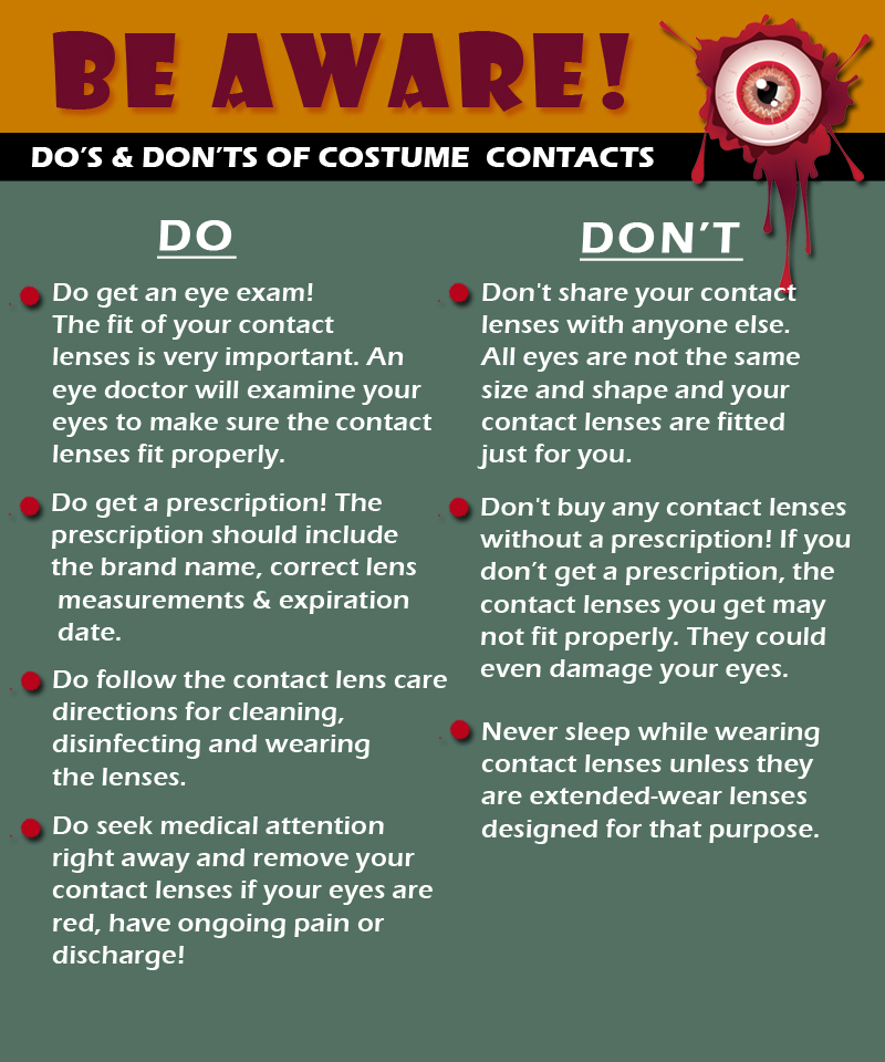
  • Rest and blink your eyes – Researchers found that over 30% of people using digital devices rarely take time to rest their eyes. Just over 10% say they never take a break, even when working from home. The eye muscles get overworked and don’t get a chance to relax and recover. Experts suggest the 20-20-20 rule; every 20 minutes, focus your eyes and attention on something 20 feet away for 20 seconds.  You can also get up and walk around for a few minutes.


  • Reduce exposure to blue light – In the spectrum of light, blue is more high energy and close to ultraviolet light. So, if you use screens throughout the day, ask your eye doctor about the value of computer glasses that block blue light. Reducing exposure to blue light may help lessen vision problems.  At home, using digital devices until bedtime can overstimulate your brain and make it more difficult to fall asleep. Eye doctors recommend no screen time at least one to two hours before going to sleep.


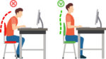
  • Sit up straight – Proper posture is important. Your back should be straight and your feet on the floor while you work. Elevate your wrists slightly instead of resting them on the keyboard.


  • Proper lighting – A setting that’s too bright (sunny backyard) or too dim (cavernous basement) can cause eye strain and headaches. Your screen should be bright enough that you don’t need to squint. A screen shield can help reduce glare.


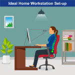
  • Set up monitor properly – Make sure your computer screen is about 25 inches, or an arm’s length, away from your face. The center of the screen should be about 10-15 degrees below eye level.  Cut glare by using a matte screen filter. You can find them for all types of computers, phones, and tablets.  Increase font size or set the magnification of the documents you are reading to a comfortable size.


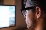
  • Consider computer glasses –For the greatest comfort at your computer, you might benefit from having your eye doctor modify your eyeglasses prescription to create customized computer glasses. This is especially true if you normally wear distance contact lenses, which may also become dry and uncomfortable during extended screen time.  Computer glasses also are a good choice if you wear bifocals or progressive lenses, because these lenses generally are not optimal for the distance to your computer screen.
  • Create Technology-Free Zones – These tips can help reduce eye strain when you’re forced to look at screens all day, creating technology-free zones in certain areas of your home, like the bedroom or bathroom. If you spend the entire day working on the computer, getting in bed and scrolling through social media until you fall asleep won’t do your eyes any favors.

Once you’re done for the day, truly unplug. Read a book or spend some quality time with family members without your phone.


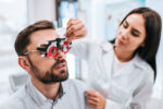
  • Get an Eye Exam – If you have tried all these tips and eye strain is still an issue, it might be time to see an eye care professional to schedule an eye exam.  The exam may even detect underlying issues before they becomes worse.


Low Vision Awareness Month

Low vision affects millions of Americans — including many older adults. People with low vision aren’t blind, but because of their vision loss, they may not be able to do everyday tasks like driving or reading even with glasses.

What is Low Vision?

When your eyesight is impaired to the degree that you struggle with daily tasks like reading or cooking – or that you have difficulty recognizing faces, you may have a medical condition referred to as low vision. Someone with low vision can’t simply put on a pair of glasses or contacts and see well; this condition is beyond the typical loss of vision that occurs with aging.

Individuals experiencing low vision can struggle with maintaining independence; hobbies, reading and even socializing may become challenging as vision loss progresses. Because of the impact low vision can have on your life, it is important to have your eyes checked by a low vision specialist if you have any concerns.

Since it is unusual to be able to restore vision once it is lost, screenings can help preserve the vision you have and help you access adaptive lenses and devices if you need them. While there is not a cure for low vision, your specialist can help you adapt and if needed, create a vision rehabilitation program designed to meet your specific needs.

Low vision is often caused by one of these conditions:

  • Age-related macular degeneration (AMD) –AMD is a progressive eye condition affecting as many as 15 million Americans. The disease attacks the macula of the eye, where our sharpest central vision occurs, affecting reading, driving, identifying faces, watching television, safely navigating stairs and performing other daily tasks. Although it rarely results in complete blindness, it robs the individual of all but the outermost, peripheral vision, leaving only dim images or black holes at the center of vision. Read More (link to DEF website)
  • Diabetes Diabetic retinopathy is the most common diabetic eye disease and a leading cause of blindness in American adults caused by changes in the blood vessels of the retina.

In some people with diabetic retinopathy, blood vessels may swell and leak fluid or blood inside the eye. In other people, abnormal new blood vessels grow on the surface of the retina. The retina is the light-sensitive tissue at the back of the eye. A healthy retina is necessary for good vision. Read More (link to DEF website)

  • Glaucoma glaucoma is a disease that causes damage to the major nerve of the eye called the optic nerve, a part of the central nervous system that carries visual information from the eye to the brain.

The eye experiences a gradual increase of intraocular pressure (IOP) due to an imbalance of the fluid produced in the eye and the amount of fluid drained. Over time, elevated IOP can cause vision loss. The most common form of glaucoma is primary open angle glaucoma which affects about 3 million Americans. However, there are other types including narrow angle, congenital, normal tension, and secondary glaucoma. (link to DEF website)

  • Injury to the eye can also be a cause

Regular eye exams are essential for all adults; it is recommended that adults over the age of 60 have eye exams each year. For older adults, diabetics and anyone with the risk factors above, a visit with a low vision specialist may be warranted as well. This specialist differs from a conventional optometrist and is trained to detect low vision and offer solutions to help you retain or regain independence and the ability to do things you enjoy again.

While Low Vision Awareness Month is the ideal time to learn about this condition and about the risk factors you could be facing, you should get in touch with a low vision specialist any time you have concern about your eyesight. Any time you notice a change in vision, if you receive a new diagnosis or have one or more of the risk factors detailed above you should schedule an appointment right away.

Low Vision Resources from the National Eye Institute

It can be hard to describe low vision. Visit our website Vision Simulation to see what life is like with common causes of low vision, like age-related macular degeneration, diabetic retinopathy, and glaucoma.

Beauty is in the Eyes of the Beholder

I love the idea that beauty is in the eyes of the beholder. It reminds us that individuals can make choices about what they perceive to be true beauty.

Let’s consider art… one person’s beautiful can be another person’s junk. If beauty is in the eyes of the beholder, the question is – are we taking the time to appreciate all that surrounds us? From Mother Nature’s unlimited panoplies of possibilities to the innocent smile of a child, beauty is the catalyst that makes being alive the greatest of all human adventures.

I am sure you’ve heard the phrase “stop and smell the roses,” so why not take the time to stop and observe the beauty your eyes can behold.

The Discovery Eye Foundation is working every day to preserve your vision and give all of us the chance to see what’s truly beautiful and treasure it.  All you have to do is open your eyes and take a look.

Louis Armstrong said it well:

I see trees of green and red roses too.
I see them bloom for me and you.
And I think to myself,
what a wonderful world.

You’re right Louis… it is a wonderful world if we all just keep appreciating the beauty that’s out there for all of us.

 
Donate today to help support the Discovery Eye Foundation! 

DonateNow

Tom Sullivan
DEF’s Ambassador of Vision

sullivanvision.com


June is Cataract Awareness Month

June is Cataract Awareness Month. This is a time to raise cataract awareness and help to educate people about one of the leading causes of treatable vision loss in the United States. There are 24 million Americans over the age of 40 who are affected by cataracts, so it seems fitting that an entire month should be dedicated to cataract education and awareness.

A cataract is a clouding of the eye’s lens, which blocks or changes the passage of light into the eye. The lens of the eye is located behind the pupil and the colored iris, and is normally transparent. Vision may become blurry or dim because the cataract stops light from properly passing through to the retina. Generally, a cataract does not cause pain, redness or tearing, but does cause increasing difficulty in seeing clearly.


Here is an overview of Cataracts:

View Video


Cataract symptoms:

  • Cloudy
  • Night vision
  • Glare
  • Halo
  • New glasses
  • Yellow tint
  • Double vision

Some Risk factors for cataracts include:

  • Older age
  • Intense heat or long-term exposure to UV rays from the sun
  • Certain diseases, such as diabetes
  • Inflammation in the eye
  • Hereditary influences
  • Long-term steroid use
  • Eye injuries
  • Eye diseases
  • Smoking

Cataracts can be easily diagnosed and visiting your eye doctor regularly is important in helping protect your eyes from further damage. An annual eye exam is recommended for everyone over the age of 60, and bi-annual exams for adults between 41-60 years to check for developing eye or vision problems.


There are a few treatment options, below is one of the newest options:
View Video


There is no proven way to prevent age-related cataracts. However, choosing a healthy lifestyle can slow the progression of cataracts. Some ways to delay the progression of cataracts include avoiding smoking, reducing exposure to UV rays, eating healthy foods, and wearing proper eye protection to avoid eye injury.

For more information and where you can find support for Cataracts click here .

OUR EYES WORK LIKE CAMERA’S!

The inner workings of the human eye are complex, but at the same time, fascinating. Have you wondered how exactly they do work or what are the major parts of the eyeball involved in creating vision? Let’s find out.

These tiny cameras spend every day processing millions of pieces of information at lightning fast speeds, and turn them into the simple images we see almost instantly.

In reality, this process is anything but simple. The eye has several distinct parts, each of which has specific responsibilities that work together like a machine.

The eyeball is just like a camera. In fact, human eyes are part of a classification known as “camera-type eyes.” And just like a camera, it can’t function without the presence of light.  As light hits the eyes, it’s focused by the eye in a way similar to a camera lens. This process allows the images we see to appear clear and sharp rather than blurry.

There are specific parts of the eye that make this focusing process possible. Each beam of light that hits the eye goes through a series of steps:

Step 1: Light passes through a thin layer of moisture

Step 2: Light hits the cornea.  The cornea is transparent, and is the first layer to begin focusing light within the eye. The cornea is connected to the sclera, which is a tough fiber on the outside of the eye that acts as protection.

Step 3: Behind the cornea is another liquid layer known as the aqueous humor, and its job is to maintain pressure levels in the front of the eye as light is passing through.

Step 4: Once light has passed through the aqueous humor, it has finally reached the pupil. The pupil is the round entryway of the colored iris.

Step 5: Once the pupil determines how much light it will let inside your eye, the job passes to the lens. The lens factors in the amount of light the pupil lets in, and figures out how far away you are from the object that the light is reflecting off of, or the object you’re trying to see. From there, the lens focuses your image into an accurate view of what you’re looking at. Part of this process is controlled by muscles in the lens called ciliary muscles, which expand and contract to pull on the lens and allow it to focus properly.

Step 6: As light reaches the center of the eye passes through another layer of moisture, called the vitreous, or vitreous humor. Then, it reaches the final stop in the process: The Retina.

The retina is the back of the eye. If the lens in your eye is most like a camera, the retina is most like its film – this is where the final product is projected. The retina has several parts:

  • Macula: The center of the retina. The center point of the macula is called the fovea, and it has the most photoreceptors and nerve endings of any part of the eye.
  • Photoreceptors: Split into two designations – rods and cones.
    • Cones are in the macula. When there is bright light, cones provide clear, sharp central vision and detect colors and fine details.
    • Rods are located outside the macula and extend all the way to the outer edge of the retina. They provide peripheral or side vision. Rods also allow the eyes to detect motion and help us see in dim light and at night.

  • Retinal pigment epithelium: Abbreviated RPE, this is a tissue layer below the rods and cones which absorbs any extra, unneeded light.
  • Choroid: The choroid is behind the retina, and is in charge of making sure the retina and RPE have enough nutrition flowing from small blood vessels.

Once the photoreceptors have converted light into an electronic signal, they send a signal to the brain’s visual command center and you have vision. It’s amazing what even small parts of our bodies can do.


How the Eye Works

January is Glaucoma Awareness Month

National Glaucoma Awareness Month reminds all of us to get regular eye exams and show support for those suffering from this conditionGlaucoma is one of the leading causes of vision loss and blindness.  Glaucoma is often called “the sneak thief of sight” because glaucoma has few symptoms or warning signs in its early stages. It can be diagnosed only by a full eye exam by an eye care practitioner.

Glaucoma is a disease where pressure builds up and damages the eye’s optic nerve. Types of this disease include the common Primary Open Angle Glaucoma, which causes peripheral eyesight to slowly diminish and is age-related. Angle Closure Glaucoma, where the fluid drainage system is narrow and closed so that the aqueous fluid remains in the front chamber of the eye and intraocular pressure rises; and Low Tension Glaucoma, where the optic nerve becomes damaged in spite of the intraocular pressures being within the normal range. There’s currently no way to restore vision lost from glaucoma because once the nerve cells become damaged, they do not regenerate.


A few important facts you should know about Glaucoma in adults:

  • More patients than ever are affected – Over 3 million people in the U.S. have glaucoma, and the number is rising.
  • Glaucoma can affect people of all ages – The most common form of glaucoma, Primary Open Angle Glaucoma, becomes more prevalent with increasing age. However, glaucoma can strike anyone, even infants and children but it is rare.
  • Demographics do play a role – Glaucoma is a leading cause of blindness among African Americans; it’s also highly prevalent in Hispanics over the age 65.
  • Is glaucoma hereditary? The risk of developing Primary Open–Angle Glaucoma is up to nine times more likely if parents or siblings have the disease.
  • Hope for future glaucoma patients – Although there is no cure for any form of glaucoma, early diagnosis and treatment help control the disease and slow the process of vision loss or blindness.

Newer Glaucoma Treatments 

Glaucoma treatment usually begins with the use of topical (eye drop) medications which lower the intraocular pressure. Within the past two years, two new topical medications have been approved for the treatment of glaucoma: VYZULTA® and Rhopressa®. VYZULTA® is a modification of a class of medications currently used to treat glaucoma – the prostaglandin analogs. This drug helps lower intraocular pressure by increasing the drainage of fluid (aqueous humor) from the eye.

Rhopressa® is part of a new class of drugs used to treat glaucoma called Rho kinase inhibitors. Rhopressa®, like VYZULTA®, also lowers intraocular pressure by increasing aqueous outflow. Both Rhopressa® and VYZULTA® are dosed once daily and pose few, if any, systemic safety concerns.
 

Laser

Using a laser to make a small opening in the iris to help with fluid drainage usually cures Angle-Closure Glaucoma.  This procedure is called a laser peripheral iridotomy.

For Primary Open Angel Glaucoma, when eye drops are not enough to reduce the pressure then a procedure called Selective Laser Trabeculoplasty (SLT) can be used. The SLT reduces intraocular pressure by stimulating increased outflow of fluid from the eye. SLT offers an improved safety profile compared to older glaucoma laser therapies and may lower eye pressure by as much as 20 to 30 percent. It is typically used as the next step in patients whose glaucoma is uncontrolled on medical therapy. Because of its excellent benefit-to-risk profile, however, SLT can sometimes be used in place of medications, especially in patients who have difficulty with their eye drops.
 

Surgery

When other treatments fail, there are many surgical therapies to lower the eye pressure. These surgical approaches, which are riskier than medical therapy or lasers, are usually employed when non-surgical means do not work well enough to stop vision loss.
 

MIGS

The goal of Minimally Invasive Glaucoma Surgery (MIGS) is to reduce intraocular pressure by enhancing the eye’s own internal aqueous humor drainage system. Some MIGS can be performed as stand-alone procedures, while others are typically done along with cataract surgery in patients with visually significant cataracts and mild to moderate glaucoma.

There are now a variety of recently FDA approved MIGS available for use in this country. MIGS typically are performed through a small incision in the eye with minimal tissue trauma and offer a favorable safety profile as well as more rapid visual recovery than traditional glaucoma surgery. There are many well qualified glaucoma specialists that perform the MIGS procedure.


Glaucoma Treatment Overview


National Glaucoma Awareness Month reminds all of us to get regular eye exams. Don’t let glaucoma steal your sight!  The best way to protect your sight is to get an annual comprehensive eye examination.


Thanks to funding from private philanthropists, DEF’s research continues to make great strides toward cures and treatment for glaucoma.  If you would like to support DEF’s sight saving research please donate today!

 DonateNow

A Vision of Christmas

A special holiday message from our beloved blind Ambassador of VisionTom Sullivan

My wife Patty loves every aspect of the Christmas season, and she begins celebrating the yule tide even before Thanksgiving.  The transforming of our house with traditional decorations brings her a marvelous sense of family and holiday season.  She loves to look at every ornament remembering how we acquired it.  As the lights turn on outside our house, she is busy making sure that every bulb on our tree is placed just so to give the room a warmth that in every way reflects her commitment to the true spirit of the season.

While I am listening to football upstairs, she’s singing along with Celine Dion as she completes the visual picture that for Patty confirms that there really are important traditions that make our home a place of love, beauty, and peace.  For the record, let me say that I really do enjoy Christmas even though I offer half-hearted complaints that Patty plays carols starting even before Turkey Day.  The truth is…I love them just as much as she does.

For over thirty years we went to Colorado every Christmas to ski, and those experiences were wonderful to just be in the mountains and feel the cold nipping at your nose as you fly down a ski slope.  We often would take a sleigh ride on Christmas Eve with hot chocolate spiced with peppermint schnapps.  Listening to a children’s choir and being with people we love always seems to make things a little more right with the world.  So I really do love Christmas, but when I consider Patty’s ebullient joy in the visuals of the holiday, I admit it makes me just a little sad to know that I’ll never be able to see them.

I realize that people go to extraordinary lengths in their decorating zeal and that certainly some of their elaborate choices that brighten the neighborhood could be considered over the top, but when Patty’s carols are playing and she’s enjoying the happiness of singing along with the decorations just so, I know that what she’s reflecting is her true commitment to her faith and the celebration of the season that she treasures. 

So open your hearts and your eyes and take in the true experience of the Christmas spirit.  It’s my hope that you’ll consider supporting Discovery Eye Foundation’s commitment to preserving vision allowing more people every year to enjoy not just the smell of a Christmas tree, or the taste of a Christmas toast, the touch of a hug from a loved one on Christmas morning, or the beautiful sounds of Silent Night sung by the voice of a child.

From all of us at Discovery Eye, we wish you a Merry Christmas and Happy Holidays!


Tom Sullivan
DEF’s Ambassador of Vision

sullivanvision.com

Your EYES will be thankful for Thanksgiving Dinner!

Thanksgiving is almost here; a meal that nourishes the family bonds and traditions.  It’s the one time of the year where you can guarantee your eyes will be bigger than your stomach. This meal also has another added bonus — almost every item on the Thanksgiving table is healthy for your eyesight!

Here are several of the most popular Thanksgiving dishes and their corresponding benefits to your eye health:


Turkey –

No Thanksgiving is complete without a turkey, roasted golden brown and stuffed with fresh vegetables and herbs. Turkey is loaded with zinc and B-vitamin niacin, which helps prevent the formation of cataracts. Cataracts are the leading cause of vision loss in the United States, so gobble up!  You are “preventing” cataracts with every bite.


Spinach, Green Bean Casserole, Asparagus and Brussel Sprouts –

These foods and other leafy greens are loaded with lutein and zeaxanthin, two nutrients that protect the retina, which may also help reduce the risk of cataracts and macular degenerationHealthy Green Bean Casserole Recipe


Sweet Potatoes –

Sweet potatoes are full of Beta-carotene, which is a carotenoid and antioxidant that promotes night vision and overall good eyesight. Sweet potatoes are also loaded with vitamins C and E. Diets that are rich in these vitamins can help prevent or delay the development of cataracts and macular degeneration. Mashed Sweet Potatoes Recipe


Cranberry Sauce –

Cranberries contain bioflavonoids, a large class of antioxidants. Bioflavonoids are found in the pulp, skin and rinds of foods that contain vitamin C. Both flavonoids and vitamin C help protect the eyes from free radical damage caused by pollution and the body’s normal metabolic processes.


Pumpkin Pie –

Pumpkin is one of the best sources of vitamin A, so pumpkin pie is an eye-healthy dessert. In fact, one cup of cooked, mashed pumpkin contains more than 200 percent of the recommended daily intake of Vitamin A. Vitamin A provides nourishment and protection to the eye’s lens, cornea and macula (part of the retina), so it improves night vision. 


A diet that’s full of the right nutrients is a great start to keeping your eyes healthy, but don’t forget that regular eye exams are equally important! We hope you enjoy a happy and delicious Thanksgiving with family and friends.

*For more eye healthy recipes click here EYE COOK