 From wearing the right eye wear and getting regular exams to eating right and caring for your lenses, there are many things you can do to protect your vision.
Healthy vision starts with you! Follow these tips to take care of your eyes.
- Get regular comprehensive dilated eye exams – You may not have any symptoms or eye problems. But visiting your eye care professional for a comprehensive dilated eye exam is the only way to be sure. Some people also don’t realize they could see better with glasses or contact lenses.
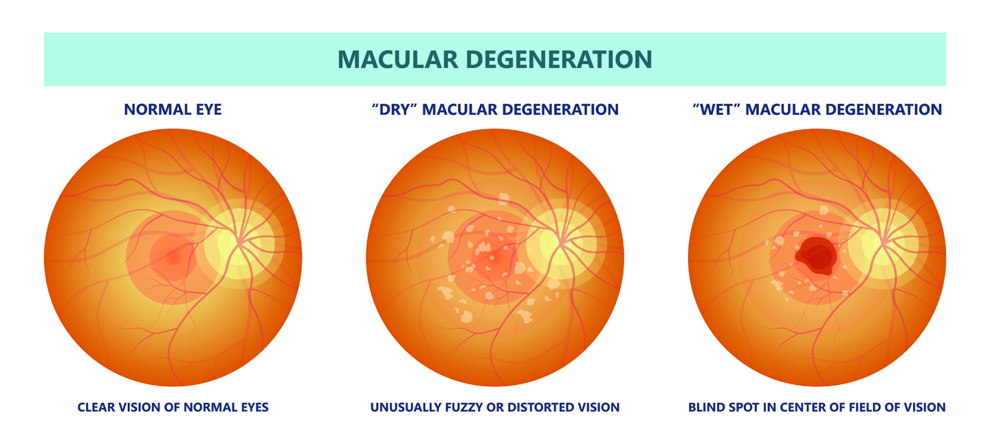
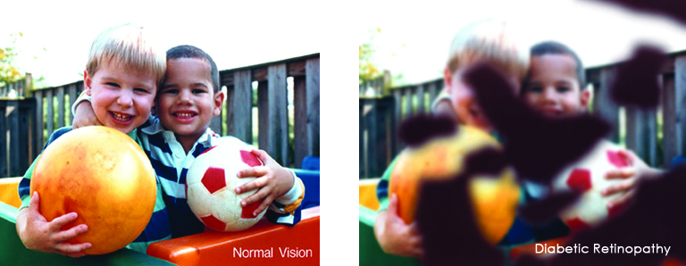
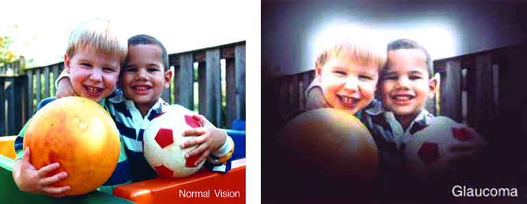
A dilated eye exam is the only way to detect some common eye diseases in their early stages. These includes conditions like glaucoma, diabetic retinopathy, and age-related macular degeneration (AMD). During a comprehensive dilated eye exam, your eye care professional places drops in your eyes to dilate them. This allows more light to enter the eye so your doctor can examine for any signs of damage or disease.
- Know your family’s eye health history – Talk to your family members about their eye health history. It’s important to know since some eye disease or condition are hereditary. This information will help to determine if you’re at higher risk for developing eye problems.
Studies show that you are at an increased risk for eye disease if you have an immediate family member with eye disease, including cataracts, glaucoma, diabetic retinopathy and age-related macular degeneration (AMD). Knowing your family history and informing your ophthalmologist or optometrist.
- Eat right to protect your sight – Research shows eye health benefits from fish high in omega-3 fatty acids, such as salmon, tuna, and halibut. Eating plenty of dark leafy greens such as spinach, kale, or collard greens as well as fruits and vegetables will help keep your eyes healthy. Visit our website for eye healthy recipes discoveryeye.org/eye-cook-delicious-food-health/
- Maintain a healthy weight – Being overweight or obese can increase the risk of developing type 2 diabetes and other chronic conditions that could damage your eyes. If you’re having trouble maintaining a healthy weight, talk to your doctor.
- Wear sunglasses – Sunglasses are a great fashion accessory, but most importantly, they protect your eyes from the sun’s ultraviolet (UV) rays. When purchasing sunglasses, look for ones that block out 99 to 100% of UV-A and UV-B radiation.
- Wear protective eyewear – Protective eyewear prevents eye injuries in the workplace (if needed), while playing sports or doing other activities that could cause damage. They include:
- Safety glasses and goggles for working and repairing.
- Safety shields for working at your hobby—gardening, home repairs.
- Eye guards designed for specific activities and sports.
- Most protective eyewear lenses are made of polycarbonate, which is 10 times stronger than other plastics. Many eye care providers sell protective eyewear, as do some sporting goods stores. Employers are required to provide a safe work environment, including protective eyewear if needed.
- Quit smoking or never start – Smoking is as bad for your eyes as it is for the rest of your body. Research has linked smoking to an increased risk of developing:
- Age-related macular degeneration
- Cataracts
- Optic nerve damage
- Heart disease, lung diseases and various cancers including lung cancer
- Clean your hands and contact lenses properly – To avoid the risk of infection, always wash your hands thoroughly before putting in or taking out your contact lenses. Disinfect contact lenses as instructed and replace them as directed by your eye care professional.
Eyes and Overall Health
Taking care of your eyes also may benefit your overall health. Some health conditions such as diabetes and high blood pressure can affect your eyes at the initial stage. The eye exam can tell you and your doctor if there are any underlying health conditions that need attention.
In addition to your comprehensive dilated eye exam, visit an eye care professional if you have:
- Decreased vision
- Eye pain
- Drainage or redness of the eye
- Double vision
- Floaters (tiny specks that appear to float before your eyes)
- Circles (halos) around light sources
- Flashes of light
 Low vision affects millions of Americans — including many older adults. People with low vision aren’t blind, but because of their vision loss, they may not be able to do everyday tasks like driving or reading even with glasses.
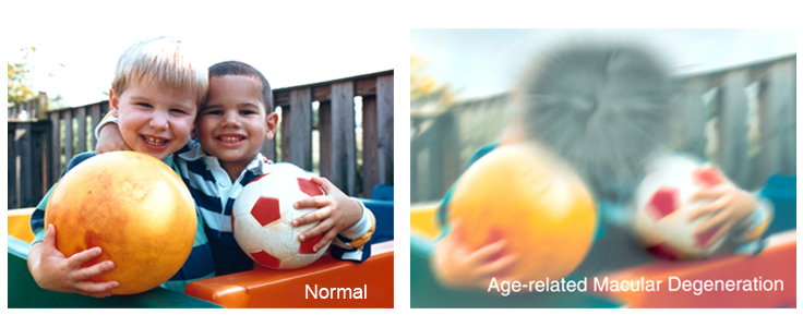

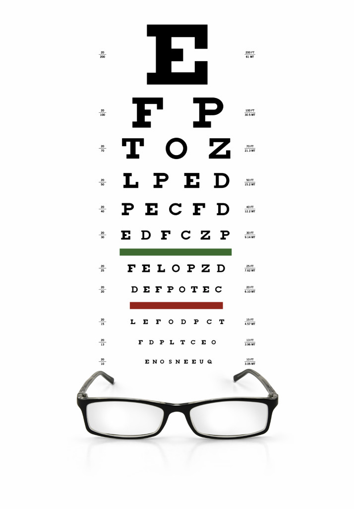 Understanding What 20/20 Vision Means