September is Healthy Aging Month

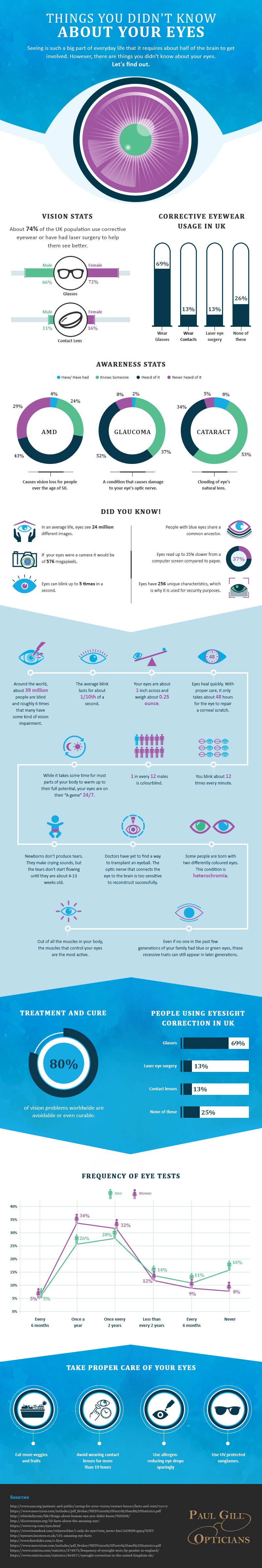
Healthy Aging Month is an annual health observance designed to focus national attention on the positive aspects of growing older.  Aging is a process that brings many changes. Vision loss and blindness, however, do not have to be one of them. There are several simple steps you can take to help keep your eyes healthy for the rest of your life.

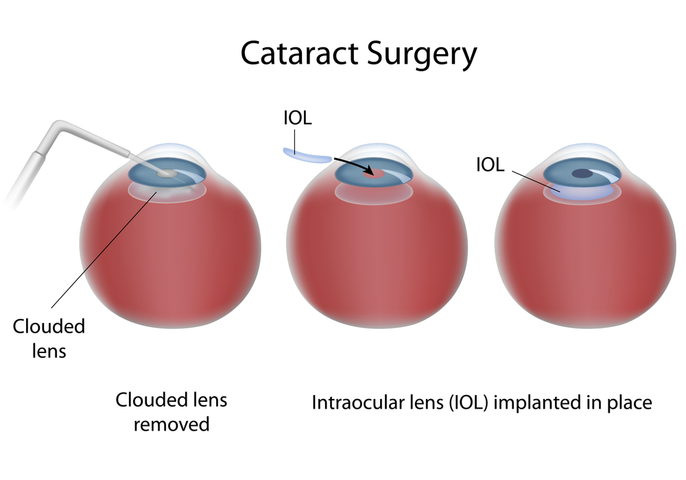
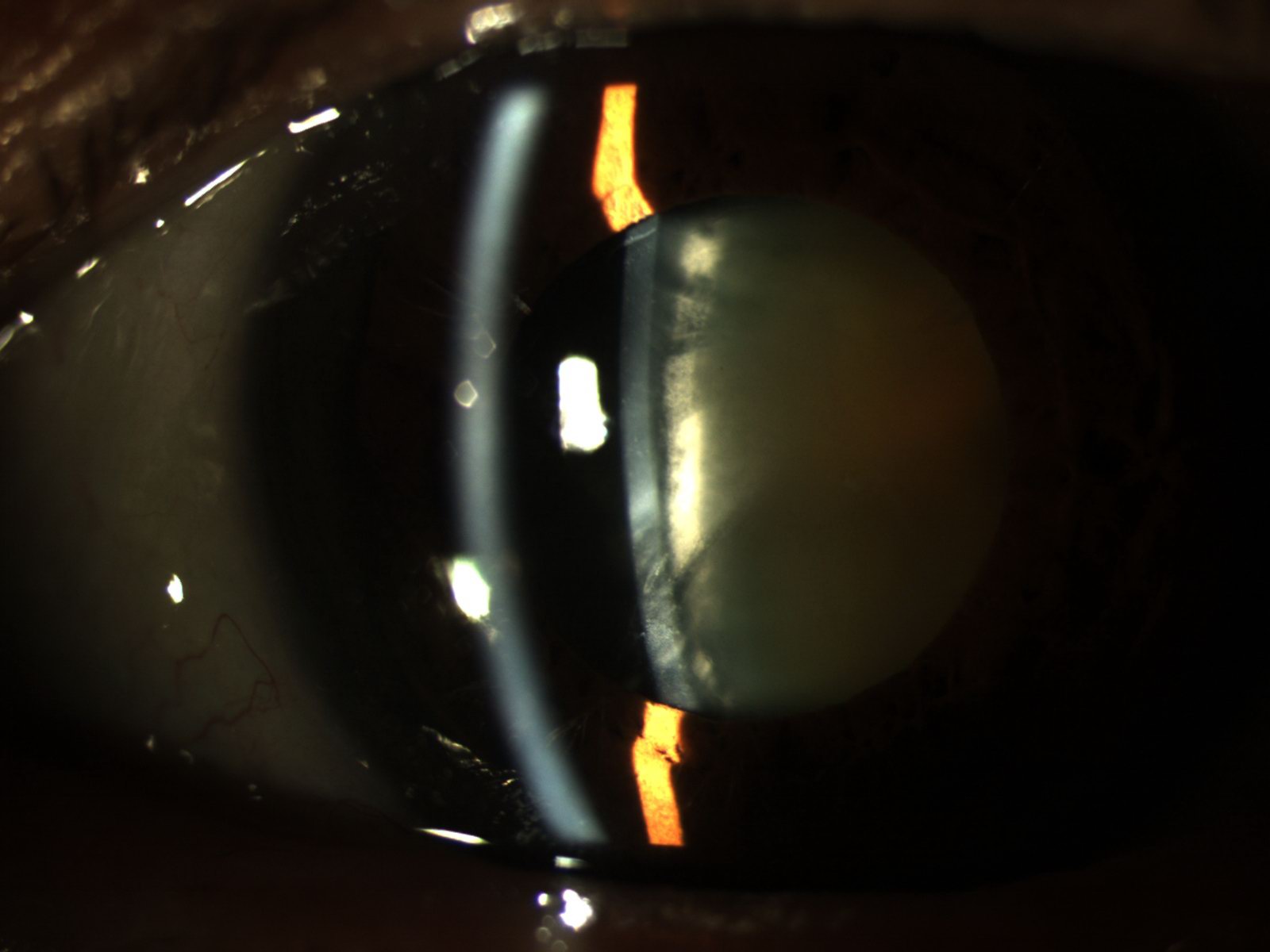
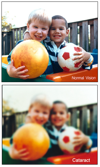
Eye diseases often have no early symptoms, but can be detected during a comprehensive dilated eye exam A comprehensive dilated eye exam is different from the basic eye exam or screening you have for glasses or contacts. By dilating the pupils and examining the back of the eyes, your eye care professional can detect eye diseases in their early stages, before vision loss occurs. By performing a comprehensive eye exam, your eye care professional can check for early signs of –

Here are some other tips to help maintain healthy vision and body now and as you age:

  • Eat a healthy, balanced diet. Loading up on fruits and vegetables can help keep your eyes healthy and disease free.
  • Maintain a healthy weight. Being overweight increases your risk for heart disease and diabetes. Complications from diabetes, such as diabetic retinopathy or glaucoma, can eventually lead to vision loss.
  • Don’t smoke. Smoking increases your risk for age-related macular degeneration, cataract, and other systemic diseases, including cancer. Wear protective eyewear when outdoors. Protecting your eyes from the sun’s ultraviolet rays when you are outdoors is important for your eye health. Choose sunglasses that block 99 to 100 percent of both UV-A and UV-B radiation.

Even if you are not experiencing vision problems, visiting an eye care professional regularly for a comprehensive dilated eye exam is the most important thing you can do to reduce your risk of vision loss as you age.


Download “Everyone’s vision can change with age”
A handout with explanation on how vision can change with age.

June is Cataract Awareness Month

I remember my father experiencing the removal of cataracts in the early 60’s, and being told by his Ophthalmologist that the recovery time would be weeks rather than days and a positive outcome was not guaranteed.  The truth is that surgery of any kind carries with it a certain amount of risk.  But now the removal of cataracts has become routine for thousands thanks to amazing technology and gifted eye surgeons.  To the doctor, the surgery might be considered routine.  But to the patient, when vision is at risk their concerns and real fears are not in any way routine.

A year ago at 96 years young, my friend and actress Betty White was worried about her vision.  She told me that she was having a great deal of difficulty reading scripts and that her eyes would tire with the strain of trying to see the pages.  Her ophthalmologist determined that the removal of cataracts was necessary and the surgery on the first eye was scheduled for the next Tuesday.  “Is it safe? she asked me.”  I assured my friend that the removal of cataracts was not something to be afraid of.  “It’s routine” I told her.  “Please don’t be frightened.” Betty’s surgery was completely successful.  In fact, she tells me she can read everything including the smallest print on any label.

At the Discovery Eye Foundation (DEF), we are committed to supporting research that we believe will make the treatment of many forms of vision loss far more predictable and successful. Whether it’s through the breakthroughs in surgery or the development of new drugs or the advancement in genetics or the use of stem-cells the major forms of vision loss are being greatly affected through the power of your contributions and the commitment of the gifted researchers DEF supports. 

Please know that the dollars you contribute to DEF will go directly into the hands of the researchers where they can do the most good.

All of us at the Discovery Eye Foundation thank you for your support and look forward to a time when most forms of blindness will no longer limit vision.


Tom Sullivan
DEF’s Ambassador of Vision

sullivanvision.com

Your Vision is Your Wealth

We see more than 24 million images in our average life span. The huge task in a human body is seeing, which requires half of one’s brain to function. Our eye lenses are equal to 576 megapixels Camera lens.

One man out of every twelve men is color blind and the chances of losing eye function increase with age. In the UK 74% of people correct their eyesight by Laser Surgery, Wearing Contact lens and glasses to have a better view. Our eyes take only 48 hrs to repair from a corneal scratch. In order to avoid these problems, restrict the continuous usage of contact lens less than 19 hours in a day.

For a better understanding, read this infographic from Paul Gill Optician.

Things-you-did-not-know-about-your-Eyes_22.11.2016

Source and Author:
Amy Lynn
Paul Gill Optician

Cataract Surgery and Keratoconus

1/8/15

The eye works like a camera, specifically a digital camera. There is the front lens of the camera (cornea), the aperture (iris), the film (retina), and a cable to take the image to the brain (optic nerve). This “camera” also has an additional lens – the natural crystalline lens, which lies behind iris. This natural lens is flexible when we are young, allowing us to focus at distance then instantaneously up close. Around age 40-45, this natural lens starts to stiffen, necessitating the need for reading glasses for most people. This stiffening is the beginning of the aging process that eventually leads to formation of a cataract. We refer to the lens as a cataract when it becomes sufficiently cloudy to affect ones quality of vision.cataract surgery and keratoconus-Cataract diagram In general, cataract surgery is one of the safest and most successful of all surgeries performed. The basics of cataract surgery in eyes with keratoconus is very similar to non-keratoconic eyes.

Keratoconus (KC) affects this “camera” by causing the front lens (cornea) to bulge. This causes the optics to be distorted. In many cases, this can be corrected for with hard contact lenses (CL) or spectacles; in other cases a corneal transplant may be necessary. When it comes time for cataract surgery in the setting of KC, there are several factors that need to be considered.

Corneal Stability
The first thing to be considered is the stability of your cornea. In general, KC progresses more in your late teens to early twenties, and then stabilizes with age. A very exciting treatment for KC is collagen crosslinking. This treatment is meant to stiffen the cornea to prevent instability that is inherent to KC. This treatment promises to stop the progression of KC at a young age. Fortunately, with age, the cornea naturally crosslinks and stiffens, therefore when it comes time for cataract surgery, there is little chance of the progression of KC. Your doctor needs to choose the appropriate intraocular lens (IOL) to refocus your eye after surgery. Two of the most important factors in IOL selection are the length of your eye and the shape of your cornea. Long term CL wear can mold your cornea. It is important to assure that you stay out of your CLs long enough for your cornea to reach its natural shape. Depending on how long you have worn your CLs, it may take several months for the cornea to stabilize. This time can be challenging as your vision will be suboptimal (because you can’t wear CLs), and will be changing (as your cornea reaches its natural shape). When your cornea does stabilize, it is important to determine whether the topography (shape) is regular or irregular. This “regularity” is also known as astigmatism. If the astigmatism is regular, light is focused as a line – generally, this distortion can be fixed with glasses. However, if the astigmatism is irregular, light cannot be focused with glasses, and hard CLs are needed to provide optimal focusing. If you have had a corneal transplant, I generally recommend all your sutures to be removed to allow your new cornea to reach its natural shape.

IOL Selection
The second thing to be considered is the type of IOL. IOLs allow your doctor to refocus the optics of your eye after surgery. In many cases, the correct choice of IOL may decrease your dependence on glasses or CLs. There are several factors that are important when considering the correct IOL for a keratoconic patient. The amount and regularity of your astigmatism plays a very significant role in IOL selection. In general, there are four types of IOLs available in the US – monofocal, toric, pseudo-accomodating, and multifocal. In general I do not recommend multifocal IOLs in patients with KC. These IOLs allow for spectacle independence by spitting the light energy for distance and near, however, with an aberrated cornea (which is what happens in KC), these IOLs do not fare well. If there is a low amount of regular astigmatism or irregular astigmatism, your best bet is a monofocal IOL. This is the “standard” IOL that is covered by your health insurance. If you have higher amounts of astigmatism that your doctor determines is mostly regular, you may benefit from a toric (astigmatism-correcting) IOL. These IOLs can significant improve your uncorrected vision and really decrease your dependence on glasses. It is important to realize that monofocal and toric IOLs only correct vision at one distance. With a monofocal IOL you still can wear a CL to fine-tune your vision, however, with a toric IOL, in general you will need glasses for any residual error. There is a pseudo-accomodating toric IOL available, and this may be a good option if you are trying to decrease your dependence on glasses and correct some of your astigmatism. These IOLs are relatively new to the US market.

If You Had A Corneal Transplant
In the setting of a corneal transplant many of the same factors need to be considered – stability of the graft, choice of IOL, etc. In addition, the health of the graft has to be judged. Prior to cataract surgery in my patients with corneal transplants, I make sure to remove all of their sutures and give the cornea time to stabilize (just as if they were a CTL wearer). If you are a CL wearer, the same rule of being out of the TL until the topography is stable applies. The health of a transplant needs to be established prior to undergoing cataract surgery. The cornea has five main layers to it –cataract surgery and keratoconus-corneal structure the back layer (inside) is called the endothelium. This layer is responsible for “pumping” fluid out of the cornea, allowing it to stay clear. In all eyes there is a loss of endothelium cells with cataract surgery. I generally perform a “specular microscopy,” which allows me to visualize and quantify the corneal endothelium prior to surgery. This allows me to risk stratify you before your surgery. It is important to realize that corneal transplants have a lifespan and may have to be repeated in the future.

Keep in mind, there is some uncertainty in biometry (the process of selecting an IOL) in all eyes – this error can be higher in keratoconic eyes. This highlights why assuring stability is important. Equally important is picking the correct IOL for your situation. Also, keep in mind that I have discussed generalities in this article. Your individual case could be different. This is a conversation best left between you and your surgeon. In general, cataract surgery and keratoconus or a corneal transplant can be a very safe and effective way in restoring vision.

Sam Garg, MDSumit (Sam) Garg, MD
Interim Chair of Clinical Ophthalmology and Medical Director
Gavin Herbert Eye Institute at the University of California, Irvine

What You Need to Know About Cataracts

6/5/14

Do you feel like your vision is getting worse? Do you feel like colors are not as vibrant as they used to be? Are you having more trouble with glare? If you have any of these symptoms, you may be experiencing the effects of cataracts.

Cataracts are a normal aging process of the crystalline lens in the eye. Well you may ask — what is the crystalline lens? It is easiest to think of the eye as a camera. The eye has a lens (actually two) – the cornea (the front window of the eye) and the crystalline lens (inside the eye). It also has an aperture (the colored iris), and film (the retina). All these structures work together to focus light and allow us to see – just like a camera. When we are young (less than 40), the crystalline lens is flexible. This is why we are able to see distance and then near without the need for reading glasses. The crystalline lens is able to change its shape depending on where one is looking.

Figure 1 – Slit-lamp photo of a visually significant cataract.

As we age, the crystalline lens becomes less flexible, thereby causing one’s near vision to be more blurry. This necessitates the need for reading glasses. As the crystalline lens become less flexible with age, the lens also starts to become more yellow and can also become cloudy. When the yellowing and/or clouding become visually significant, we refer to this as a cataract (figure 1).

Are cataracts dangerous? The simple answer is no. In the vast majority of cases, a cataract can be monitored until it becomes visually significant (drop in vision, glare, decreased contrast, vision related difficulties with day to day activities, etc). However, there are a few instances in which cataract removal is a medical necessity. Routine examinations by your eyecare provider can help you determine if you are at risk for these less common instances.

Figure 2 – Intraoperative photo during cataract surgery (prior to cataract removal).

What can I expect during cataract surgery? Do you have to replace the crystalline lens with anything? Cataract surgery involves removing the clouded crystalline lens (figure 2) and replacing it with an artificial lens known as an intraocular lens (IOL) (figure 3). Surgery generally takes 10-15 minutes under a mild sedative, and you don’t have to stop any of your current medications. Anesthesia is achieved with drops and you will only feel mild pressure during the surgery. IOLs come in different styles – Standard IOLs grossly correct your vision and you can fine tune your vision (distance and near) with glasses post-operatively;

Figure 3 – Intraoperative photo during cataract surgery (after implantation of an IOL).

Toric (astigmatism correcting) IOLs allow for increased spectacle independence, but glasses will still be needed for distance or near; Accommodating IOLs “flex” within the eye to decrease your dependence on distance and near glasses; Multifocal IOLs allow spectacle independence for distance and near. I always counsel patients that there is no perfect IOL and you have to determine which IOL is best for your particular situation. Your doctor can help you decide which IOL is best for you. Generally, cataract surgery is extremely safe. Your doctor will discuss particular risks specific to your eye.

How do I know if cataract surgery is right for me? The best way to know if you have a cataract and if it time to consider surgery is to consult with your local ophthalmologist. If you have experienced a recent drop in vision, that is not correctable with glasses, cataract surgery may be able to restore your vision!

Garg feb 2014 thumbSumit “Sam“ Garg, MD
Medical Director
Vice Chair of Clinical Ophthalmology
Assistant Professor of Ophthalmology
Gavin Herbert Eye Institute – UC, Irvine