
June is Cataract Awareness Month. This is a time to raise cataract awareness and help to educate people about one of the leading causes of treatable vision loss in the United States. There are 24 million Americans over the age of 40 who are affected by cataracts, so it seems fitting that an entire month should be dedicated to cataract education and awareness.
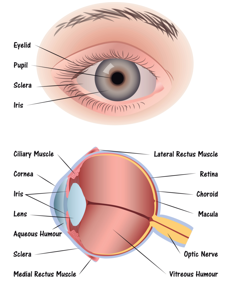
A cataract is a clouding of the eye’s lens, which blocks or changes the passage of light into the eye. The lens of the eye is located behind the pupil and the colored iris, and is normally transparent. Vision may become blurry or dim because the cataract stops light from properly passing through to the retina. Generally, a cataract does not cause pain, redness or tearing, but does cause increasing difficulty in seeing clearly.
Here is an overview of Cataracts:
Cataract symptoms:
- Cloudy
- Night vision
- Glare
- Halo
- New glasses
- Yellow tint
- Double vision
Some Risk factors for cataracts include:
- Older age
- Intense heat or long-term exposure to UV rays from the sun
- Certain diseases, such as diabetes
- Inflammation in the eye
- Hereditary influences
- Long-term steroid use
- Eye injuries
- Eye diseases
- Smoking
Cataracts can be easily diagnosed and visiting your eye doctor regularly is important in helping protect your eyes from further damage. An annual eye exam is recommended for everyone over the age of 60, and bi-annual exams for adults between 41-60 years to check for developing eye or vision problems.
There are a few treatment options, below is one of the newest options:
There is no proven way to prevent age-related cataracts. However, choosing a healthy lifestyle can slow the progression of cataracts. Some ways to delay the progression of cataracts include avoiding smoking, reducing exposure to UV rays, eating healthy foods, and wearing proper eye protection to avoid eye injury.
For more information and where you can find support for Cataracts click here .


 Step 1: Light passes through a thin layer of moisture
 National Glaucoma Awareness Month reminds all of us to get regular eye exams and show support for those suffering from this condition.
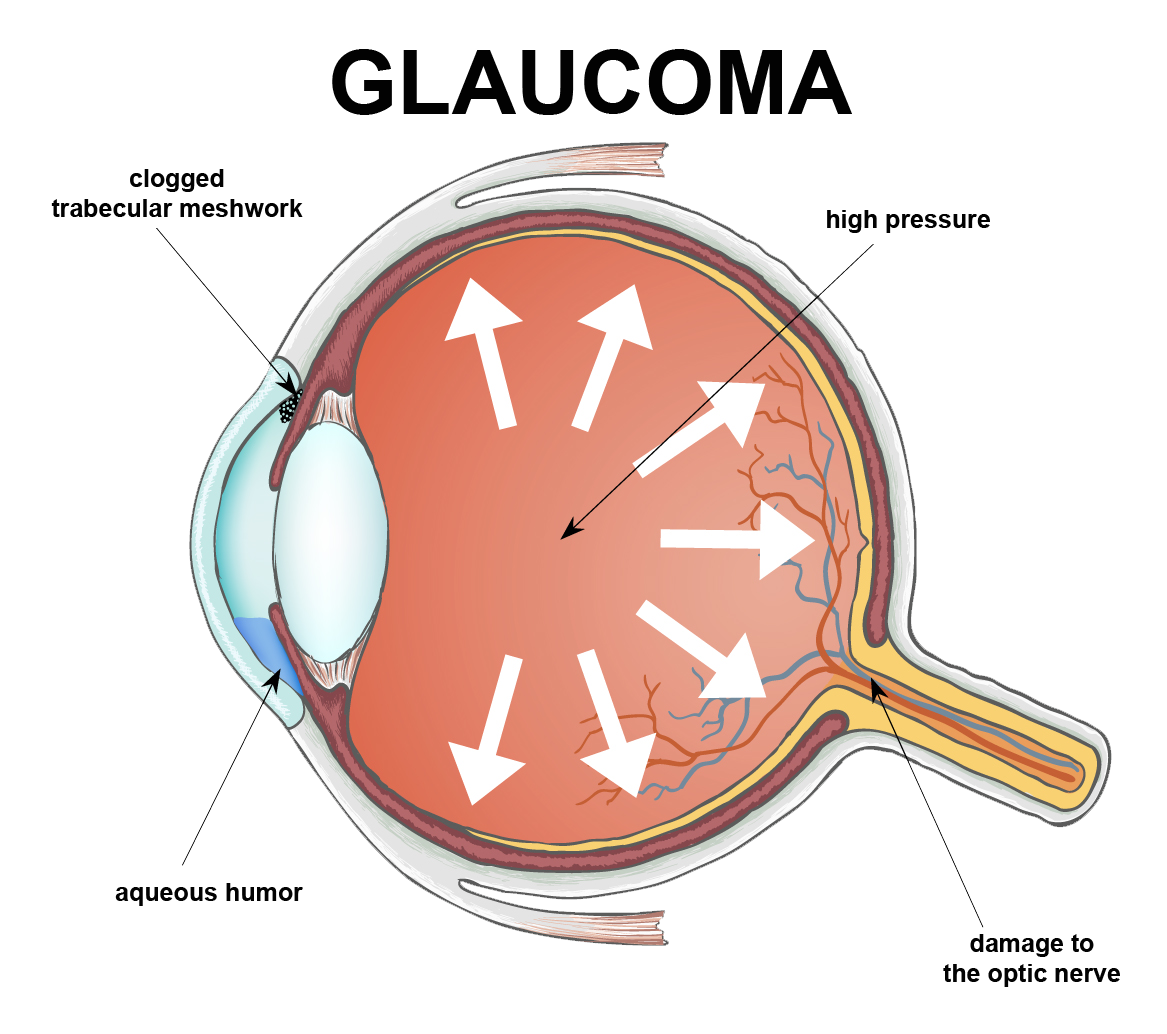

 Healthy Aging Month is an annual health observance designed to focus national attention on the positive aspects of growing older. Aging is a process that brings many changes. Vision loss and blindness, however, do not have to be one of them. There are several simple steps you can take to help keep your eyes healthy for the rest of your life.
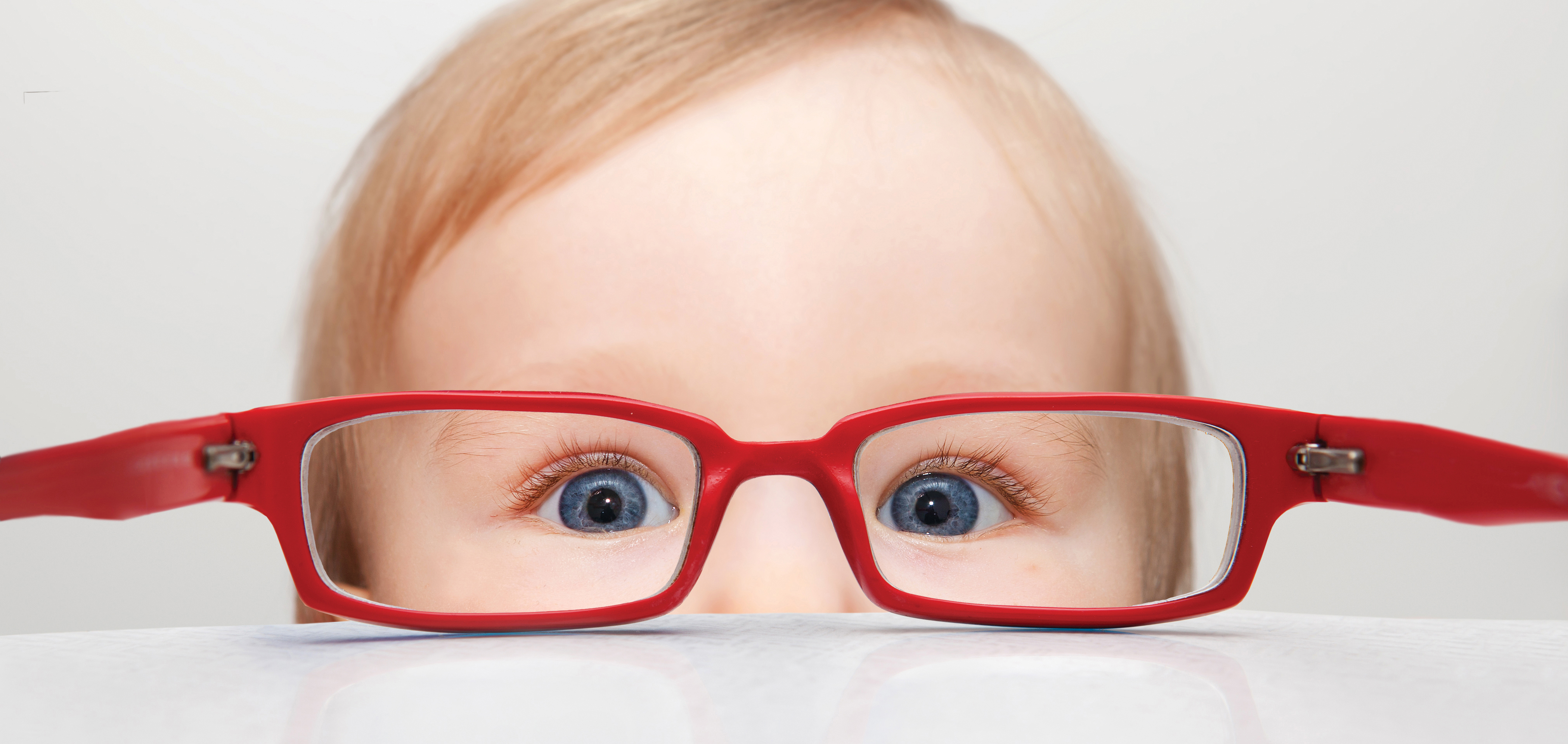

 When you think of cancer, most of us do not think about the eye or vision. Though rare, cancer can start inside or outside of the eye. If cancer starts inside the eyeball it’s called intraocular and if it starts outside the eye (eyelid or in the eye socket) then it’s called extraocular tumor. It can occur in both children and adults. Most major eye centers have specialists who are trained in the diagnosis and treatment of eye cancers.
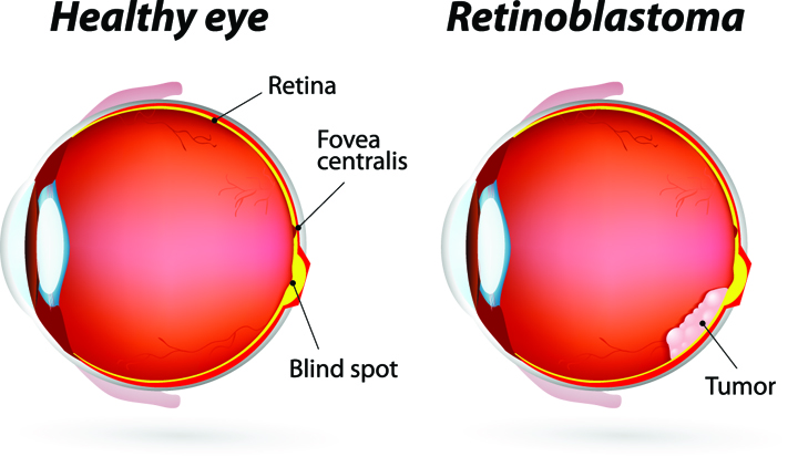 At the later stage of this cancer, the only one way to survive is to remove the eyeball (enucleation). Like many of other types of cancer, retinoblastoma has a genetic component so genetic testing needs to be done. The tumor begins with the RB1 gene mutation that stimulates retinal cells to develop into a tumor called a retinoblastoma. The RB1 mutation can be inherited from the parents, but in some cases it is sporadic and not inherited. There are various treatments such as surgery, chemotherapy, radiotherapy etc. to cure retinoblastoma cancer. Rarely it can spread beyond the eye.
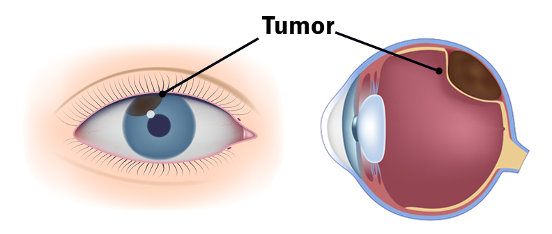

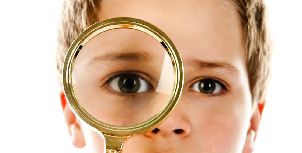
 UV Protection – Sunglasses for children should block 100% of UV radiation as well as between 75 – 90% of visible light. Any sunglasses you buy should have this information provided in the packaging
 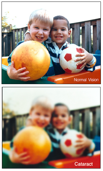 At the Discovery Eye Foundation (DEF), we are committed to supporting research that we believe will make the treatment of many forms of vision loss far more predictable and successful.