Having Trouble with Your Scleral Lenses?

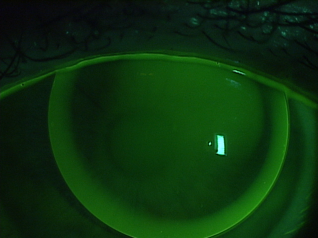
Scleral lens have become very popular and while many enjoy the comfort and vision correction they provide, some find it difficult to apply (insert) and remove these large diameter RGP lenses. If you are one of the many who are having difficulty managing to get your scleral lenses in or out, there are a number of tools available to help.

The Scleral Lens Education Society website provides a wealth of information about the care and handling of these lenses trouble with your Scleral Lenses as well as an excellent video. There are photos demonstrating various ways to hold the lens while applying it …some you may not have tried! If you have trouble with your schleral lenses, see below.

Troubleshooting tips and tricks:

    • If you are unable to maintain fluid in the bowl of the lens as you bring it towards your eye, make sure that your face is fully parallel to the floor. It may seem like you are nearly standing on your head when you’re in the correct position to apply the lens.
    • Lid control is essential; use one hand to hold lids completely out of the way, and don’t release the lids until the lens is actually fully in place and the plunger (or your finger tripod) has been removed.
    • If you are unable to successfully apply a solid lens with saline, you could practice applying the lens after filling the bowl of the lens with Celluvisc™ or another non-preserved viscous lubricant. These viscous lubricants will blur your vision compared to saline, however, so you may simply want to use them to practice lens application. Once you’ve mastered this step, switch to saline to give you better vision.
    • Try to keep both eyes open as you apply your lenses. This may also help you to position the lenses correctly.
    • If you are using a bulbed (DVM) plunger, and can see the opening in the center of the suction cup, look directly at the hole as you bring the lens into position. This will help you to position the lens correctly.

    Many find the DVM plungers helpful. They are readily available at your doctor’s office and online. These are just a few of the places to find them: DMV Corp, Dry Eye Zone, and Amazon.

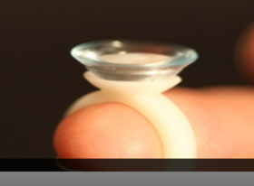
    trouble with your schleral lensesAnother variation to the standard lens inserters is a ring-style lens applicator by EZI Scleral Lens. It was designed by a post-transplant patient who like so many, had trouble inserting his scleral lens without getting a bubble. Read Tim’s story.

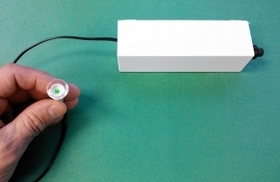
    If you have tried the above techniques and still have trouble applying scleral lenses there are a number of devices available that may help. Dalsey Adaptives has developed the See-Green devicetrouble with your schleral lenses that can be used to help successfully apply scleral contact lenses. The See-Green system comes with a stand that holds a lighted plunger (Figure 2). Using this system, you don’t hold the lens, you lower your eye onto it, which leaves both hands free for improved lid control. The light at the center of the plunger is used as a target to help you position the lens centrally on the eye. Click here to see the detailed instruction sheet.

    Scleral lenses offer good vision and comfort but can be challenging to manage. Discuss these options with your eye care professional to get his or her recommendation for your specific situation.

    1/28/16


    CathyW headshotCathy Warren, RN
    Executive Director
    National Keratoconus Foundation

Top 10 Articles of 2015

eye facts and eye disease
In looking at the many articles we shared with you in 2015, we found that your interests were varied. From the science of vision, eye facts and eye disease to helpful suggestions to help your vision.

Here is the list of the top 10 articles you read last year. Do you have a favorite that is not on the list? Share it in the comments section below.

    1. Rods and Cones Give Us Color, Detail and Night Vision
    2. 20 Facts About the Amazing Eye
    3. Understanding and Treating Corneal Scratches and Abrasions
    4. 32 Facts About Animal Eyes
    5. 20 Facts About Eye Color and Blinking
    6. When You See Things That Aren’t There
    7. Posterior Vitreous Detachment
    8. Can Keratoconus Progression Be Predicted?
    9. Winter Weather and Your Eyes
    10. Coffee and Glaucoma: “1-2 cups of coffee is probably fine, but…”

Do you have any topics you would like to see discussed in the blog? Please leave any suggestions you might have in the comments below.

1/7/16


Susan DeRemerSusan DeRemer, CFRE
Vice President of Development
Discovery Eye Foundation

The Importance of An Eye Exam

Why You Need An Eye Exam

The end of the year is fast approaching – when was the last time you had an eye exam? Was it a comprehensive eye exam?
eye exam
To keep your eyes healthy and maintain your vision, the American Optometric Association (AOA) recommends a comprehensive eye exam every two years for adults ages 18 to 60, and annual exams for people age 61 and older. However, if you have a family history of eye disease (glaucoma, macular degeneration, etc.), diabetes or high blood pressure, or have had an eye injury or surgery, you should have a comprehensive exam every year, unless otherwise indicated by your doctor.
Also, adults who wear contact lenses should have annual eye exams.

An important part of the comprehensive eye exam is the dilated eye exam to look inside your eye. Drops are placed in each eye to widen the pupil and allow more light to enter the eye. This gives your doctor a clear view of important tissues at the back of the eye, including the retina, the macula, and the optic nerve. This allows for early diagnosis of sight-threatening eye diseases like age-related macular degeneration, diabetic retinopathy, glaucoma, etc.

To better understand the importance of the dilated eye exam, here is a video from the National Eye Institute (NE) that explains what to expect.

At the end of your comprehensive eye exam your doctor should raise any concerns he has with you. But it is up to you to be prepared to react and ask questions for peace of mind and to help save your vision.

Questions To Ask After Your Eye Exam

It is always important to know if anything about your eyes have changed since your last visit. If the doctor says no, then the only thing you need to know is when they want to see you again.

If the doctor says the have been some minor changes, you need to know what questions to ask, such as:

  • Is my condition stable, or can I lose more sight?
  • What new symptoms should I watch out for?
  • Is there anything I can do to improve or help my vision?
  • When is the next time you want to see me?

If the doctor sees a marked change in your vision or give you a diagnosis of eye disease, you would want to ask:

  • Are there treatments for my eye disease?
  • When should I start treatment and how long will it last?
  • What are the benefits of this treatment and how successful is it?
  • What are the risks and possible side effects associated with this treatment?
  • Are there any foods, medications, or activities I should avoid while I am undergoing this treatment?
  • If I need to take medication, what should I do if I miss a dose or have a reaction?
  • Are there any other treatments available?
  • Will I need more tests necessary later?
  • How often should I schedule follow-up visits? Should I be monitored on a regular basis?
  • Am I still safe to drive?

Your vision is a terrible thing to lose, but with proper diet, exercise and no smoking, along with regularly scheduled eye exams, you improve your chances of maintaining your sight.

11/5/15


Susan DeRemerSusan DeRemer, CFRE

Laser Refractive Surgery: LASIK, LASEK, PRK and PTK

The introduction of the excimer laser to eye surgery in the early 1990’s represented a revolutionary innovation in the treatment of refractive errors: nearsightedness, farsightedness, and astigmatism. The development of this technology allows the safe and dependable correction of vision in many patients. For the most part, however, keratoconus (KC) patients are not candidates for such procedures for 2 reasons. First, the inherent biomechanical weakness of the keratoconic cornea could worsen if tissue is removed from the already thin cornea. Second, in addition to standard nearsightedness, farsightedness, and astigmatism, keratoconic vision is also impeded by higher order aberrations, which can be thought of as static in the eye’s optical system. However, recent advances may make variations of such procedures applicable to selected patients with KC.
Laser Refractive Surgery: LASIK, LASEK, PRK and PTK

LASIK, LASEK, PRK and PTK

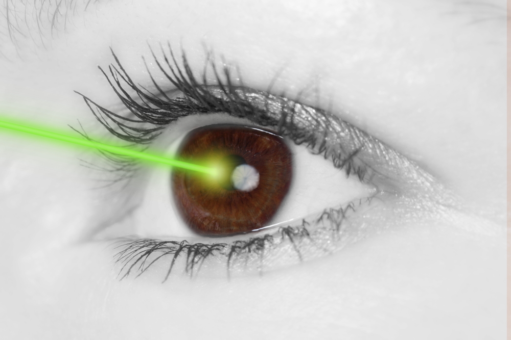
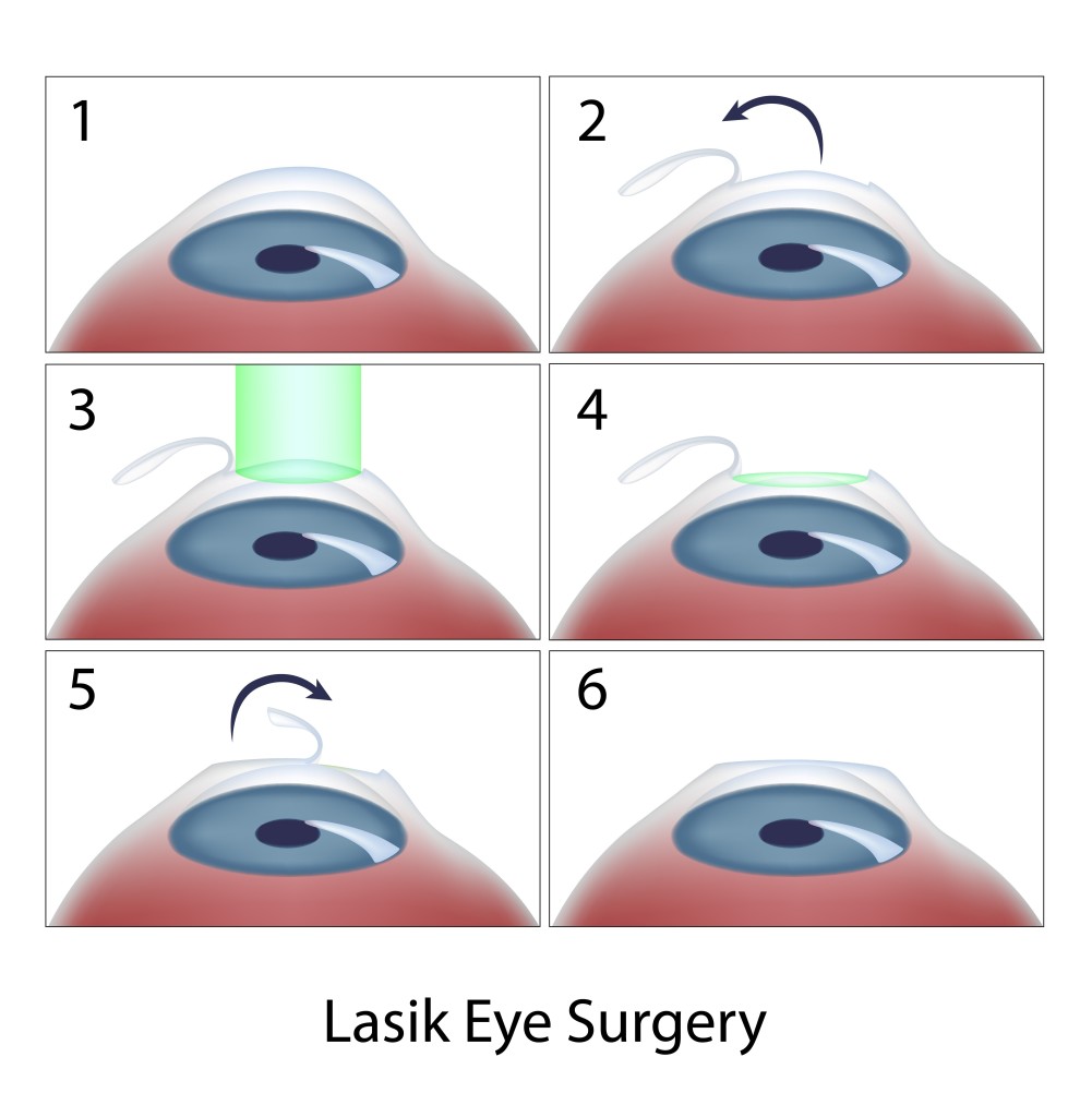
Today, laser in situ keratomileusis (LASIK) is the most popular method of laser eye surgery. LASIK uses an excimer laser to correct nearsightedness, farsightedness, or astigmatism by removing a thin lenslet of tissue from the surface of the cornea (the clear, front “watch crystal” of the eye). This is analogous to removal of a “tissue contact lens”. In LASIK, which is now an “all-laser” technique, a pancake-like thin flap of the cornea is first prepared with a high speed femtosecond laser. The flap acts to preserve the surface epithelial cells (which are like tiles on a floor) to promote quick healing and recovery of vision. Next, the excimer laser is used to remove a small amount of tissue from the corneal surface beneath the flap. The excimer laser used in LASIK produces a beam of invisible ultraviolet light energy, which when applied via an eye tracking mechanism, results in meticulous removal of this “tissue contact lens”. After corneal reshaping, the LASIK procedure is finished when the corneal flap is repositioned. When the flap is replaced, it lies in the bed of excimer laser removed tissue, causing the surface to change shape with the effect of decreasing nearsightedness, farsightedness, or astigmatism.

Laser vision correction can also be performed without a LASIK flap. These procedures, which are also perfomed with the excimer laser, go by a number of names – PRK (photorefractive keratectomy), LASEK (laser epithelial keratomileusis), epi-LASIK, or ASA (Advanced Surface Ablation). Although LASEK and LASIK sound the same, unlike traditional LASIK, LASEK does not require the preparation of a corneal flap. This has two potential advantages. First, risks of making the corneal flap in LASIK are avoided. This may be important in some patients in whom there is an additional risk in making the flap, such as patients with corneal scars or irregularities. Second, since laser treatment is done on the surface, LASEK/PRK preserves more corneal tissue. In particular, patients who have thinner corneas may be more safely treated with a no flap technique rather than LASIK.

At the beginning of the LASEK / PRK eye surgery procedure, the surface cells of the cornea are loosened and removed. The laser treatment then is applied, just as in LASIK, removing the properly shaped “tissue contact lens” for the desired optical correction. At the end of the procedure, a contact lens bandage is applied. Topical drops are used for a few week afterwards to avoid infection and control wound healing.

Vision after LASEK/PRK takes a little while longer to completely improve and stabilize than after LASIK because the epithelium needs to grow and smooth. Substantial improvement usually is noticed the day after the procedure and fluctuates over the next 2 weeks. The contact lens is removed in 5 days in most patients. Driving vision in the days after the procedure can be variable and take up to 2 weeks in some patients.

For both LASIK and LASEK/PRK, there are two basic types of possible side effects. Because patients may respond and heal differently, it is possible that the entire refractive error may not be fully corrected. In this case, vision will be clearer without glasses, but may not be as good as desired. In these situations, patient often can undergo a re-treatment procedure to further improve their vision. In addition, optical side effects include halos around lights and glare, especially at night, and some patients may experience dry eye sensations. Other, more rare, complications include infection or scarring.
LASIK, LASEK, PRK, PTK
Recently, there has been much talk in the keratoconus community about combining corneal collagen crosslinking with topography-guided LASEK/PRK. Topography-guided PRK uses information gained from your corneal map to program the laser to help make your cornea more optically regular. The goal of topography-guided PRK, like Intacs, is to improve corneal contour in the KC patient to improve glasses corrected vision and contact lens tolerance. In general, you will still need contacts and glasses afterwards.

Typically, LASEK/PRK procedures for keratoconus are combined with corneal collagen crosslinking, which has the goal to strengthen the weak keratoconic cornea and decrease progression of corneal mishapening over time. It is important to note that such treatments are not FDA-approved and are not generally available in the U.S. However, a number of international surgeons have been exploring the potential role of combined LASEK/PRK with crosslinking to improve keratoconus outcomes. In our practice, we have also had the opportunity to use Intacs and other procedures to further improve corneal shape in patients who have undergone topography-guided treatments with crosslinking with encouraging results.

In addition to LASIK and LASEK/PRK, the excimer laser may provide a novel therapeutic modality in the treatment of a number of superficial corneal disorders. This treatment is known a phototherapeutic keratectomy or PTK. Whether PTK eye surgery is used alone or as an adjunctive strategy in traditional corneal surgical techniques, a number of disorders affecting the corneal surface may be successfully treated by taking advantage of the excimer laser’s ability to meticulously remove superficial corneal tissue. These include a variety of corneal degenerations and dystrophies, corneal irregularities, and superficial scars, such as surface nodules found at the apex of the keratoconic cone. While some of these conditions, heretofore, could be treated by mechanical superficial keratectomy techniques, PTK may minimize tissue removal and surgical trauma.

So, for patients with keratoconus, it is important to know that, although LASIK type procedures are generally not indicated, research using these advanced technologies continues. A tailored therapeutic approach over time may combine a variety of procedures to optimize the corneal shape and ultimate visual outcome for the patient with keratoconus.

9/15/15


Peter Hersh, MD - Laser Refractive Surgery: LASIK, LASEK, PRK and PTKPeter S. Hersh, MD
Cornea and Laser Eye Institute – Hersh Vision Group
CLEI Center for Keratoconus

Which Eye Care Specialist Do You Need?

It’s time to get your eyes checked – do you go to an ophthalmologist, optometrist or optician? Your optometrist sees the beginnings of age-related macular degeneration, but is sending you to see and ophthalmologist, why?

One of the most confusing things about taking care of your eyes can be differentiating between an ophthalmologist, optometrist and optician. Each eye care specialist has a very important part to play in the health of your eyes and here is a quick synopsis of what each does so you can choose the best one for your vision issues and treatment.

Ophthalmologist
These specialists are fully trained medical doctors that have completed the eight years of training beyond a bachelor’s degree. Their training has included a full spectrum of eye care, from prescribing glasses and contact lenses and giving eye injections, to carrying out intricate eye surgeries. Many doctors may also be involved eye research to better understand vision, improve eye disease treatments or potentially find a cure. They are easily identified by the MD following their name.

Optometrist
These medical professionals have completed a four-year program at an accredited school of optometry. They have been trained to prescribe and fit glasses and contact lenses, as well as diagnose and treat various eye diseases. They provide treatments through topical therapeutic agents and oral drugs, and are licensed to perform certain types of laser surgery, such as Lasik. They are easily identified by the OD following their name.

Optician
These eye care professionals are not licensed to perform eye exams, medical tests or treat patients. Their purpose is to take the prescription from the ophthalmologist or optometrist and work with you to determine which glasses or contact lenses work best for you. If you suffer from an eye disease like keratoconus, these specialists can make the difference between a relatively normal life, or one that is dictated short periods of vision because of contact lens pain. These eye care professionals may hold and associate optician degree or have apprenticed fore required number of hours.

While each one of these eye specialists has their own area of expertise, they can form a team whose only concerns are your eye health and the ability to see as clearly as possible.

8/11/15


Susan DeRemerSusan DeRemer, CFRE
Vice President of Development
Discovery Eye Foundation

Eye Issues For Every Age Recap

Vision is something we take for granted, but when we start to have trouble seeing it is easy to panic. This blog has covered a variety of eye issues for every age, from children through older adults. Here are a few articles from leading doctors and specialists that you may have missed and might be of interest.
Artistic eye 6
Bill Takeshita, OD, FAAO – Visual Aids and Techniques When Traveling

Michelle Moore, CHHC – The Best Nutrition for Older Adults

Arthur B. Epstein, OD, FAAO – Understanding and Treating Corneal Scratches and Abrasions

The National Eye Health Education Program (NEHEP) – Low Vision Awareness
Maintaining Healthy Vision

Sandra Young, OD – GMO and the Nutritional Content of Food

S. Barry Eiden, OD, FAAO – Selecting Your Best Vision Correction Options

Suber S. Huang, MD, MBA – It’s All About ME – What to Know About Macular Edema

Jun Lin, MD, PhD and James Tsai, MD, MBA – The Optic Nerve And Its Visual Link To The Brain

Ronald N. Gaster, MD FACS – Do You Have a Pterygium?

Anthony B. Nesburn, MD, FACS – Three Generations of Saving Vision

Chantal Boisvert, OD, MD – Vision and Special Needs Children

Judith Delgado – Driving and Age-Related Macular Degeneration

David L. Kading OD, FAAO and Charissa Young – Itchy Eyes? It Must Be Allergy Season

Lauren Hauptman – Traveling With Low Or No Vision  /  Must Love Dogs, Traveling with Guide Dogs  /  Coping With Retinitis Pigmentosa

Kate Steit – Living Well With Low Vision Online Courses

Bezalel Schendowich, OD – What Are Scleral Contact Lenses?

In addition here are few other topics you might find of interest, including some infographics and delicious recipes.

Pupils Respond to More Than Light

Watery, Red, Itchy Eyes

10 Tips for Healthy Eyes (infographic)

The Need For Medical Research Funding

Protective Eyewear for Home, Garden & Sports

7 Spring Fruits and Vegetables (with some great recipes)

6 Ways Women Can Stop Vision Loss

6 Signs of Eye Disease (infographic)

Do I Need Vision Insurance?

How to Help a Blind or Visually Impaired Person with Mobility

Your Comprehensive Eye Exam (infographic)

Famous People with Vision Loss – Part I

Famous People with Vision Loss – Part II

Development of Eyeglasses Timeline (infographic)

What eye topics do you want to learn about? Please let us know in the comments section below.

7/21/15


Susan DeRemerSusan DeRemer, CFRE
Vice President of Development
Discovery Eye Foundation

Driving With Vision Loss

Challenges Faced by Individuals Driving With Vision Loss

Overview
People of all ages often view driving as the key to independence. Individuals with vision loss are no exception. Three groups of people with vision loss who wish to acquire or maintain the privilege of driving include teenagers with a congenital or acquired visual impairment who have never driven, adults with the same who have never driven, and adults with an acquired visual impairment who have driven in the past but may lose their license because of their vision loss. driving with vision lossHowever, vision standards for driving vary from state to state, and this variation persists despite decades of research demonstrating that there is no absolute cutoff criteria in visual acuity or peripheral vision for safe versus unsafe driving. The fact that states have variable standards results in people with visual impairments not being able to be licensed in some states, including perhaps their own, while being able to be licensed in a neighboring state. Clearly, the ability of these individuals to safely operate a motor vehicle does not change when they cross a state line. Yet, to maintain at least some driving privileges, they may find themselves having to move to a different state.

It is well known that many older drivers modify their driving norms to help keep themselves and others safe. For example, many older drivers voluntarily reduce or stop driving at night, in hazardous weather conditions, or on super highways. By limiting their driving, older drivers, particularly those with visual impairments, are able to continue operating their automobiles safely and efficiently in spite of reduced vision. This is important, considering the vast majority of older adults live in the suburbs or in rural areas where automobiles are required for transportation.

Maximizing Visual Capabilities
It is important for all individuals, but particularly for drivers who are visually impaired, to make sure their spectacle correction is up-to-date. Contrast enhancement and glare control with filtering lenses can also be of great benefit. Most drivers have experienced driving into the glare of the sun, while looking through a dirty windshield. Although wearing sunglasses and keeping windshields clean is not mandatory, they certainly help drivers see more easily and feel more comfortable when driving.

Maximizing Visual Attention
Human factors research has found that inattention blindness and the cost of switching contribute to or directly cause automobile mishaps. Inattention blindness refers to when a person’s attention to one activity undermines his or her attention to other activities. For example, when drivers focus on directional signs, their attention is not on what is happening on the road in front of them. The cost of switching refers to the time it takes a person to switch attention between different activities. A common example that causes driving mishaps is when drivers text while driving. When people focus on texting while driving, their response to the traffic around them is delayed.

Useful Field of View testing research has shown that the time it takes a person to process visual information, especially the complicated visual environment experienced each time a person drives, increases with age. With this in mind, decreasing or eliminating the time it takes older drivers or drivers with visual impairments to look for and visually process signage should help them maintain their concentration on the road ahead and the traffic around them.

A simple way to reduce or eliminate the need to look for directional signage is with the use of a Global Positioning System (GPS) device that uses spoken directions. Older drivers and drivers with visual impairments in particular should consider using a GPS device with spoken directions so that they are freed from the distraction of looking for/at road signs and can keep their attention on the traffic around them.

Finally, with the technology, such as adaptive cruise control and lane alert warnings, currently available in cars, it is expected that all drivers will be safer behind the wheel.

Final Considerations
A good driver is someone who has the ability to perceive change in a rapidly changing environment; the mental ability to judge and react to this information quickly and appropriately; and the motor ability to execute these decisions, along with the compensatory skills to compensate for some loss of ability in the other areas. Additionally, a driver’s familiarity with the driving environment and his or her past driving record should be taken into account when considering limiting driving activities or retiring from driving altogether.

For many drivers with vision loss, a limited driver’s license that allows them to drive during daylight hours, within a restricted radius of their home, and at lower rates of speed may be all they desire. However, there are times when an individual will need to retire from driving altogether because of vision loss or a combination of vision and cognitive changes. When this time comes, the individual needs to understand that retiring from driving is for his or her safety and the safety of others.

Finally, it is well known that vision loss in general, as well as the loss of driving privileges, can lead to feelings of hopelessness and depression. Fortunately, there are many things that can enhance the functional abilities of individuals with vision loss. To learn about available resources for individuals with vision loss, visit the National Eye Health Education Program low vision program page at www.nei.nih.gov/nehep/programs/lowvision.

7/9/15

Dr. Wilkinson - driving with vision lossMark Wilkinson, OD
University of Iowa Carver College of Medicine
Chair of the National Eye Health Education Program Low Vision Subcommittee

Scleral Lens Education Society

Scleral Contact Lenses have taken over a century to evolve into one of the best options for managing eye diseases such as keratoconus. This evolution began in the late 1800’s, with blown glass lenses. However, until the advent of highly oxygen permeable plastics, scleral lenses had very limited application. Now, with current technology and materials, scleral lenses have become a mainstream and rapidly growing lens option.
SLES_Logo_final
Scleral lenses are becoming more popular due to the exceptional comfort they can provide even to the most unusual eye shape. This comfort is attributable to their large size that allows them to tuck behind the eyelids, their relative lack of movement with eye blinks, and their fluid reservoir that keeps the cornea hydrated and does not actually touch the fragile corneal tissue in individuals with keratoconus.

As utilization of and demand for scleral lenses began to grow last decade, it became apparent that there was a need for more professionals trained in fitting scleral lenses, as well as someone to provide a consensus opinion for the eye care world on what the standard of care should be for these lenses. In addition, a process for providing a credential for those that attained a level of expertise in scleral lens fitting would allow those seeking experts in the field of fitting sclerals to find an experienced professional.

The Scleral Lens Education Society (SLS) was established in 2009 as an organization to help bring professional consensus to the suddenly rapidly growing area of scleral lenses. The mission statement of the SLS reads: “The Scleral Lens Education Society (SLS) is a non-profit organization 501(c)(3) committed to teaching contact lens practitioners the science and art of fitting all designs of scleral contact lenses for the purpose of managing corneal irregularity and ocular surface disease. SLS supports public education that highlights the benefits and availability of scleral contact lenses.”

Beginning with the founding board which included world renown experts in scleral lens fitting such as Greg DeNaeyer, OD, Christine Sindt, OD, and Bruce Baldwin, OD, PhD, the SLS has worked to spread the word about the potential benefits of scleral lens wear to both providers and patients alike. Professional education has included scleral lens webinars, workshops, and lecture series that are always standing room only events.

Currently, the SLS has over 2000 member contact lens practitioners as well as over 50 fellows, or certified scleral lens fitters that have demonstrated their expertise through a peer reviewed process of case reports, publications, and lectures. Many of these members and fellows are international, with SLS fellows from 11 countries, 5 different continents, and 20 different states in the US. Members hail from all 50 states, 6 continents, and over 40 countries.

In addition, the SLS has numerous industry sponsors that support the mission of the society to provide patient access to experienced fitters across the world. The sponsors provide the resources that allow the educational opportunities for practitioners as well as the website and patient resources that are available.

SLS board members are elected to serve in various capacities, including fellowship, public education, and international relations, and are elected to one year terms. The current board consists of:
President, Muriel Schornack, OD, Mayo Clinic, Rochester, MN
Vice President, Melissa Barnett, OD, University of California, Davis
Secretary, Michael Lipson, OD, University of Michigan
Treasurer, Mindy Toabe, OD, Metrohealth, Cleveland, OH
Immediate Past President, Jason Jedlicka, OD, Indiana University
Fellowship Chair, Pam Satjawatcharaphong, OD, University of California, Berkeley
Public Education Chair, Stephanie Woo, OD, Havasu Eye Center, Lake Havasu, AZ
International Chair, Langis Michaud, OD, University of Montreal

For more information about scleral lenses and the Scleral Lens Education Society, please visit the website at www.sclerallens.org. If you or someone you know might benefit from scleral lenses, you can locate a fitter in your area through the website as well. If you are unable to locate a fitter near you on the website, please contact the SLS and we will try to locate options in your local area.

7/2/15


Dr. JedlickaJason Jedlicka, OD
Clinical Associate Professor, Chief of Cornea and Contact Lens Service
Indiana University, School of Optometry

Vision Recap Of Previous Articles of Interest

Besides the comments that we get, one of the best parts of putting together this blog is the wonderful group of guests who share their expertise and personal stories. I want to thank all of the eye care professionals and friends that have contributed to make this blog a success.
Vision Recap
Here is a quick vision recap of some of the articles we had in the past that you may have missed.

Jullia A. Rosdahl, MD, PhDCoffee and Glaucoma and Taking Control of Glaucoma

David Liao, MD, PhDWhat Are A Macular Pucker and Macular Hole?

Kooshay MalekBeing A Blind Artist

Dan Roberts15 Things Doctors Might Like Us To Know

Jennifer VilleneuveLiving With KC Isn’t Easy

Daniel D. Esmaili, MDPosterior Vitreous Detachment

Donna ColeLiving With Dry Age-Related Macular Degeneration

Pouya N. Dayani, MDDiabetes And The Potential For Diabetic Retinopathy

Robin Heinz BratslavskyAdjustments Can Help With Depression

Judith DelgadoDrugs to Treat Dry AMD and Inflammation

Kate StreitHadley’s Online Education for the Blind and Visually Impaired

Catherine Warren, RNCan Keratoconus Progression Be Predicted?

Richard H. Roe, MD, MHSUveitis Explained

Sumit (Sam) Garg, MDCataract Surgery and Keratoconus

Howard J. Kaplan, MDSpotlight Text – A New Way to Read

Gerry TrickleImagination and KC

In addition to the topics above, here are few more articles that cover a variety of vision issues:

If you have any topics that you would like to read about, please let us know in the comments section below.

6/23/15


Susan DeRemerSusan DeRemer, CFRE
Vice President of Development
Discovery Eye Foundation

What Are Scleral Contact Lenses?

In the beginning…of contact lenses…there were scleral lenses…only.

In the year 1887 a great gift was given to the world of sufferers of distorted vision resulting from corneal tissue that was irregular in shape from disease or trauma.
scleral lenses

The contact lens was invented nearly simultaneously by physicians working separately in Germany and in France. Working from drawings of Leonardo da Vinci (1508) and ideas of the British astronomer Sir John Herschel (1828), August Müller and separately Adolph Fick and Eugene Kalt blew glass shells to fit the outer eye and to some extent remedy their visual difficulties. These lenses rested on the conjunctiva of the eye above the sclera or white of the eye and were thus the first scleral contact lenses — the first contact lenses of any sort.

What are Scleral Contact Lenses?

The design and manufacture of scleral lenses has been a story of technological development significant for improvement in comfort, material, and affinity for the ocular surface.

For many years the lenses were partially molded and partially ground from the material of which hard contact lenses are made: PMMA (poly-methyl methacrylate) known as Plexiglas or Perspex. To form these lenses, like tooth implants, a plaster cast is made from a negative mold prepared from dental impression putty. The plastic would be heated and given the shape of the fitting surface of the lens from the plaster cast and then the power and edge curves would be ground onto the outside surface of the lens. Later came preformed trial sets not unfamiliar to those which we use today.

The current generation of scleral lens fitting began sometime in the last fifteen years with the mating of advanced corneal topography measurements, computer driven lathes and the observations of some very clever contact lens scientists. Proprietary designs of scleral lenses offering a variety of diameters, fitting philosophies, and multiple parameters are filling the gaps contact lens specialists have been wrestling with using smaller corneal contact lens designs for decades with less than optimal results. Most recently a firm has begun manufacturing lenses with a 3-D printer from an image generated from the eye. One eye…one lens, the lens is meant to fit like a fingerprint.

When discussing contact lens treatment, experts are experts because they agree that, any sort, size, or design of lens will have both positive and negative effects on the eyes and the tissues surrounding them. While it is true that many of the fitting and comfort issues confronted with corneal lenses of any size and design can be managed well with scleral designs, the scleral lens can also be difficult for some patients; for some eyes; for some conditions.

From the outset the larger size of the today’s scleral lens provides comfort on par with soft contact lenses for exactly the same reason: their size. Also, like soft contact lenses the scleral will not move around on the surface of the eye allowing the wearer a much more relaxed contact lens experience — there is no necessity to balance small corneal lenses between tense eyelids – vision can be enjoyed in any direction of gaze. The lenses will not fall off the eye and the increased size is a clear plus in finding a dropped contact lens.

On the other hand the quality of vision gained with scleral lenses specifically in cases of distorted corneae is far more comparable to that achievable with corneal GP lenses than with soft contact lenses in most cases.

Over the years my keratoconus patients have benefited from a series of contact lens breakthroughs that have variously improved the quality of their vision, their comfort with contact lenses, or in some other way the health of their eyes. Some years ago I “re-invented” the piggy-back system of contact lens wear which I summarized in 2008 in an article published in the Contact Lens Spectrum. Piggy-backers would place their vision restoring firm contact lens on top of a disposable daily wear lens of minimal focusing power. The soft lens would reduce the sensation of the firm lens while in many cases preventing the contact lens from abrading the cornea. More recently I have been successfully moving patients to scleral lenses because there is certainly less bother (only one lens per eye) and far less worry over corneal abrasions as the lens rests on the conjunctiva over the sclera and maintains a fluid cushion over the cornea itself.

Scleral lenses are finding their place in the world of contact lens fitting primarily to remedy vision problems from very irregular or otherwise damaged corneae both those caused by developing disease and trauma through injury or surgery. More and more, these lenses are requested by patients with normal eyes who want to enjoy the benefits provided by these lenses while participating in sports or other activities.

Scleral lenses are renowned for their greater comfort. In many cases a correctly fitted lens can be worn for many waking hours. Many patients have found that they benefit from exchanging the fluid from the reservoir from time to time throughout the day. The fluid that fills the lens-cornea space is sterile, non-preserved normal saline or in some cases saline with a non-preserved tear substitute added when needed for improved comfort.

A proper care regimen for scleral lenses is not different from that for any other contact lens manufactured from a firm oxygen permeable material. The lenses require cleaning upon removal, soaking in a recommended solution appropriate to the material of the lens and a periodic treatment to remove protein deposits. Of course, the exact care specifications will vary from patient to patient according to the evaluation of their contact lens specialist.

Just like any lens modality, the fitting requires expertise. Many who fit and dispense contact lenses rely on boxed soft lenses for their patients. When corneae become distorted those lenses will hardly fill the need. Greater expertise is required to fit rigid corneal lenses needed for these more problematic surfaces. The decision of the corneal lens expert to move on to the world of larger lenses is not of the same magnitude as that from boxes to corneal GP lenses. The investment is more a matter of time spent in discussion with the manufacturer’s fitting consultants, some reading, a webinar or two and keeping up to date with the lens designs that are available.

I was not among the first to use the current generation of scleral lenses, but when the opportunity knocked some years ago, I realized the importance of this form of contact lens and I believe I have positively influenced the quality of life of many of my patients.

5/28/15

Bezalel Schendowich - scleral lensesBezalel Schendowich, OD
Medical Advisory Board of the National Keratoconus Foundation
Fellow of the International Association of Contact Lens Educators
Clinical Supervisor & Specialty Contact Lens Fitter, Sha’are Zedek Medical Center, Jerusalem, Israel