Your Comprehensive Eye Exam

Your Comprehensive Eye Exam

Being able to see clearly is important to all of us. But it is also something we have a tendency to take for granted until we notice changes in our vision. The point of the yearly comprehensive exam is to monitor your eyes before any problems arise, and address any concerns that could affect your vision later. Here is what to expect at your next comprehensive eye exam.

An eye exam involves an external examination of your eyes followed by a series of tests designed to evaluate your vision and check for eye diseases. Each test evaluates a different aspect of your vision and includes specific tests for visual acuity, pupil function, muscle function, visual fields, eye pressure and viewing the back of the eye through a dilated pupil.
cross section of eye - eye exam
External Exam
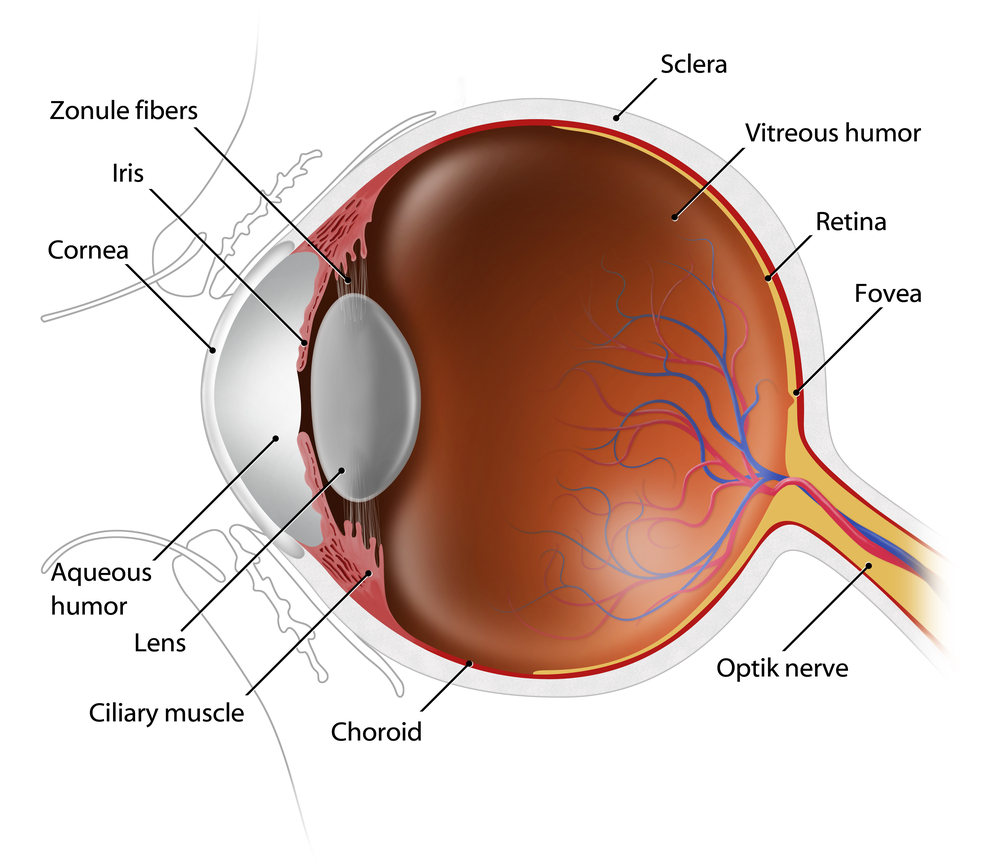
The external examination consists of inspecting the eyelids, surrounding tissues and the eyeball including the sclera (white part of the eye), iris and cornea.

Visual Acuity & Fields
Visual acuity is your eye’s ability to detect fine details and see an in-focus image at a certain distance. A Snellen chart and a phoropter are used. The standard definition of normal visual acuity is 20/20. The term 20/20 comes from even sized objects that can be seen by a “person of normal vision” atvisual acuity eye exam 
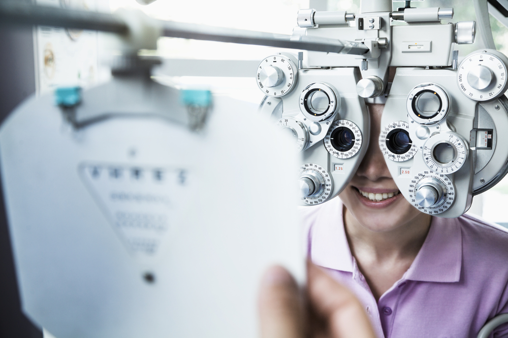
the specified distance. For example, if a person can see at a distance of 20 feet an object that normally can be seen at 20 feet, then they have 20/20 vision. If they can see at 20 feet what a normal person can see at 40 feet, then they have 20/40 vision. For the visual acuity test each eye is tested separately to gauge your side or peripheral vision.

Pupil Functionpupil function eye exam
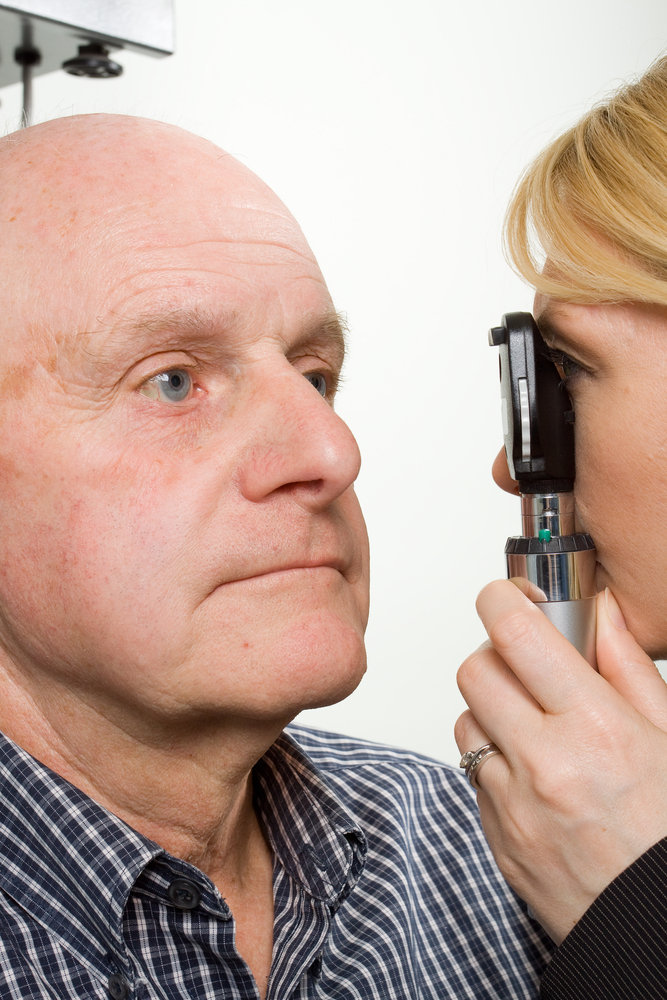
An examination of the pupil begins with inspecting your pupils for equal size, regular shape, reaction to light, and direct and consensual reaction (meaning the pupil of one eye constricts when the other eye is exposed to light).

Eye Muscle Function
Eye movement is assessed two ways. First by having you move your eye quickly to a target at the far right, left, top and bottom. Then by slow tracking which uses the ‘follow my finger’ test, which tests all the muscles that move your eye.

tonometer eye exam
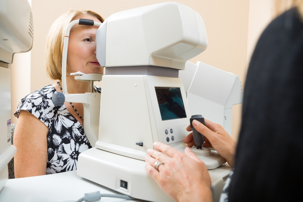
Eye Pressure Measurement
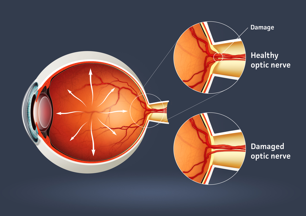
Intraocular pressure, or IOP, is measured using a tonometer to determine the fluid pressure inside your eye. This test provides information regarding your potential for glaucoma.

Viewing the Back of the Eyedilated eye - eye exam
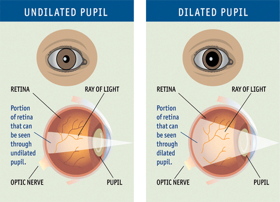
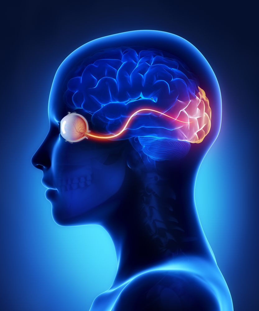
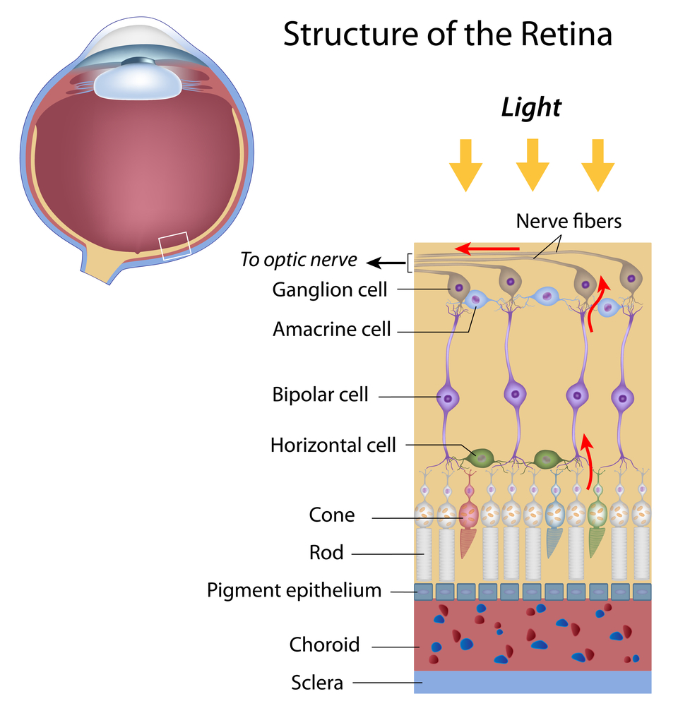
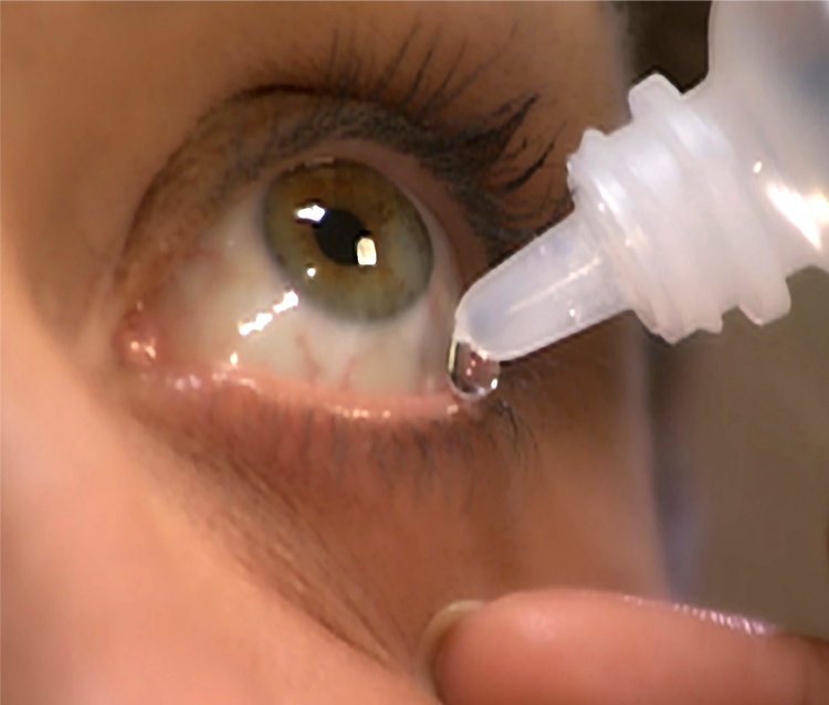
Increasing the size of your pupil with eye drops (known as dilating your eyes) allows the doctor to have a larger view of the back of your eye, including the retina, as demonstrated by this diagram. This is very important for diagnosis and tracking of macular degeneration and diabetic retinopathy.

Eye Structureslit lamp eye exam
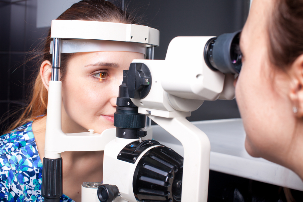
A special, high-powered microscope, called a slit-lamp, is used to view the structures of your eye clearly and in detail, enabling early diagnosis of a variety of eye conditions such as cataracts, presbyopia and corneal injury.

Taking care of your eyes, especially as you become older, is very important. Changes in vision may be gradual, or fast, but in both cases, early diagnosis is key to successful treatment and retaining your vision. It is suggested that individuals 40 and over have a comprehensive eye exam every one to two years. Exceptions would be if you have diabetes, in which case you should see your eye doctor yearly; or there is a family history of eye diseases such as glaucoma, macular degeneration, or corneal diseases, which may require more frequent visits to your eye doctor. If you have any degree of sudden vision loss, eye pain, or significant irritation, contact your eye doctor immediately.

4/23/15

Susan DeRemerSusan DeRemer, CFRE
Vice President of Development
Discovery Eye Foundation

Do I Need Vision Insurance?

With the rising costs of health insurance, many people are looking for ways to reduce their costs. Since not all insurance packages include vision insurance, many people wonder, do I need vision insurance?
vision insurance

Standard Vision Insurance

Vision insurance is a type of health insurance that entitles you to specific eye care benefits such as routine eye exams and other procedures, as well as a specified dollar mount or discount for the purchase of eyeglasses and contact lenses. It only supplements regular health insurance and is designed to help reduce your costs for routine preventative eye care and eyewear.

You can get vision insurance as part of a group, such as your employer, an association, etc., through a government program such as Medicare or Medicaid, or as an individual. It is often a benefit linked to your regular HMO (health maintenance organization) or PPO (preferred provider organization) health insurance.

There are two primary vision insurance plans available:

  • Vision Benefits Package – provides free eye care services and eyewear within a fixed dollar amount for which you pay an annual premium or membership fee and a small co-pay. It may also include a deductible.
  • Discount Vision Plan – provides eye care and eyewear at a discounted rate after you pay an annual premium or membership fee.

Both insurance plans generally include:

  • Annual eye exams
  • Eyeglass frames (usually once every 24 months)
  • Eyeglass lenses (usually once every 24 months)
  • Contact lenses (usually once every 24 months)
  • Discounted rates for LASIK and PRK

Here is where you can check for a list of some vision insurance providers.

Medicare and Medicaid

Different kinds of vision care are included in the US government programs, Medicare and Medicaid. These programs are for qualifying American age 65 and older, individuals with specific disabilities and people with low income.

The Types of Medicare For Vision:

    • Medicare Part A (Hospital Insurance) –Medical eye problems that require a hospital emergency room attention, but routine eye exams are NOT covered.
    • Medicare Part B (Medical Insurance) – Visits to an eye doctor that are related to an eye disease, but routine eye exams are NOT covered.
    • Medicare Part D (Prescription Drug Coverage) – Will help pay for prescription medications for eye diseases.

If you have Medicare Parts A & B you are generally eligible for the following vision coverage, however, there is a deductible before Medicare will start to pay, at which point you will still be paying a percentage of the remaining costs.

  • Cataract surgery – covers many of the cost including a standard intraocular lens (IOL). If you chose a premium IOL to correct your eyesight and reduce your need for glasses, you must pay for this added cost out-of-pocket.
  • Eyewear after cataract surgery – one pair of standard eyeglasses OR contact lenses.
  • Glaucoma screening – an annual screening for people at high risk for glaucoma, including people with diabetes or a family history, and African-Americans whom are 50 or older.
  • Ocular prostheses – costs related to the replacement and maintenance of an artificial eye.

There is also Medicare Supplement Insurance (Medigap) which is sold by private insurance companies to supplement only Medicare Parts A & B. It is intended to cover your share of the costs of Medicare-covered services including coinsurance, co-payments and deductibles. For more details about Medicare plans and coverage check their website or call 800-633-4227.

Medicaid is the US health program that gives medical benefits to low-income people who may have no or inadequate medical insurance. A person eligible for Medicaid may be asked to make a co-payment at the time medical service is provided. Vision benefits for children under the age of 21 include eye exams, eyeglass frames and lenses. Each state determines how often these services are provided and some states offer similar vision services to adults. To learn more about Medicaid eligibility requirements and vision benefits call your state’s Medicaid agency or visit their website.
vision dial - vision insurance

Defined Contribution Health Plans

A way to lower your vison care costs is to take part in a defined contribution health plan (DCHP). You are given a menu of health care benefits to choose from where a portion of the fees you receive for health coverage come from money that is deducted from our paycheck before federal, state and social security taxes are calculated. Four types of DCHP are:

Cafeteria Plans – your employer takes a portion of your salary and deposits it into a non-taxable account for health care spending. The amount taken depends on the number and costs of the benefits you select.

Flexible Spending Accounts (FSA) – your employer takes a predetermined portion of your pre-tax salary and deposits it into health care account for you to pay medical expenses. But generally preventative care such as routine eye exams and are not reimbursable. Nor are eyeglasses and contact lenses reimbursable. You would need to verify with your employer. If you do not use all the money at the end of a 12 month period, the money goes back to your employer.

Health Reimbursement Arrangement (HRA) – this is similar to an FSA except you can use it for preventative care like eye exams and you do not lose the money if it isn’t spent within a certain time period as it can be carried from year to year.

Health Savings Account (HSA) – it can be employer-sponsored of you can set up one independently; however you must purchase a high-deductible health insurance plan to open an HSA and you cannot exceed the annual deductible of your health insurance plan. You cannot be enrolled in Medicare of be a depended on someone else’s tax return. You can use it for preventive care such as eye exams. You can learn more about HSAs by visiting the US Treasury’s website.

There are a variety of options when it comes to vision insurance. You just need to determine your needs and ask providers the correct questions.

4/14/15


Susan DeRemerSusan DeRemer, CFRE
Vice President of Development
Discovery Eye Foundation

6 Ways Women Can Stop Vision Loss

Women account for 53% of the world’s population. However, 2/3 of the blind and visually impaired people in the world are women. While 80% of these women are in developing countries, women in developed countries like the US are still more likely to face vision loss than men.
women can stop vision loss

Why are women more prone to eye disease than men?

Women are the caregivers in families, taking care of the health of family members over themselves. In addition, with many having jobs outside the home, they don’t feel they have the time to go to the doctor until something major happens, especially related to vision.

Women live longer than men are at greater risk for age-related eye diseases such as age-related macular degeneration (AMD), diabetic retinopathy, glaucoma and cataracts.

Women are more likely to develop several autoimmune diseases that can affect their eyes including, multiple sclerosis, lupus, rheumatoid arthritis and Sj?gren’s syndrome.

75% of new breast cancer diagnosed each year is estrogen-sensitive. A common part of estrogen-sensitive breast cancer treatment includes the prescription of tamoxifen. More studies are being done, but cataracts due to tamoxifen have been identified in about 10% of the patients taking the drug.

What can women do to lessen their chances for eye disease?

Know your family history as genetics play and important role in your eye health, so know what eye diseases run in your family. Let your eye doctor know so he can look for early warning signs that can help prevent of lessen the conditions in you.

Get routine comprehensive, dilated eye exams starting at the age of 40, to create a baseline for your doctor to work from. After that you can go every 2-4 years until the age of 60. At 60+ have a compressive, dilated exam every two years if you are symptom-free and low risk.

Eat healthy and exercise. It is important to maintain a healthy weight to reduce the risk of some eye diseases. Eating fresh fruits and vegetables is also important as they can contain carotenoids an some antioxidants that appear to help with vision retention. It should also be noted that in many studies, supplements did not show the same beneficial effects as whole foods.

Stop smoking! You not only increase your risk for cancer and heart disease, but smoking is the only thing besides advancing age that has been proven to be directly related to AMD.

Avoid ultraviolet light by wearing sunglasses (with wide-brimmed hats) and indoor glasses with UV protection. While everyone knows the sun is a source of UV light, so are electronic screens such as your TV, computer, tablet or smartphone. Prescription glasses and readers can have a clear UV coating put on them that will not distort your color vision. If you don’t need vision correction, there is eyewear with no correction that is coated to protect your eyes to avoid dry eye and retinal damage.

Use cosmetics and contacts safely. Always wash your hands first. Throw away old makeup and lens solutions. Do not share cosmetics or apply while driving. Make sure to clean your lenses thoroughly before putting them in your eyes.

Because women are relied upon to take care of the family, vision loss that can impact that responsibility can be devastating to the entire family. And later in life, when they may have outlived a spouse, the isolation and depression can destroy their quality of life as they try to cope on their own.

Reach out to women you know and remind them to take an active part in their own healthcare. Especially with regards to their vision, when women are at a higher risk of vision loss than men.

4/7/15


Susan DeRemerSusan DeRemer, CFRE
Vice President of Development
Discovery Eye Foundation

Medical Research Funding Needs Individual Donors

The Need for Medical Research Funding

About 1.75 million U.S. residents currently have advanced age-related macular degeneration with associated vision loss, with that number expected to grow to almost 3 million by the year 2020.

About 8.4 million individuals worldwide are blind from primary open-angle glaucoma, with that number expected to grow to almost 11 million by the year 2020.

About 22 million Americans have cataracts affecting their vision, with that number expected to grow to more than 30 million by the year 2020.

The economic impact of this increase of people with vision loss will be tremendous.  But right now scientist are working on ways to treat and eventually cure many eye diseases.  The only problem is the funding necessary to support this sight-saving research. Here is a look at the decline of medical research funding in the US and what you can do to help.
medical research funding

3/17/15


Susan DeRemerSusan DeRemer, CFRE
Vice President of Development
Discovery Eye Foundation

The Optic Nerve And Its Visual Link To The Brain

The optic nerve, a cable–like grouping of nerve fibers, connects and transmits visual information from the eye to the brain. The optic nerve is mainly composed of retinal ganglion cell (RGC) axons. In the human eye, the optic nerve receives light signals from about 125 million photoreceptor cells (known as rods and cones) via two intermediate neuron types, bipolar and amacrine cells. In the brain, the optic nerve transmits vision signals to the lateral geniculate nucleus (LGN), where visual information is relayed to the visual cortex of the brain that converts the image impulses into objects that we see.
Optic Nerve
In the retinal tissues of the eye, more than 23 types of RGCs vary significantly in terms of their morphology, connections, and responses to visual stimulation. Those visual transmitting RGCs are the neuronal cells. They all share the defining properties of:

  1. possessing a cell body (soma) at the inner surface of the retina
  2. having a long axon that extends into the brain via the optic chiasm and the optic tract
  3. synapsing with the LGN. The RGCs form multiple functional pathways within the optic nerve to mediate the visual signal

Human beings can see three primary colors: red, green, and blue. This is due to our having three different kinds of color sensitive cone cells: red cones, green cones, and blue cones.

The RGCs connecting to the red and green cones are midget RGCs. They are mainly located at the center of the retina (known as fovea). A single midget RGC communicates with as few as five photoreceptors. They transmit red-green color signals to the parvocellular layer in the LGN (see Figure). The midget-parvocellular pathway responds to color changes, but has little or no response to contrast change. This pathway has center-surround receptive fields, and slow conduction velocities. Because of this pathway, we can see objects precisely in detail and in full color.
retina and optic nerve
The bistratified RGCs are likely involved in blue color vision. Bistratified cells receive visual information input originally from an intermediate numbers of cones and rods. The bistratified RGCs connect to the koniocellular layers in the LGN (see Figure). The koniocellular neurons form robust layers throughout the visual hemifield and have moderate spatial resolution, moderate conduction velocities, and can respond to moderate-contrast stimuli. They have very large receptive fields that only possess on-center regions (no off-surround regions).

Objects can be seen in the dark with motion and coarse outlines accentuated due to the parasol RGCs. At the periphery of the retina, a single parasol RGC connects to many thousands of photoreceptors (many rods and few cones). The parasol RGCs project their axons to the magnocellular layers of the LGN (see Figure) and are primarily concerned with visual perception. They have fast conduction velocities, can respond to low-contrast stimuli, but are not very sensitive to changes in color.

Finally, humans can see objects in three-dimension courtesy of the crossing over of optic nerve fibers at the optic chiasm. This anatomic structure allows for the human visual cortex to receive the same hemispheric visual field from both eyes (see Figure), thus making it possible for the visual cortex to generate binocular and stereoscopic vision.

Recently, a new type of RGC, called photosensitive RGCs, was discovered. The photosensitive RGCs contribute minimally to our vision, but play a key role in vision regulation. Photosensitive RGCs axons do not have connections to the LGN, but form the retino-hypothalamic tract, and synapse to three other locations in the brain for specific vision regulation functions:

  1. Pretectal nucleus: involved in reflexive eye movements, thereby helping to target what we want to see
  2. Midbrain nuclei: involved in controlling the size of the pupil, thus helping to adjust the brightness of objects; and coordinating movement of the eye for focusing
  3. Suprachiasmatic nucleus: involved in regulating the sleep-wake cycle

A fully functional optic nerve is essential for vision. Obviously, any damage of the optic nerve will sever the precise transmission of visual information between the retina and brain, directly leading to vision distortion and/or vision loss. Damage to the optic nerve can result from:

  1. Direct/indirect physical damage (e.g. ocular trauma)
  2. Acute/sub-acute physiological lesion (e.g. infection or inflammation, or malignancy (cancer))
  3. Chronic neuronal degeneration (e.g. glaucoma, a most common cause of optic nerve damage)

Moreover, the optic nerve is also a very important vivo model for studying central nervous protection and regeneration. At the cell biology level, the RGC axons are covered with myelin produced by oligodendrocytes (rather than Schwann cells of the peripheral nervous system) after exiting the eye on their way to the LGN and thus part of the central nervous system. Scientists have recently acquired more and more evidence that certain types of damage to the optic nerve may be reversible in the future. Therefore, the optic nerve provides a potential window to explore more complicated neuronal degenerative diseases, such as Alzheimer’s disease and Huntington disease.

3/12/15

Jun Lin, MD, PhD
Assistant Professor,
Department of Ophthalmology
New York Eye and Ear Infirmary of Mount Sinai
Icahn School of Medicine at Mount Sinai

—–

James Tsai, MD, MBA
Chair, National Eye Health Education Program Glaucoma
Subcommittee President, New York Eye and Ear Infirmary of Mount Sinai Chair
Department of Ophthalmology
Icahn School of Medicine at Mount Sinai

10 Tips For Healthy Eyes

In honor of Save Your Sight Week, here are 10 tips for healthy eyes. There are some simple things you can begin to do today to help your retain your vision. Also there are no warning signs for many of the leading eye diseases (age-related macular degeneration, glaucoma, cataracts), yet most people don’t see an eye doctor until they have a problem seeing. Unfortunately, by that time, irreparable damage has been done.
10 Tips For Healthy Eyes

3/3/15


Susan DeRemerSusan DeRemer, CFRE
Vice President of Development
Discovery Eye Foundation

February Is Low Vision Awareness Month

Help for People With Vision Loss

Here’s eye-opening news: Currently, 4.2 million Americans ages 40 and older are visually impaired. Of these, 3 million have low vision. By 2030, when the last baby boomers turn 65, the number of Americans who have visual impairments is projected to reach 7.2 million, with 5 million having low vision. For the millions of people who currently live or will live with low vision, the good news is there is help.
Low vision awareness
But first, what is low vision? Low vision is when even with regular glasses, contact lenses, medicine, or surgery, people have difficulty seeing, which makes everyday tasks difficult to do. Activities that used to be simple like reading the mail, shopping, cooking, and writing can become challenging.

Most people with low vision are age 65 or older. The leading causes of vision loss in older adults are age-related macular degeneration, diabetic retinopathy, cataract, and glaucoma. Among younger people, vision loss is most often caused by inherited eye conditions, infectious and autoimmune eye diseases, or trauma. For people with low vision, maximizing their remaining sight is key to helping them continue to live safe, productive, and rewarding lives. The first step is to seek help.

“I encourage anyone with low vision to seek guidance about vision rehabilitation from a low vision specialist,” advises Paul A. Sieving, MD, PhD, director of the National Eye Institute (NEI), one of the National Institutes of Health (NIH) and the federal government’s principal agency for vision research.

What is a low vision specialist? A low vision specialist is an ophthalmologist or optometrist who works with people who have low vision. A low vision specialist can develop a vision rehabilitation plan that identifies strategies and assistive devices appropriate for the person’s particular needs. “A vision rehabilitation plan helps people reach their true visual potential when nothing more can be done from a medical or surgical standpoint,” explains Mark Wilkinson, O.D., a low vision specialist at the University of Iowa Hospitals and Clinics and chair of the low vision subcommittee for the National Eye Health Education Program (NEHEP).

Vision rehabilitation can include the following:

  • Training to use magnifying and adaptive devices
  • Teaching new daily living skills to remain safe and live independently
  • Developing strategies to navigate around the home and in public
  • Providing resources and support

There are also many resources available to help people with low vision. NEI offers a 20-page, large-print booklet, titled What You Should Know About Low Vision, and companion DVD, featuring inspiring stories of people living with low vision. This booklet and DVD, among other resources, are available at
www.nei.nih.gov/lowvision.

With the aging of the population, eye diseases and vision loss have become major public health concerns in the United States. NEI is committed to finding new ways to improve the lives of people living with visual impairment. Aside from making information and resources readily available, NEI has dedicated more than $24 million to research projects on low vision, including learning how the brain adapts to vision loss; strategies to improve vision rehabilitation; and the development of new technologies that help people with low vision to read, shop, and find their way in unfamiliar places. Research like this will help people with low vision to make the most of their remaining vision and maintain their independence and quality of life.

2/17/15

NEHEPsq75The National Eye Health Education Program (NEHEP) of the National Eye Institute (NEI), a part of NIH, offers a 20-page, large-print booklet, titled What You Should Know About Low Vision, and a series of videos featuring patient stories about living with low vision. These and other resources are available at http://www.nei.nih.gov/lowvision.

Taking Control of Glaucoma

Taking control of glaucoma: The importance of adherence to glaucoma treatment


Glaucoma is known as the “silent thief of sight”: people with glaucoma usually have no symptoms. The only intervention that has been proven to reduce the risk of vision loss in glaucoma is lowering the eye pressure. The most common way to lower eye pressure is with eye drop medications. Glaucoma treatments do not improve vision, but they do to help prevent vision loss from happening.
taking control of glaucoma - eyedrops
What does it mean to be “compliant”?

Adherence (the newer term that is replacing “compliance”) with glaucoma treatment usually means taking your eye drops everyday and at the right times and coming to your glaucoma check ups. Your doctor needs to check your eye pressure regularly, as well as look at your optic nerves and measure your visual fields, to monitor your disease.

Why is it difficult to take eye drops everyday?

Some people with glaucoma only need 1 or 2 eye drops everyday to control their glaucoma, but some may need as many as 4 glaucoma medications, taken multiple times throughout the day. Imagine using 2 eye drops in the morning, 1 eye drop at noon, 1 at dinnertime, and 2 more in the evening, and doing this everyday for years and years, to help protect your sight. It is easy to see how people could miss some drops. Reasons can include forgetting them (or falling asleep before that bedtime drop), the cost of the medications, the side effects from the eye drops, and many others.

Why is adherence important for glaucoma patients?

In one word: blindness. The eye drops lower the eye pressure, which helps protect the eye from loosing vision from glaucoma. If a patient does not put in their eye drops, then the eye pressure will not be as low as it needs to be during that time and eye can be damaged. The damage from glaucoma is not reversible, so prevention is the goal.
Eyedrops2
What can you do?

If you have glaucoma and you take eye drops, use them everyday and as close to the right time as possible. Also, see your doctor for your glaucoma check-ups. Ask about your eye pressure readings, your optic nerve appearance, and your visual fields, so that you know what’s going on with your disease. If you are having any trouble getting your drops in (for example, increased cost due to change in insurance, always forgetting the morning drop, red eyes drawing attention at work), then tell your doctor about it, so you can make changes in your treatment plan.

Friends and family members can help, too. If someone you care about has glaucoma, think about asking them if they need any help with their drops. Some ways you might help: look at videos to see eye drop techniques, put in the drops for them, provide gentle reminders, or go with them to doctor’s visits to be a “second set of ears” on instructions and recommendations.

Taking eye drops for glaucoma is not easy, but it does work. Most people who are treated for glaucoma do not go blind. Take control of your disease, by taking your eye drops and going to your glaucoma check-ups.

For more information about glaucoma and treatment, visit www.nei.nih.gov/health/glaucoma.

1/29/15


Julia Rosdahl - coffee and glaucomaJullia A. Rosdahl, MD, PhD
National Eye Health Education Program Glaucoma Subcommittee
Duke Eye Center, Duke University

Glaucoma Awareness Month

In recognition of Glaucoma Awareness Month, here is a list of the top 20 things you should know in order to help save your vision as you get older.
Glaucoma - glaucoma awareness
What is Glaucoma?

1. Glaucoma is not just one eye disease, but includes a group of eye conditions that are a result of damage to the optic nerve thus causing vision loss. While unusually high pressure inside your eye (known as intraocular pressure – IOP) is often the cause, this may not always be the case.

2. It is one of the leading causes of vision loss in the US, and left untreated can result in blindness. It is estimated that 2.2 million people in the US have glaucoma, but only half of them have been diagnosed. While it primarily affects those over 60, it affects all ages with 1 in 10,000 babies born with glaucoma in the US.

3. The two most common types of glaucoma are primary open-angle glaucoma and closed angle glaucoma. Fluid in the eye flows through and area between the iris and the cornea and drains through the trabecular meshwork – this area is the “angle.”
optic nerve - glaucoma awareness
Symptoms

4. Often call the “silent thief of sight,” open angle glaucoma, which affects 90% of those diagnosed, is not indicated by eye pain. There is a gradual loss of peripheral vison, generally in both eyes, and in the advanced stages there is tunnel vision.

5. The symptoms of closed angle glaucoma are easier to recognize and include eye pain, blurred vision, nausea and vomiting, vision issues in low light, halos around light sources and red eyes.

What to Expect From a Glaucoma Exam

6. Tonometry to measure your intraocular pressure. Your eyes will be numbed with eye drops making the procedure painless.

7. Dilated eye exam to look through your pupil to the back of your eye and the optic nerve.

8. Visual field test to check your peripheral vision.

9. Visual acuity to test your ability to see at a distance.

10. Pachymetry to determine the thickness of your cornea. Your eyes are numbed so this will be painless.

11. Gonioscopy to check the angle in the eye where the iris meets the cornea to help determine between open angle and closed angle glaucoma.

Treatment Options

12. Eyedrops are a common treatment options and may include more than one type. The importance here is to let your doctor know your complete medical history and comply completely with your doctor’s instructions to get the desired result. All include side effects and your medical history will allow your doctor to select the safest option. Some examples include:

  •  Prostaglandins – they increase the outflow of the fluid in your eye and reduce internal pressure.
  • Beta Blockers – they reduce the production of fluid in the eye.
  • Carbonic anhydrase inhibitors – they reduce the production of fluid in the eye.
  • Cholinergic or miotic agents – they help increase the outflow of fluid from the eye.
  • Alpha-adrenergic agents – they reduce the production of fluid in the eye and increase the outflow of fluid.

13. Oral medications, such as carbonic anhydrase inhibitors, will be used if the eye drops cannot bring your eye pressure down on their own.

14. Surgery is an option if the medications or don’t work or you can’t tolerate them. In some cases you may need to continue using eyedrops. Surgeries include:

  • Trabeculoplasy – laser is used to unblock clogged drainage canals.
  • Viscocanalostomy – laser used to remove a small piece of the trabecular meshwork.
  • Aqueous shunt implant – small tube is inserted into the eye to improve drainage of the fluid.

Risk Factors

15. Age – you are 6 times more likely to get glaucoma if you are over 60.

16. Family history – you are 4-9 times more likely to get open angle glaucoma if someone else in your family has it.

17. Ethnicity – it plays a big factor in being diagnosed with glaucoma:

  • African Americans are at a higher risk than Caucasians of developing glaucoma; develop it earlier and experiencing permanent blindness.
  • Mexican-Americans have a greater incidence than Caucasians.
  • Asians are at increased risk for closed angle glaucoma, with people of Japanese descent being at higher risk for normal tension glaucoma.

18. Steroid use – long-term use increases the risk by as much 40%.

19. Medical conditions – such as diabetes, high blood pressure and hypothyroidism.

20. Other eye conditions – blunt injuries that “bruise” they eye (most commonly sports-related), retinal detachment and eye tumors, eye inflammation and certain eye surgeries are examples that increase the risk.

You can work to prevent, or at least lessen the effects of glaucoma on your vision by getting regular comprehensive eye exams, use any eye drops prescribed by your doctor to treat eye pressure according to their instructions, eat a healthy diets and wear eye protection to prevent eye injury.

1/20/15


Susan DeRemerSusan DeRemer, CFRE
Vice President of Development
Discovery Eye Foundation

Spotlight Text – A New Way to Read

For People With Low Vision There Is Spotlight Text – A New Way To Read

Spotlight Text is a new e-reading app specifically designed to address the needs of patients with eye disorders. Dr. Howard J. Kaplan, a retina surgeon in the Hudson Valley, started developing the app five years ago. Dr. Kaplan states, “When the first Amazon Kindle came out, a light bulb went off. If books are now digital, you can make the text of the book adapt to the reader instead of forcing the reader to adapt to the text. My patients were extremely frustrated with low vision devices such as desktop readers. Most found them very difficult to use and affordable.” Working with low vision experts at the Lighthouse Guild International, various text presentation methods were evaluated. “The app is based on real visual science and was built with the input of the top low vision specialists in the country, “ says Dr. Kaplan.
spotlight text - a new way to read
The greatest difficulty in creating the app proved to be getting access to e-books. Initially Dr. Kaplan approached the major e-content providers such as Amazon, Google, and Barnes & Noble. All of them considered the low vision market too small to address. During the 5 years, Bookshare, a Silicon Valley nonprofit, began to expand exponentially. Bookshare is dedicated to creating an accessible library for the print disabled.

How Spotlight Text Works, and What Makes it Different
The app is seamlessly tied into the e-book library of Bookshare. The library currently has 300,000+ titles including all current and recent NY Times bestsellers. It has a very extensive collection of textbooks for K-12 children. E-book downloads are free and unlimited for children, and Vets. There is a minimal joining/maintenance fee for adults. All patients that have any visual deficit that prevents them reading standard print are eligible to join. A physician, optometrist, therapist, or even librarian has to certify a patient by checking a single box on the form and signing their name. Bookshare then does the rest by contacting the patient and giving them an account. Bookshare functions due to an exception in US copyright law that allows the free distribution of copyrighted material in formats that are unique for patients with visual disabilities. The books are coded in DAISY, which is a sound file format. The App takes these files and renders them back to written text.

The user interface is designed such that an 80-year-old technophobe or a five-year-old child can easily use it (Apple-like minimalism). The app also synchronizes with Bluetooth Braille readers that convert the text to Braille. It can be connected to the HDMI port of any TV for unlimited screen size (hardwire or wireless through Apple TV). As you will see when you demo the app, text is now dynamic: in both teleprompter and marquee modes the text will move so that ocular movements are minimized. Marquee mode was specifically designed and tested to work for end-stage Retinitis Pigmentosa patients and any patient with only a remaining very narrow central visual field. Using VoiceOver all books are now audible books.

Social Entrepreneurship
Special iTunes links are created for vision nonprofits. If a patient clicks on those links and purchases the Spotlight Text App, 50% of sales profits are donated to the organization, including the Discovery Eye Foundation or the American Academy of Ophthalmology Foundation. Prior to being placed on the AAO’s website the app was evaluated by its Low Vision Rehabilitation Committee. It is the only app that the American Academy of Ophthalmology has ever endorsed.

The Future
Dr. Kaplan hopes to return to the major providers of e-content and persuade them that low vision and blind users are a viable market for them.

“I believe universal accessibility is achievable, but it will take a coordinated and combined effort. Reading is such a vital part of all our lives, with e-books, everyone should be able to enjoy a good book.”

Howard J. Kaplan MDHoward J. Kaplan, MD
Retina Surgeon
Hudson Retina

1/15/15