Silent Thief of Sight – Glaucoma

January is National Glaucoma Awareness Month. The National Eye Institute, through their education program NEHEP, have created this infographic to provide you with information you need to know about this blinding eye disease.

When adults reach their 40s, they often begin to notice small changes in their vision that can affect their daily lives and jobs. It could be difficulty in reading a book or working on a computer. This can be annoying, but it can often be addressed by seeing an eye care professional for comprehensive dilated eye exam. This allows the doctor to detect diseases and conditions that can cause vision loss and blindness and yet have no symptoms in their early stages.

Silent Thief of Sight – Glaucoma

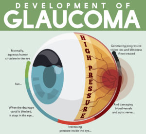
Glaucoma is one of these age-related eye diseases that has no early symptoms, which is why it is called the silent thief of sight. It is actually a group of diseases that can damage the eye’s optic nerve and result in vision loss and blindness. Open-angle glaucoma is the most common form disease.

In addition to an eye exam, you can reduce your chances of losing your vision to glaucoma by also:

  1. Live a healthy lifestyle that includes maintaining a proper weight, eating healthy foods, and not smoking.
  2. Know your family history to determine if you are at a higher risk for some eye diseases.
  3. Protect your eyes against harmful UV rays from the sun or your computer by wearing sunglasses when you are outdoors or computer glasses when using the computer for extended periods of time.
Silent thief of sight glacoma
Courtesy of NEI/NEHEP

1/5/16


Susan DeRemerSusan DeRemer, CFRE
Vice President of Development
Discovery Eye Foundation

5 Elements of Accessible Web Design

A common misconception is that all you need to have a successful blog or website is good content. While content people want to read is important, if you can’t see it, or it is difficult to read, very few people are going to take the time to try. There is lots of good content to choose from.

Making your content easy to scan and read and using great graphics that can tell a story are just as important. Especially if you want to reach the millions of potential readers that are blind or visually impaired.

While a person with low vision (due to age-related macular degeneration, cataracts,glaucoma or other eye diseases that are related to aging) can increase font size or graphics by enlarging them with a pinch of the fingers or scroll of the mouse, the result is often blurry and still difficult to see.

If a person is blind and using a screen reader, what they hear may not match what is written or displayed. A picture without underlying descriptive text is worthless. And when a blind person is using a screen reader to read a web site, they will often tab from link to link to scan your article, skipping over your text, to get a sense of what the options are. “Click here” says doesn’t tell the reader anything.

Here are 5 essential elements of accessible web design.

5 elements of accessible web design

12/15/15

Susan DeRemerSusan DeRenmer, CFRE
Vice President of Development
Discovery Eye Foundation

Low Vision Aging Adults at Higher Risk for Falls

Low vision in aging adults varies as do the occupational therapy techniques that might help older adults in becoming more independent. Mr. P has glaucoma resulting in a narrowed field of view. He is light sensitive, and keeps his blinds closed, darkening the house. He is responsible for doing his laundry, yet his washer and dryer are in the basement, causing a safety concern. Mrs. K has macular degeneration with 20/400 visual acuity. She has severely reduced contrast sensitivity, and can no longer drive or recognize faces easily. She no longer gets to the senior center for regular exercise sessions, which is concerning to her and her OT. Ms. T was diagnosed with diabetic retinopathy. She has struggled for years to accurately see her glucometer and insulin. She has recently developed peripheral neuropathy, leading to wheelchair dependency. Each of these patients is likely to leave their home less often because of their low vision, eventually leading to a decline in lower extremity weakness, balance, endurance and confidence. The fear of falling leads to more isolation, which can cause even more lower extremity weakness, problems with balance, decreased endurance, and even depression. The irony is that this cycle, which began with caution and a fear of falling, lends itself to exactly that. . .a fall.

Why are low-vision aging adults at higher risk for falls?

According to the CDC, “Each year, millions of older people-those 65 and older-fall. In fact, one out of three older people fall each year, but less than half tell their doctor. Falling once doubles your chances of falling again.” The typical aging adult with low vision faces challenges that others do not. Difficulty seeing details combined with reduced contrast sensitivity leads to a decline in mobility and socialization.
risk for falls
risk for falls
So how do we help older adults with low vision lower their risk for falling? When considering safety within the home, persons with low vision must make it a priority to add lighting & contrast whenever possible. Consider all rooms of the house, including entrances, hallways and stairways.

  • Placement of night lights in hallways, stairway, etc.
  •  

  • Keep flashlights in accessible places throughout the home where night lights are not possible.
  •  

  • Instead of closing blinds, keep them open & wear appropriate colored filters to manage glare/light sensitivity.
  •  

  • Small battery operated stick-on lights or rope lighting is inexpensive, and easy to place on steps to light up a stairwell.
  •  

  • Line edge of steps, or stairway railing with brightly colored duct tape to increase visibility.
  •  

  • Make sure grab bars, tub benches, shower chair are high contrast, to be most visible.
  •  

  • Remove throw rugs, with the exception of those providing function, such as the one at the entrance and bathtub. Their purpose is for providing dry shoes and feet, but they should have a non-skid back and a high contrast color to “stand out.”
  •  

  • Reduce clutter in rooms to increase safety by removing items from floors, walkways and stairwells.
  •  

  • Consider investing in a Medical Alert System to provide added piece of mind, confidence, especially when living alone.

Persons with low vision can decrease their frequency of falls by staying social and walking in the community. Unfortunately, many low vision adults become more house bound when they can no longer drive. Locating sources of alternative transportation may be helpful. Seeing faces is difficult, making socializing a challenge, which can lead to depression. The following recommendations take into consideration the challenges of not seeing details or across the visual field normally, while improving lower body strength, endurance, coordination, confidence, and hopefully reducing the risk of a fall.

  • Encourage regular trips to the grocery stores and the mall to keep physically fit. Malls are safe environments to walk around because of wide, straight hallways. Use magnifiers to see price tag/label details, or take advantage of personal shoppers who assist with locating items.
  •  

  • Participate in regularly scheduled exercise sessions (videos, groups, etc). Sit/stand in the front row, ask instructor to provide clear verbal instruction, instead of only demonstration. Consider hiring a personal trainer in order to get 1:1 instructions for how to use exercise equipment. If watching a video, move closer to your largest TV screen. Home exercise equipment can be labeled with high contrast markings to increase visibility of its details.
  •  

  • Consider joining a senior center in the community. Some have low vision support groups.
  •  

  • Access driving alternatives, such as the Smart Bus, Senior Centers, Local Volunteer or Church groups. Consider using money previously spent for a car and its expenses to hire a private driver.
  •  

  • Use appropriate colored filters in bright outside conditions or darker inside environments (i.e. Amber outside in the sun; Yellow inside a dark restaurant or outside with overcast weather).
  •  

  • Consider using a walker or support cane when walking longer distances in the community or neighborhood. Many individuals decline using an assistive device, not realizing how active and fit it can make them. Rolling walkers are even available with seats, allowing for rest breaks wherever necessary.

While the fear of falling is great amongst all seniors, those with low vision need to consider adding strategies that specifically benefit them. Making changes within the home may be as simple as improving lighting and contrast. Remaining physically fit outside of the home may be done with shopping or exercise equipment, but either way staying active will improve confidence, the fear of falling, and hopefully decrease the chance of a fall. If you are unsure of what approach is best for you, consult with your ophthalmologist about scheduling a low vision eye exam and occupational therapy.

Reference:
Centers for Disease Control and Prevention, National Center for Injury Prevention and Control-CDC 24/7: Saving Lives, Protecting People

12/3/15

Annette Babinski's headshot thumbnailAnnette M. Babinski, OTR/L CLVT
Henry Ford Health System
Vision Rehabilitation Center


Michelle Buck's headshot thumbnailMichelle Buck, MS, OTR/L CLVT
Henry Ford Health System
Vision Rehabilitation Center

#GivingTuesday – Giving For Eye Research

Medical research funding from the US government, the medical device and pharmaceutical industries, biotech and foundations has dropped 0.8% per year, every year from 2007 to 2012. Even with the economic recovery, the funding of eye research from the NIH in 2014 decreased by 17% from 2012.
giving for eye research
What makes this particularly disturbing is that the need for eye research is more important now than ever, as demonstrated by the infographic below.

Giving to eye research
This infographic by the National Institute of Health (NIH) is used with their permission.

Giving For Eye Research

The eye diseases in the above graphic are related to aging. The number of people being affected is more than doubling for each eye diseases, except for diabetic retinopathy which will increase by 47%, by 2050.

While we are more health conscious than in previous years, knowing the importance of exercise, healthy eating and not smoking, the US population is aging.

Aging baby boomers, the largest population group in US history, are creating a dramatic shift in the age composition of the U.S. population. It is projected that the entire senior population, including the pre-boomer silent generation, will reach 71.4 million people by 2029. This means that those people 65 and older will make up about 20%of the US population by 2029, up from almost 14% in 2012.

We need your help to find treatments and cures for these sight-threatening eye diseases.

December 1st will be the fourth year of #GivingTuesday. It is a global day of giving using combined power of social media and collaboration. Always the Tuesday following Thanksgiving, it has become as widely recognized as Black Friday and Cyber Monday and kicks off the philanthropic season, when many focus on their holiday and end-of-year giving.

We are asking that you support eye research through the Discovery Eye Foundation this philanthropic season with a gift on #GivingTuesday. It is easy to do; you can do it from the comfort of your home – just click here.

Also share this post with family and friends so they can also give the gift of sight this holiday season.

Thank you for reading our blog and for your support.

11/24/15

Susan DeRemerSusan DeRemer, CFRE
Vice President of Development
Discovery Eye Foundation

Technology for Vision

In just over 10 years, the technology that has been developed to help people see has been amazing. While medical research continues to move forward to find treatments and potential cures of the future, those with the technological know-how have created ways for people to see NOW. Here is a very brief look at some of these technologies.

Technology for Vision

Second Sight and the Argus II

One of the pioneers in the field of vision technology, Second Sight started in 1998, and they are now currently producing the second version of their device. It is made up of two parts:

The Implant: Requiring a 4-hour surgery, a device is surgically implanted in the eye on top of the retina, and along the outside of the eye. It includes an antenna, an electronics case, and an electrode array.
technology for vision
The External Equipment: It includes glasses, a video processing unit (VPU) and a cable.
technology for vision
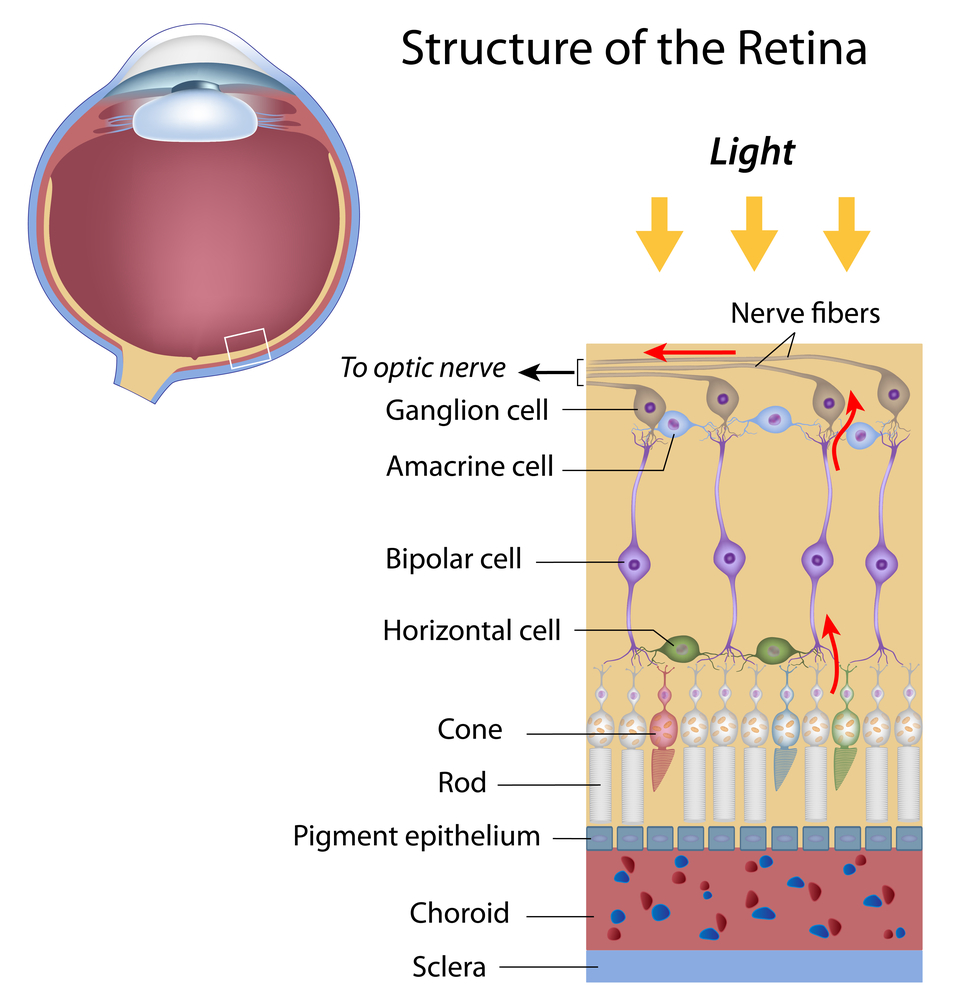
In a healthy eye, photoreceptors (rods and cones) in the retina convert light into tiny electrochemical impulses that are sent to the brain, where they are translated into images. If the photoreceptors don’t function correctly your brain can’t produce images. The Argus II Retinal Prosthesis System (“Argus II”) is designed to bypass the damaged photoreceptors.

A miniature video camera housed in the patient’s glasses captures an object. The video is sent to the small VPU that the patient wears. It is then processed and transmitted back to the glasses via a cable. This information is sent wirelessly to the antenna in the implant and signals are sent to the electrode array, which emits small pulses of electricity. These pulses bypass the damaged photoreceptors and stimulate the retina’s remaining cells. The visual information is then sent to the brain to create the perception of patterns of light which patients can learn to interpret as objects. Significant training is required to use the system.

The implant is designed to give you a visual field of about 3.5 inches by 6.5 inches at arm’s length; however, the actual size of light you see may be larger or smaller. Since it is strictly based on light, there is no color perception.

eSight

Another system that relies upon the user wearing glasses is eSight which started in 2012. While the Argus II is for people that have very little or no vision, the eSight is for people with low vision or that are legally blind. eSight glasses require the individual to have a certain degree of sight remaining to be successful. If you can only see shadows you probably don’t have enough remaining sight for the glasses to work for them.

This system is also composed of two parts, but it does not require any surgery:

The Headset: It contains a high-definition camera, OLED screens, and the ability to capture and display a real-time video feed. The headset is mounted on carrier frames, which enables eSight’s “bioptic tilt” feature so the user can shift between viewing modes and engage their peripheral vision.
technology for vision
The Controller: A small, lightweight processing unit that adjusts every pixel of the video in real time. It also houses the battery, which powers eSight.
technology for vision
Because most legally blind individuals retain limited sight concentrated in their peripheral vision, their eyes do not receive an adequate signal for the brain to recognize what is being seen. This can create blind spots, blurriness, inability to detect contrast, and other symptoms that reduce vision. eSight is able to significantly corrects these issues by using a high-speed camera, video processing software, a computer processor and the high quality video OLED screens to project a real-time image on the inside of the glasses, allowing people to see.

eSight requires considerably less training than the Argus II. It is intuitive, but as with learning anything new, the more you use it the easier it is to use. It is best to practice on a daily basis.

CentraSight from VisionCare Ophthalmic Technologies

The CentraSight uses a tiny telescope that is implanted inside the eye. The telescope implant was created to improve for people with end-stage age-related macular degeneration (AMD). The tiny telescope – about the size of a pea – is implanted inside one eye, behind the iris and is barely noticeable in your eye.
technology for vision
In end-stage AMD, the macula, where central vision occurs, is degenerated in both eyes without any healthy macular areas left for detailed central vision. There is no way for the individual to see around the central blind spot in their vision. It does not affect peripheral vision, which is blurry so you can’t use it to read, but you can use it to detect objects and movement.
technology for vision
Once implanted inside the eye, the tiny telescope works like a telephoto lens of a camera. It magnifies images onto the healthy areas of the retina to help improve central vision. Because the image is enlarged it reduces the effect the blind spot has on central vision. The surgical procedure is only performed on one eye because the peripheral (side) vision will be restricted in the eye with the telescope implant. This means the peripheral vision in the untreated eye will need to work in conjunction with the implanted eye. “A person uses the eye with the telescope implant for detailed central vision (such as reading “WALK” signs at a crosswalk). The other eye is used for peripheral vision (such as checking to see if cars are coming from the side).

Training with a CentraSight low vision specialist will be needed to develop the skills you need to use your new vision, such as how to switch your viewing back and forth between the eye with the telescope implant and the eye without the implant. You will still need to wear eye glasses and may need to use a hand-held magnifier with the telescope-implanted eye to read or see fine details clearly. However, in general, less magnification will be needed.

Ocumetics Bionic Lens

After eight years of research, a Canadian optometrist, Dr. Gareth Webb, has invented a tiny bionic lens that is able to enhance eyesight so that an individual can see three times better than the sharpness of 20/20 vision. The Ocumetics Bionic Lens is a button-shaped lens that can be injected into the eye in eight minutes in a procedure identical to cataract surgery.

As people get older, the lens inside the human eye becomes cloudy over time, causing blurred vision, known as cataracts. The Bionic Lens would be inserted, replacing the person’s clouded lens, similar to the intraocular lenses currently used in cataract surgery.

The Bionic Lens features a patented Ocumetics camera optics system, which is a tiny bio-mechanical camera that is able to shift focus from a close range object to optical infinity – as far as the eye can see – much faster than the human brain.

This device is still not available to the public, but Webb is hopeful that clinical trials will start soon. Depending on regulatory processes in each country, Webb hopes the Bionic Lens will be commercially available by 2017.

11/10/15


Susan DeRemerSusan DeRemer, CFRE
Vice President of Devlopment
Discovery Eye Foundation

The Importance of An Eye Exam

Why You Need An Eye Exam

The end of the year is fast approaching – when was the last time you had an eye exam? Was it a comprehensive eye exam?
eye exam
To keep your eyes healthy and maintain your vision, the American Optometric Association (AOA) recommends a comprehensive eye exam every two years for adults ages 18 to 60, and annual exams for people age 61 and older. However, if you have a family history of eye disease (glaucoma, macular degeneration, etc.), diabetes or high blood pressure, or have had an eye injury or surgery, you should have a comprehensive exam every year, unless otherwise indicated by your doctor.
Also, adults who wear contact lenses should have annual eye exams.

An important part of the comprehensive eye exam is the dilated eye exam to look inside your eye. Drops are placed in each eye to widen the pupil and allow more light to enter the eye. This gives your doctor a clear view of important tissues at the back of the eye, including the retina, the macula, and the optic nerve. This allows for early diagnosis of sight-threatening eye diseases like age-related macular degeneration, diabetic retinopathy, glaucoma, etc.

To better understand the importance of the dilated eye exam, here is a video from the National Eye Institute (NE) that explains what to expect.

At the end of your comprehensive eye exam your doctor should raise any concerns he has with you. But it is up to you to be prepared to react and ask questions for peace of mind and to help save your vision.

Questions To Ask After Your Eye Exam

It is always important to know if anything about your eyes have changed since your last visit. If the doctor says no, then the only thing you need to know is when they want to see you again.

If the doctor says the have been some minor changes, you need to know what questions to ask, such as:

  • Is my condition stable, or can I lose more sight?
  • What new symptoms should I watch out for?
  • Is there anything I can do to improve or help my vision?
  • When is the next time you want to see me?

If the doctor sees a marked change in your vision or give you a diagnosis of eye disease, you would want to ask:

  • Are there treatments for my eye disease?
  • When should I start treatment and how long will it last?
  • What are the benefits of this treatment and how successful is it?
  • What are the risks and possible side effects associated with this treatment?
  • Are there any foods, medications, or activities I should avoid while I am undergoing this treatment?
  • If I need to take medication, what should I do if I miss a dose or have a reaction?
  • Are there any other treatments available?
  • Will I need more tests necessary later?
  • How often should I schedule follow-up visits? Should I be monitored on a regular basis?
  • Am I still safe to drive?

Your vision is a terrible thing to lose, but with proper diet, exercise and no smoking, along with regularly scheduled eye exams, you improve your chances of maintaining your sight.

11/5/15


Susan DeRemerSusan DeRemer, CFRE

Diabetic Eye Diseases

November is National Diabetes Month. Diabetes is a controllable condition that is growing in the US. In adults 20 and older more than one in 10 people have diabetes, while in seniors (65 and older) that number increases to more than one in four.

Diabetic Eye Diseases

One of the eye diseases that can result from diabetes is diabetic retinopathy, which will affect approximately 11 million people by 2030. Check the infographic below to learn more about diabetic eye diseases.

diabetic eye diseases

11/3/15

NEI LogoCourtesy of the National Eye Institute (NEI), a part of NIH.

Increased Awareness for Saving Vision

The following is a survey done by Essilor (a French company that produces ophthalmic lenses along with ophthalmic optical equipment) and a large marketing research firm in the UK, YouGov. While the focus in on people living in the UK, the results would probably be similar to the US population. Even with increased access to the Internet, many people are still not aware of the risks associated with eye disease and what they can do to help retain their vision. Increased awareness of informational resources are important for saving vision.
saving vision
There are a number of websites with easy to understand information about taking care of your vision that I have listed under Resources to Help Save Vision at the bottom of this article. And while there are eye diseases that are hereditary, you can slow the onset and progression by making good lifestyle choices about smoking, diet and exercise. Your eye care specialist is also an excellent source of information about what you can to do reduce your risk of vision loss, at any age.

Increased Awareness for Saving Vision

A YouGov poll conducted with Essilor reveals that most Britons are unaware of damage to their eyes by surrounding objects, activities, and devices. This widespread lack of awareness means fewer people seeking methods of prevention and avoidance, and for those that are aware of risks, most are not informed of existing preventative measures.

The poll has shown* that many British people remain uninformed about the various ways in which eyes are damaged by common daily factors, despite evidence that eye health is affected by blue light, UV rays (reflected from common surfaces), diet, obesity, and smoking.
Of the 2,096 people polled, the percentage of respondents aware of the link between known factors affecting and eye health were:

  • Poor diet – 59%
  • Obesity – 35%
  • Smoking tobacco – 36%
  • UV light, not just direct from the sun but reflected off shiny surfaces – 54%
  • Blue light from low energy lightbulbs and electronic screens – 29%

More than one in ten people were completely unaware that any of these factors could affect your eyesight at all.
saving vision
72% of respondents own or wear prescription glasses but only 28% knew that there were lenses available (for both prescription and non-prescription glasses) to protect against some of these factors; specifically, blue light from electronic devices and low energy light bulbs, and UV light from direct sunlight and reflective surfaces.

76% admitted they haven’t heard of E-SPF ratings – the grade given to lenses to show the level of protection they offer against UV.

Just 13% have lenses with protection from direct and reflected UV light, and only 2% have protection from blue light (from screens, devices, and low energy bulbs).

Poll results showed that younger people were most aware of the dangers of UV and blue light, yet least aware of how smoking tobacco and obesity can affect your eye health. Within economic sectors, middle to high income people are more aware of the effects of smoking & obesity on eyesight than those with low income –

  • 39% of people with middle to high income compared to 33% of people with low income are aware of the impact of smoking tobacco.
  • 38% of people with middle to high income compared to 31% of people with low income are aware of the impact of obesity.

Awareness of the impacts of smoking and obesity on eye health is significantly higher in Scotland (47% & 49% respectively) than anywhere else in the UK (35% & 33% in England and 40% & 38% in Wales).
Essilor’s Professional Relations Manager, Andy Hepworth, has commented: “The lack of awareness about these common risks to people’s eyes is concerning. Not only would many more glasses wearers be better protected, but also many people who do not wear glasses would likely take precautions too, if made aware of the dangers and the existence of non-prescription protective lenses.”

To see the full results of the poll, please visit the Essilor website.

For more information on the protection offered from blue light and UV through specialist lens coatings, for both prescriptions and non-prescription glasses, please see here for UV & Blue Light Protection options.

*All figures, unless otherwise stated, are from YouGov Plc. Total sample size was 2,096 adults. Fieldwork was undertaken between 21st and 24th August 2015. The survey was carried out online. The figures have been weighted and are representative of all GB adults (aged 18+).

Resources To Help Save Vision
All About Vision
Macular Degeneration Partnership
National Eye Institute (NEI)
Prevent Blindness

10/16/15


Susan DeRemerSusan DeRemer, CFRE
Vice President of Development
Discovery Eye Foundation

Vision Loss and Depression

On Tuesday, September 29, 2015, the National Eye Institute (NEI) hosted a Twitter chat on vision loss and depression. Here are some highlights of that discussion, along with some great resources to learn more about dealing with vision loss and depression.
vision loss and depression

  • Many studies show that people with vision loss or low vision are at risk for depression, although not everyone with vision loss gets depressed.
  •  

  • A person with low vision is defined as someone who finds it difficult to do daily tasks even with regular glasses, contacts, medications or surgery.
  •  

  • The number of Americans with low vision will increase over 70% by 2030. Broken down by ethnicity, African Americans with low vision will increase 93% and Hispanics with low vision will increase 190% during the same period. This is due to the rapidly aging Boomer population. 88% of Americans with low vision are age 65 and older.
  •  

  • Symptoms of depression include persistent feelings of sadness, anxiety, irritability and fatigue. It is a common and serious illness that interferes with daily life. Each year, about 6.7% of American adults experience major depressive disorder. Women and men experience depression differently, with women 70% more likely to experience depression than men.
  •  

  • People 65 and older are at particular risk for developing serious depression related to vision loss and yet it is often underdiagnosed and undertreated. Older adults may have other, less obvious symptoms of depression or they may not be willing to talk about their feelings. Many overlooked because sadness is not their main symptom. It is important to remember that while depression is a common problem among older adults, it is not a normal part of aging.
  •  

  • An estimated 29-58% of those who suffer significant vision loss have major depressive disorder one year later. People with vision loss are 2x more likely to be depressed than someone without vision loss. Depression can be very disabling and may reduce the effectiveness of low vision rehabilitation interventions.
  •  

  • A recent study confirmed age-related macular degeneration (AMD) is a big contributor to depression risk, as it accounts for about 45% of low vision cases.
  •  

  • Older adults w/ vision loss are also 3X more likely to report difficulty in 1) walking, 2) managing medications, and 3) preparing meals. In fact about 39% of people with severe vision loss experience activities of daily living ADL limitations, compared to 7% of those with better vision. ADLs include eating, bathing, dressing, toileting, walking and continence.
  •  

  • A link between depression and vision loss was also found in people as young as 20 according to a recent study. It looked at over 10,000 adults in the US and found they were approximately 2x more likely to be depressed.
  •  

  • A decline in vision can also be associated with lower emotional, physical, and social functioning. To help those with low vision avoid depression it is important that they remain active and engaged in the world around them. And while people may become depressed because of vision loss, other causes of depression may also be present.
  •  

  • An integrated approach to depression management in older adults with impaired vision may be the best course of action. Behavioral activation helps people recognize that loss of the activities they enjoyed that have led to depression and encourages them to find ways to re-engage with these activities. After 4 months, behavioral activation reduced the risk of depression by 50% compared to the control group. Behavioral activation can be used alone, or as part of psychotherapy called cognitive-behavioral therapy (CBT).
  •  

  • Cognitive behavioral therapy (CBT) helps people with depression restructure negative thought patterns and to correct distorted thinking that is often part of depression. But it is important to remember that the best approach to treating depression is to personalize it for each individual.
  •  

  • Often, the combination of pharmacotherapy and psychotherapy is a very effective option for depression treatment. Other time-limited psychotherapies, including interpersonal therapy (IPT) are effective in treating depression in people of all ages.
  •  

  • Collaboration between eye care and mental health professionals can help people with vision loss improve their quality of life.

Resources
Understanding depression

How to live with low vision

Living with Low vision – How you can help webinar

Update on depression and AMD

Association of vison loss and depression in those over 20

Sadness impairs color perception

Rehab helps prevent depression from age-related vision loss

10/6/15

Susan DeRemerSusan DeRemer, CFRE
Vice President of Development
Discovery Eye Foundation

Primary Congenital Glaucoma

What is primary congenital glaucoma?

Glaucoma in children includes a variety of disorders in which drainage system of the eye does not function adequately, leading to abnormally high pressure inside of the eye (the intraocular pressure, or IOP), and resulting in damage to many different structures of the child’s eye. If not treated promptly and successfully, pediatric glaucoma can lead to severe vision loss or even blindness in one or both eyes. In primary childhood glaucoma, the drainage system usually has not formed properly (often resulting from a genetic abnormality) while in secondary childhood glaucoma, the abnormal fluid outflow problem results from other problems with the eye(s), sometimes accompanied by other medical problems outside the eyes.

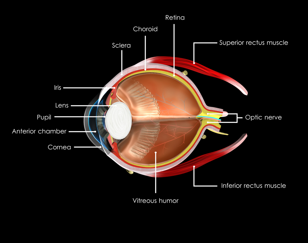
Primary congenital glaucoma is the most common of the primary childhood glaucoma types, although it is still rather rare. Let’s take a moment now to review the parts of the eye, and eye’s drainage system, sometimes also called the “aqueous outflow pathway”, since it drains the fluid within the eye (the aqueous humor), which is separate from the tears that flow on the outside of the eye’s surface and then into the nose or down a child’s cheeks.

The aqueous outflow pathway of the eye (comprising both the trabecular meshwork and Schlemm canal), situated at the junction (or “angle”) between the iris (the colored portion of the eye) and the sclera (the white part of the eye), has not formed correctly (Figure 1).

primary congenital glaucoma
Figure1. Schematic eye shows different structures of the ocular globe. Note that the Schlemm canal is part of the drainage system of the eye . Modified from National Eye Institute.

The aqueous humor therefore builds up within the front portion of the eye, causing abnormal elevation of the IOP.

In contrast to the eyes of adults and older children, the entire eye in infants and young children is distensible and the high IOP in primary congenital glaucoma often causes stretching and damage to several parts of the eye; this most often results in enlargement, clouding and scaring of the cornea (the front window of the eye) as well as severe nearsightedness, damage to the optic nerve, and resulting poor vision.

Primary congenital glaucoma (also called PCG) is almost always genetic, although usually there is no one else in the family with the condition. It is not related to anything that the parents did (or did not do) during the pregnancy or afterwards, and does not have any relationship to the baby’s sex or racial background. It occurs in about 1 every 10,000 to 20,000 births in western countries, but may be more common in certain populations of the world. Most babies with this disease are otherwise normal.

How is primary congenital glaucoma diagnosed?

Most cases present within six months of birth, with nearly 80% presenting before one year of age. In 70- 80 % of cases both eyes are affected. Most cases present for medical attention due to the size or cloudy appearance of the cornea in one or both eyes (Figure 2).

primary congenital glaucoma
Fig 2. Left eye of child with congenital glaucoma. The eye is enlarged and the front part of the eye is cloudy (corneal edema).

In cases where only one eye is affected, a difference in size can be seen between the two eyes and this sometimes brings the baby to the ophthalmologist (Figure 3).

primary congenital glaucoma
Fig 3. Different size of eyes in a child with congenital glaucoma. Note the increased size of the right eye. The brown area (iris) and the transparent part in front of the color part (cornea) are significantly larger in the right eye.

The diagnosis of PCG is based on clinical findings and there are three classic signs that the child can present with:

  • abnormal sensitivity or intolerance to light (photophobia)
  • excessive blinking or squinting of the eyelids (blepharospasm)
  • excessive tearing (epiphora)

The exam in clinic can be challenging for infants and young children and most require an exam under anesthesia, to allow detailed examination of the eye(s) that would not be possible in the clinic. Often the ophthalmologist will be able to follow the examination under anesthesia with the most appropriate surgery for the glaucoma, if surgery is indeed required.

How is primary congenital glaucoma treated?

PCG is almost always treated with surgery, although medications are often needed to help in addition to the surgery. Medications are very useful before initial surgery to help reduce the IOP and decrease the clouding of the cornea. In addition, medications may be recommended to keep the IOP to a safe level after surgery has been performed. If the IOP is not controlled successfully, or if damage has been substantial prior to diagnosis and treatment, PCG causes severe vision loss and can even cause blindness. Sometimes the damage from PCG is uneven between a child’s two eyes, leading to amblyopia (“lazy eye”) in the more severely affected size.

The initial surgical procedure of choice is usually aimed at opening the trabecular meshwork and Schlemm canal (the aqueous outflow pathway) of the affected eye(s). This so-called “angle surgery” can be performed either from inside of the eye (goniotomy) or externally (trabeculotomy), and may need to be repeated more than once in some cases.

If angle surgery fails, other procedures are available to allow the aqueous humor fluid to exit the eye (glaucoma drainage device or filtration surgery), or even to decrease the amount of fluid the eye makes (cycloablation procedures). For these more difficult procedures, the child is usually referred to an ophthalmic surgeon with expertise in treating childhood glaucoma.

What is the prognosis for children with primary congenital glaucoma?

While vision loss can be severe, prompt diagnosis and effective treatment and follow-up for children with PCG usually allows affected children to have best-corrected vision of at least 20/50 vision in their better-seeing eye. Children with PCG require continued careful follow-up and treatment their lifetime, and may require more than one surgery, eye drops, and spectacles.

Successful care for children with PCG takes a dedicated team including the family, ophthalmologist, teacher and community support, and the child him/herself.

10/1/15

primary congenital glaucomaElena Bitrian, MD
Assistant Professor of Ophthalmology, Division of Glaucoma
Mayo Clinic


primary congenital glaucomaSharon F Freedman, MD
Professor of Ophthalmology and Pediatrics
Chief of Pediatric Ophthalmology
Duke Eye Center, Duke University