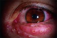
7 Healthy Eating Tips for the Holidays

12/16/14

Every year the holidays come around to tempt us with a variety of wonderful foods. It doesn’t make any difference if its Christmas or Hanukah, food is an integral part of the celebration. And the celebrations are pretty non-stop from Thanksgiving through New Year’s. Here are seven healthy eating tips for the holidays to help you handle all the delicious temptations of the season (the average person says they gain around five pounds).
healthy eating tips for the holidays
1. Eat before you go – Have a small healthy snack before you go to the party, to help control your appetite once you get there. Some great snacks include, cut up fresh vegetables, small handful of nuts (remember to make it small as they have quite a few calories, but they are healthy and filling), low-fat string cheese, etc.

2. Select wisely – When confronted with an open bar ask for club soda, tonic water or even tomato juice. Still want that glass of wine? Add some club soda to make it a wine spritzer and cut some of the calories. Once you have your drink, walk as far away from the bar as possible. Another good reason beside calories to cut down on alcohol – you will be able to drive home safely.

3. Strive to be the last in line – This works well in two ways, first being that once the food is picked over it doesn’t have the visual impact of the buffet when everything is presented in its perfection, making your mind go into overload as you try to decide what all to pile on your plate. Second, by the time you go through not everything might be available, and what’s left might not let you take too large of a helping.

4. How to use your plate – If you have more than one size plate to choose from, select the smallest one. Also know that filling your plate does not mean you have to cover every square inch with food, nor do you have see how high you can layer food. You are better off making more than one trip. On the first trip start with vegetables and salads while you scan what else is available. Then you can go back for small portions of what you think are the three best offerings.

5. Lend a helping hand – If the pull of the buffet becomes too strong, help out your hosts by pouring drinks, taking dirty dishes to the kitchen, even helping clean dishes.

6. Leave the party empty-handed – No matter how grateful your hosts are for you attending, do not take home any of the leftovers. No matter where you hide them in the refrigerator, you will still know they are there.

7. Elastic is not your friend – As you socialize and catch up with old friends you can get distracted, making it hard to watch what you eat. You are better off wearing something fitted, or at least with a regular waistband, to remind you that you might be eating too much. Belts are great as long as you don’t adjust them after getting to the party.

Just because a food is healthy, it doesn’t mean that it is low in calories. As you fill your plate, select foods that “bright and colorful.” These will generally be healthier options with a lower calorie count, such as fresh fruits and vegetables. But remember that fruits, because of their higher sugar content, will have more calories. Also the “bright and colorful” approach won’t work at the dessert table with brightly-decorated cookies, cakes and pies.

Here is a color palate to get you started:

RED – tomatoes, red peppers, kidney beans, strawberries, cranberries

ORANGE/YELLOW – pumpkin, butternut squash, yellow peppers, citrus fruit

GREEN – kale, spinach and other green leafy produce, broccoli, asparagus, apples, green peppers

BLUE/PURPLE – purple cabbage, eggplant, blueberries, plums, blackberries

Two bonus tips:

1. Shopping is stressful – Even when you are shopping you will be surrounded by food, especially at the mall where food courts and food kiosks offer a variety of “quick” snacks and meals. Plan ahead. You know what your shopping area has for you to eat. Find a healthy option that offers you good fiber and protein and plan to go there for lunch. Taking a lunch break will keep you from snacking all day and help you power through an afternoon of shopping. If you need snacks, pack something healthy in your purse.

2. Moms and baking go together – There is something about the holidays that brings out the baker in every mom. So when you go home, or even if mom arrives at your house, you will be presented with baked goods filled with love . . . and calories. Here is where avoidance won’t work, you need to rely on strategic moderation. Choose just a few samples to keep and enjoy, then either take the rest to the office, or make goodie gift bags for the mail or UPS delivery person, your manicurist, paperboy, etc. Mom will be pleased that you want to share her baking expertise.

And if you are the mom making these baked goods – try making fewer in December and spread them throughout the year. Less stress for you, and whom wouldn’t want to eat a spritz cookie in July?

Susan DeRemerSusan DeRemer, CFRE
Vice President of Development
Discovery Eye Foundation

Diabetes And The Potential For Diabetic Retinopathy

11/20/14

Defining Diabetes

Diabetes mellitus is a group of metabolic disorders in which a person has high blood sugar levels. It occurs either because the pancreas does not produce enough insulin, or because the body does not respond appropriately to the insulin that is produced. Insulin is the hormone that converts sugar into energy for the body. There are several forms of diabetes.

Type 1 diabetes occurs when the pancreas does not produce adequate levels of insulin. It usually develops during childhood or adolescence; however, it can also occur in adults. Those with type 1 diabetes must take insulin injections. About 10 percent of those with diabetes have type 1.

Type 2 diabetes is the most common type of diabetes, affecting about 90 percent of those with diabetes. It is caused by the combination of the body’s resistance to insulin (not using its own insulin efficiently) and the pancreas not producing enough insulin. As a result, there is an increase in the level of sugar, or glucose, in the blood.

Gestational diabetes occurs in pregnant woman who have not previously had diabetes but whose blood sugars become elevated during pregnancy. In many patients, their blood sugars will return to normal after the pregnancy is over. Placental hormones make the mother resistant to insulin, causing a buildup of blood sugars. These women are at high risk for type 2 diabetes later on in their lives.

Diabetic Retinopathy: An Overview

diabetic retinopathy
Anyone with uncontrolled diabetes is at risk for developing diabetic retinopathy. According to the National Eye Institute, between 40 to 45 percent of Americans with diabetes have some form of diabetic retinopathy, the most common eye condition associated with diabetes.

In the United States, the Centers for Disease Control and Prevention reports between 12,000 and 24,000 new cases of blindness each year due to diabetic retinopathy, making it the leading cause of vision loss among American adults, ages twenty to seventy-four. The Center also projects that by 2050, the number of Americans age forty and older with diabetic retinopathy will grow from a current 5 million individuals to about 16 million. Although these statistics are alarming, you can prevent or delay damage to your vision by controlling your diabetes, along with obtaining regular eye evaluations and treatment.

Defining Diabetic Retinopathy
Diabetic retinopathy is a disease affecting the retina, caused by elevated blood sugar levels. It usually affects both eyes and occurs when uncontrolled blood sugar levels damage the small vessels of the retina, the light-sensitive tissue in the back of your eye. The retina is responsible for processing images that make vision possible. To produce clear, distortion-free vision, the retina must lie completely flat. If the delicate retinal tissue is damaged, images that you see may be blurred or distorted.

Diabetic retinopathy is a progressive disease, and thus worsens over time. Although some effects, such as blurriness and distortions, may be mild or subtle, the long-term consequences can cause severe vision loss.

Symptoms of Diabetic Retinopathy

Because diabetic retinopathy rarely causes pain, symptoms are not always apparent in the earliest stages. In fact, damage to your retina could be occurring long before you have noticeable signs. When symptoms do occur, they’re often caused by retinopathy affecting the macula, the area at the center of the retina. Symptoms may include the following:

• blurred vision

• seeing dark spots or “floaters” (small specs in your field of vision)

• decreased night vision

• vision loss

• problems seeing colors

It’s important that you see your eye specialist immediately if you have any such symptoms. Diabetic retinopathy cannot be cured, but with careful monitoring, it can be diagnosed and treated, before your vision is impaired. The treatment is typically less invasive and more effective when diagnosed at an earlier stage, before permanent damage has occurred.

How the Retina Works

The retina is made up of specialized nerve tissue that contain microscopic receptors (called cones and rods) and other nerve cells that line the back of the eyeball.

These cells carry signals (images that we see) along the optic nerve to a special area of the brain, where they’re interpreted into what we perceive as sight. You might compare the retina to film in a camera—the film delivers the photo image that the camera captures.

There are two main areas of the retina that can be damaged by diabetic retinopathy:

• The macula, the center of the retina. The macula allows you to read and see fine details and recognize colors. At the very center of the macula is a dimple known as the fovea. It is the most sensitive portion of the macula and makes sharp vision possible.

• The peripheral retina, which is the portion of the retina that is outside the macula. It’s responsible for your side vision and also makes night vision possible.

The retina lies on a nutrient-rich flat “carpet” of vessels that nourish it with necessary oxygen and nutrients. To reach the retina, however, nutrients must pass through two buffers—a thin membrane called Bruch’s membrane and a single layer of specialized cells called the retinal pigment epithelium. Waste products are also transported away from the retina through these two membranes. Diabetic retinopathy can interfere with this constant import of necessary nutrients and export of waste products.

Risk Factors for Diabetic Retinopathy

There are many factors that can raise your risk for diabetic retinopathy. However, you’ll note that many of these risk factors can be controlled.

Duration of Diabetes and Glucose Control

The longer you’ve had poorly controlled blood glucose levels, the higher your risk for diabetic retinopathy. Most diabetic individuals develop eye problems overtime, making duration of their diabetes one of the strongest predictors that they will develop this eye disease. Research has shown that nearly all type 1 diabetics and 60 percent of type 2 diabetics develop the condition within the first two decades of their diabetes diagnosis.

The American Diabetes Association (ADA) recommends fasting glucose levels between 70 and 120 mg/dL and less than 180 mg/dL two hours after meals. They also recommend a hemoglobin A1c of 7 percent of less. Hemoglobin A1c is a protein in red blood cells that bonds with blood sugars. Since red blood cells can live from 90 to 120 days, the hemoglobin A1c stays in the blood for that length of time. Accordingly, it is effective in measuring the average blood sugars over a period of time. This test tells doctors how well your treatment plan is working. You should always know what your hemoglobin A1c values are, as they may affect the interval between your retinal examinations.

Obesity

The more fatty tissue you have, the more resistant your cells are to insulin. Obesity increases your risk for diabetes as well as other serious conditions such as heart disease. Estimates suggest that 65 percent of Americans may be overweight. Being overweight aggravates high blood pressure and cholesterol. Achieving a healthy weight is important in controlling blood sugars and diabetes related complications.

Lifestyle Choices

A sedentary lifestyle, especially if you are overweight, contributes to many diseases, including diabetes, heart disease, high blood pressure, and high cholesterol levels. On the other hand, physical exercise improves circulation, lowers blood sugars, and improves your body’s use of insulin. This results in improved blood sugar levels. This benefit of increased sensitivity to insulin continues for hours after you stop exercising.

Exercise also promotes weight loss. A sedentary lifestyle contributes to insulin resistance, and makes it more difficult to keep weight off. Even light or moderate physical activity can help lower blood sugars.

Smoking is another major risk factor for developing diabetic retinopathy. Smoking also causes diabetic retinopathy to progress faster. The nicotine in tobacco not only contributes to higher blood pressure and higher cholesterol levels, but it also impairs insulin activity. Even though quitting can be difficult, it is critical to heart health and diabetes control.
Unlike smoking, alcohol consumption doesn’t have a direct influence on diabetic retinopathy. Yet because it can affect diabetes control, drinking in excess can affect the health of your eyes. Your doctor can tell you what constitutes drinking in moderation for you.

High Cholesterol Levels

Diabetes puts you at risk for chronically high cholesterol or blood fats that promote the buildup of plaque in your arteries. Although the tiniest vessels of the retina are too small for such build-up, uncontrolled cholesterol can contribute to macular edema and the development of hard exudates, the small yellow spots or lipid deposits that may form in the macula. Both conditions are associated with a higher risk of vision loss.

Doctors advise keeping “bad” or low density cholesterol (LDLs) less than 70 mg/dL. Good cholesterol or high density lipoproteins (HDLs) should be greater than 40 mg/dL in men and 50 mg/dL in women. Both men and women should strive for triglycerides, another type of fat, at levels less than 150.

High Blood Pressure

If you have both diabetes and high blood pressure (also called hypertension), you may be at higher risk for a number of eye-related problems, including retinopathy, glaucoma and optic nerve damage. Seriously elevated blood pressure not only stresses your heart, it also raises the risk for eye problems, particularly macular edema and bleeding. Chronic hypertension combined with long-term diabetes also increases the chance that your retinopathy will be more destructive and progress more rapidly. Research has consistently shown that keeping your blood pressure below 130/80 mmHg is important in minimizing the risk of hypertension related complications.

Race/Ethnicity

Diabetic retinopathy is more common in some ethnic and racial groups than others. African Americans, Asian Americans, Hispanic/Latino Americans, American Indians and Alaskan Natives are at higher risk for type 2 diabetes than non-Hispanic whites.

African Americans and Mexicans are almost twice as likely as whites to have eye problems, according to the American Diabetes Association. Native Americans also have an increased for diabetic retinopathy. Researchers aren’t sure why some ethnic groups have higher rates of diabetes, which increase the risk for retinopathy and other problems.

Age and Gender

As mentioned earlier, the longer you have diabetes, the greater your risk for diabetic retinopathy. Not surprisingly, this complication is rare among children but common among older diabetic adults. A recent study by Prevent Blindness America and the National Eye Institute, demonstrated that older adult Americans are facing a bigger threat of all age-related eye diseases (diabetic retinopathy, age-related macular degeneration, cataracts and open angle glaucoma) today than at any other time.

Genetics

Our genetic make up has an important effect on our predisposition for many health issues such as diabetes. Scientists believe that many genes or combinations of genes either promote diabetes in certain individuals or protect them from developing it.

Scientists have yet to identify every gene involved in type 1 and type 2 diabetes, but they have shown that genetics are a factor. Research studies of identical twins, for instance, have demonstrated that if one twin has type 1 diabetes, the other twin has a 50 percent change of developing the disease. If one twin has type 2 diabetes, the other twin has a 75 percent chance of developing it.

Pregnancy

Gestational diabetes is a type of diabetes linked to pregnancy; however, diabetic retinopathy is usually not a complication in these women. However, if you’re already a diabetic and become pregnant, you are at an increased risk of developing diabetic retinopathy. This is a result of the hormonal and metabolic changes that occur during pregnancy, making the disease and its complications progress more rapidly. It is recommended that you see a retinal specialist for evaluation and monitoring.

In Summary

• Diabetic retinopathy is a serious complication of diabetes that results from high glucose levels damaging the retinal blood vessels. This can cause loss of vision.

• Between 40 and 45 percent of diabetic Americans have some form of diabetic retinopathy.

• The earliest form of the disease is called background diabetic retinopathy. With time it progresses to mild, moderate, or severe nonproliferative diabetic retinopathy.

• Without proper diagnosis and treatment of nonproliferative diabetic retinopathy, the condition can advance to proliferative diabetic retinopathy, which is a serious sight-threatening stage of the disease.

• Macular edema is due to build up of fluid and thickening of the macula and can occur with any type of diabetic retinopathy. It is the most common cause of vision loss in those with diabetes.

• The duration of your diabetes and how well blood glucose is controlled are major risk factors for the development and progression of diabetic retinopathy.

• Other risk factors that play a significant role in the development of retinopathy, include high blood pressure, high cholesterol, and smoking.

• As an individual with diabetes, you’re also at increased risk for other eye diseases, especially glaucoma, cataracts, retinal vein occlusion and optic nerve damage.

• Good blood sugar control, regular eye examination, and timely treatment are the key factors in reducing the damage to the eye and keeping your vision.

Pouya Dayani - Diabetic RetinopathyPouya N. Dayani, MD
Retina-Vitreous Associates Medical Group

Photophobia

11.4.14

Photophobia is a severe sensitivity to bright light which can result in discomfort and even pain. It can vary from person to person with discomfort coming from lights that don’t ordinarily affect others, including natural outdoor light, overhead lighting (such as fluorescent light), reflected light or flames of a fire.

Although in most cases photophobia is harmless, it is NOT an eye disease, but may be a symptom of an underlying problem. These could include an infection or abrasion that irritates the eye, a viral illness, medications, severe headache or migraine. sky

It could also be the result of your unique physical makeup. A person’s eye color can also affect ones sensitivity to light. People with lighter colored eyes can be more sensitive to light as opposed to people with darker eyes. An examination by an eye doctor can help determine the cause of your sensitivity.

The best treatment option for relieving the discomfort of photophobia is to address the underlying cause. The answer may be as simple as changing a medication or treating a corneal abrasion. Or your photophobia could be the clue that alerts doctors to another treatable condition. If you are naturally sensitive to light, wearing sunglasses with UV protection, brimmed hats or avoiding bright light situations are the only alternatives. In most cases, if you treat the underlying cause, the sensitivity will decrease or disappear.

Susan DeRemerSusan DeRemer, CFRE
Vice President of Development

Night Blindness

10/28/14

As the number of daylight hours decrease and daylight savings time is about to end, many of us feel that the days are getting much shorter. If you suffer from night blindness, your days are shorter, because getting around or driving at night, are sometimes impossible.

Night blindness is a condition that makes it difficult for a person to see in low-light situations or at night. Some types are treatable, while others are not. You will need to consult your eye doctor to determine the underlying cause of your night blindness to determine what can or cannot be done.

night blindness
Courtesy of wikipedia
There are several things that could cause night blindness:
•Cataracts
•Genetic eye disease
•Vitamin A deficiency
•Diabetes
•Aging eye
•Sunlight exposure

Here is a brief look at each.
Cataracts – This is when the lens of the eye becomes gradually becomes clouded, reducing vision. Besides reducing vision at night you may also experience halos around lights. This is a treatable condition requiring cataract surgery and replacing your clouded lens with a clear artificial lens. Your vision should improve considerably.

Genetic Eye Disease – Both retinitis pigmentosa or Usher syndrome are progressive genetic eye diseases where the rods that regulate light, and cones that control color perception and detail die. Progressive night blindness is one of the first visual symptoms of these two diseases. Currently there is no treatment for them as there is no way to treat or replace the dying rods.

Vitamin A Deficiency – While rare in the US, it can be a result of other diseases or conditions such as Crohn’s disease, celiac disease, cystic fibrosis or problems with the pancreas. Options to help with the deficiency include vitamin supplements suggested by your doctor, or increasing your intake of orange, yellow or green leafy vegetables.

Diabetes – People with diabetes are at higher risk for night vision problems because of the damage to the blood vessels and nerves in the resulting in diabetic retinopathy. Not only can it cause poor night vision, it may also take longer to see normally after coming indoors from bright light outside. There is no cure, but controlling blood sugar levels with medicine and diet can help prevent developing retinopathy or help slow the progression.

Aging Eye – As we age several things happen to our eyes. Our iris, which regulates the amount of light going into the eye, gets weaker and less responsive. This can make adapting from light to dark more difficult and slower. Our pupils shrink slightly allowing less light into the eye. The lens of the eye becomes cloudier, as explained above in cataracts, limiting the amount of light into the eye. We also have fewer rods for light perception. Aside from cataract surgery there is no treatment for age-related night blindness. However, eating a diet rich in fruits and vegetables and low in saturated fat is the best way to slow the progression. Here is more information on how the aging eye is affected.

Sunlight Exposure – If your night vision seems temporarily worse after a trip to the beach or a day on the ski slopes, it probably is. Sustained bright sunlight can impair your vision, especially if you fail to wear sunglasses or goggles.

Night blindness due to genetic conditions or aging cannot be prevented. However if you protect your eyes from extreme sunlight, eat a healthy diet, and monitor blood sugar levels if needed, you can reduce your chances for night blindness.

As we head into the holiday season, you should know that some great sources of vitamin A include sweet potatoes, butternut squash and … pumpkins!

Susan DeRemerSusan DeRemer, CFRE
Vice President of Development

15 Things Doctors Might Like Us To Know

10/16/14

The Doctor Patient Relationship

Doctors are human. Professional decorum may not let them speak their minds and for that reason, I have gathered comments heard from eye doctors with whom I have been privileged to associate as a so-called “expert patient”. These may not speak for all, but I do think they represent the majority. Here are 15 of the most important, which we might benefit from hearing.15 - doctor patient relationship

1. Your eyes are growing older, and I can’t turn back the years in an aging retina. I can, however, help you try to maintain your current vision for as long as possible.

2. Your appreciation means a lot. Don’t hesitate to compliment me if I have earned it.

3. I have quite a few patients who need my help and don’t want to be kept waiting. So I don’t have to keep repeating myself, please take notes, or bring someone with you who can refresh your memory later.

4. It is important that you comply with my instructions and show up for appointments if you want the best results.

5. My specialty is eye care. I will, however, try to provide you with resources for other ailments you may have.

6. Please be concise with your questions and stay on topic, so I’ll have time to treat other deserving patients.

7. Don’t believe everything you see in the media unless the source is reliable. If the information isn’t based on good evidence, my response to you may be disappointingly negative.

8. My services may seem expensive, but I have so many expenses and debts resulting from my profession that my bottom line may not be much different than yours.

9. An ethical physician will not choose a particular drug or treatment by how much of a profit he can make prescribing it.

10. I might not agree with everything you say, but rest assured that I will never let it affect the level of care I will provide.

11. Don’t expect me to keep all eye research in my head. I do know where to look it up, so allow me some time to get back to you on some questions.

12. Conferences I attend are sometimes in exotic places, but most of the time is spent going from one event to another, so it is really not a vacation.

13. Even if there is no current effective treatment for your eye disease, I still need to see you regularly to monitor your condition. Regular check-ups are important to your health and preventative care and treatment options are changing all the time.

14. I may not remember you personally, especially in the beginning or if I see you only see you once or twice a year, but your records contain everything I need to know to care for your vision.

15. I may not agree with your decision to try an alternative treatment, but I respect your right to do so. All I ask is that you seriously consider my opinion, and if you decide to go ahead with the treatment on your own, please keep me in the loop so I can monitor your condition.

We should try to understand that doctors are human beings who truly care about our welfare. Doing so could turn a top-down doctor-patient relationship into one of mutual respect and understanding. We might even get a few extra minutes of precious time in the chair, just because we’re a pleasure to have around.

dan robertsDan Roberts
“Expert Patient”

Being A Blind Artist

10/14/14

One thing that was always nurtured in me as a child was art. My father was an architect, and my mother is an art-lover. I remember they would collect all my artwork from preschool and elementary school. I used to have trouble sleeping as a kid, so I started drawing on the wall next to my bed; it would comfort me until I fell asleep. Most parents would probably scold a kid for drawing on the wall, but my parents loved it! In fact, my father would bring his architect friends into the room to “see my daughter’s mural.” Art has always been one area where I don’t feel inadequate. Art is so subjective, and anything can be art, so there is no judgment, and it’s very liberating. I’ve done abstract painting, mosaic tile work, ceramics and, currently, sculpture.

Becoming a Blind Artist

When I became blind, maybe five or six years in, I started going to Braille Institute, and I rediscovered art as a blind person. I saw people there creating art, and I thought, “This is amazing.”
Kooshay scuplture - blind artist
I always wanted to learn how to sculpt, but every time I called art schools about classes, when I told them I’m blind, they told me I had to take private lessons. Fear came up, and they did not know where to put me. Finally, two years ago, I started taking private lessons. My first piece was a portrait of a man, and the school was so amazed, they not only offered to put me in the sculpting class, but they gave me a full scholarship.

My private-lesson teacher wasn’t even a sculptor; she was a painter, but she was so tickled by the idea of a blind person wanting to sculpt that she wanted to be involved. She was a true artist and wanted to try something new. It was a wonderful experience for both of us. My second teacher would blindfold herself to figure out how to teach me. She really went out of her way, and I learned some really great techniques. I started my current class, which is a figure-sculpting class, by sculpting shoes — I have a shoe fetish — but I starting getting jealous of everyone else sculpting figures. I can’t touch the naked models, so now I touch other people’s sculptures and use that as a study to make my own.

I love it. I love the feel of clay. I’m a very tactile person, and I love the sensation of it in my hands. It’s so malleable. I use my hands more than sighted people; I don’t really use tools. I need to feel the clay to shape it, and I think there’s more emotion involved for me. It’s me, the piece and nothing else. Maybe it’s because I can’t see it — it’s like a meditation for me. I get lost in my piece. There is so much emotion — that’s where I get my inspiration. It’s a way to fantasize or fulfill an emotion or need. For me, it’s more about the process — I don’t see the finished product. It’s a very fulfilling way of expressing myself that words can’t; it’s more about expressing what I feel.

Kooshay Malek - seeingKooshay Malek
Marriage and Family Therapist
Los Angeles

What Are A Macular Pucker and Macular Hole?

10/9/14

What is the macula?
The eye is very much like a camera, taking light from the outside world and converting it into picture information that our brains perceive as vision. The retina is the light sensitive layer in the back of the eye that is very much like the film in that camera. The central retina, also known as the macula, is essential for crisp, high definition vision. Conditions that damage or distort the macula can therefore result in blurred or distorted vision. Two common conditions that affect the macula are macular puckers and macular holes.

What is a macular pucker or macular hole?
A macular pucker is a thin layer of scar tissue that forms on top of the retina. The amount of scar tissue can range from mild to severe. Mild macular puckers may be barely noticeable during an eye exam and resemble a fine layer of cellophane resting on the macula. More severe macular puckers can cause wrinkling or distortion of the macula. In contrast to a macular pucker, a macular hole is a small gap that extends through the entire thickness of the macula.

What are the symptoms of a macular pucker or a macular hole?
At first, a macular pucker may lead to mild blurring of the central vision. Because the problem involves the back of the eye, glasses will not completely restore vision. More severe macular puckers may result in wavy or distorted vision. For instance, objects that normally appear straight, such as venetian blinds or a printed line of text, might appear to have a dip or bend in the center. Small macular holes can cause similar symptoms of blurring or distortion. Larger macular holes often result in a central blind spot. This can also result in straight lines appearing broken or having a piece missing in the middle. Patients with a macular pucker or hole do not normally experience difficulty with peripheral vision.

What can cause a macular pucker or macular hole?
Recall that a macular pucker is a scar tissue. Anything that causes scar tissue, such as trauma or inflammation in the eye, can result in scar tissue and hence a macular pucker. Certain diseases that affect the retinal blood vessels such as diabetes can also cause a macular pucker to form. However, one of the most common causes of macular pucker is simple aging of structures within the eye. As the eye ages, the clear jelly that fills it, called the vitreous gel, shrinks. When enough shrinkage occurs, the vitreous gel detaches from its normal position adjacent to the retina. This process of vitreous detachment can cause microscopic damage or inflammation leading to macular pucker formation. In some cases, the vitreous gel does not detach cleanly from the retina. Instead it can put traction on the macula, pulling its delicate structures apart in the center, resulting in a macular hole.

How are macular puckers and macular holes diagnosed?
A simple examination from an ophthalmologist or retina specialist is often enough to diagnose a macular pucker or hole. However, additional testing is often useful in diagnosing subtle cases or monitoring eyes for changes. An optical coherence tomography (OCT) scan is a specialized photograph that allows your physician to look for microscopic changes in the contour of the macula. The following figures show an OCT of a normal macula, a macular hole, and a macular pucker. Note that the normal macula has a central dip known as the fovea, shown in Figure 1. In Figure 2, the dip is replaced by a gap which is a macular hole. Finally, Figure 3 shows a macular pucker where the dip is no longer visible. This is because the macular pucker, seen as a thin white line is distorting the normal shape of the macula.

Normal - Macular Pucker and Macular Hole
Figure 1: Normal Macula
Hole - Macular Pucker and Macular Hole
Figure 2: Macular Hole
Pucker - Macular Pucker and Macular Hole
Figure 3: Macular Pucker

What treatments are available for macular puckers and macular holes?
Macular puckers can be quite mild. For mild cases in patients with minimal symptoms, periodic monitoring may be all that is required. When blurred vision due to a macular pucker begins to affect activities such as driving or reading, treatment in the form of surgery can be considered. Surgery for a macular pucker is known as a vitrectomy. Vitrectomy surgery is usually done under local anesthesia and as an outpatient procedure. During the surgery, fine instruments are used to remove the scar tissue from the surface of the macula. After surgery, patients usually experience an improvement in the blurring and distortion as the eye recovers gradually over a period of months. Some residual waviness can be normal. Vitrectomy is generally very safe although there is a chance of increased cataract growth and a small risk of infection or retinal detachment.

For patients with small macular holes, close monitoring can also be an option since some macular holes can close on their own. For larger holes, there are two options. In select cases where the vitreous gel is actively pulling on the macula, an injection of medication into the eye may cause the gel to release cleanly, allowing the hole to close. In other cases, vitrectomy is recommended. During the surgery, any pulling on the macula is relieved and a gas bubble is placed in the eye to help the hole close. After surgery, patients are asked to look down for a several days to allow the bubble to float up against the hole. Once the body absorbs the bubble, vision is usually significantly improved.
In summary, both macular puckers and holes are common causes of blurry or distorted central vision. If treatment or surgery by a retina specialist is needed, the results are generally quite good and lead to significant restoration of vision.

Liao - Macular Pucker and Macular HoleDavid Liao, MD, PhD
Retina-Vitreous Medical Group

Our Thanks to Guest Bloggers Continues

10/7/14

More Amazing Guest Bloggers

Last week I took the opportunity to thank our very first guest bloggers for helping us launch the Discovery Eye Foundation Blog. We are pleased that so many people appreciate the wide range of eye-related information from eye care professionals, as well as the stories from people that live with eye disease on a daily basis.
Thank you part 2
Here is a round-up of guest bloggers since June 2014 that shared their time, experience and/or expertise to provide you with the best eye-related information.

Sumit “Sam“ Garg, MDwhat you should know about cataracts

Randall V. Wong, MDfloaters, causes and treatments

Roy Kennedyhis personal experiences with the miniature telescope implant

Sandra Young, ODthe importance of getting vitamins and minerals from your food and not just supplements

Jeanette Hassemanliving with keratoconus

Greg Shanetheater for the blind

Caitlin Hernandezblind actress and playwright

Jullia A. Rosdahl, MD, PhDlasers for glaucoma and genetics and glaucoma

Maureen A. Duffy, CVRTways to reduce harmful effects of sun glare

Kooshay Malekwhat is it like to lose your vision and being a blind therapist

Jeffrey J. Walline, OD PhDchildren and contact lenses

Robert Mahoneychoosing a home care agency

Robert W. Lingua, MDnystagmus in children

Buddy Russell, FCLSA, COMTcommon pediatric eye diseases, treatment options for children and pediatric contact lenses

NIH (National Institute of Health)telemedicine for ROP diagnosis

Harriet A. Hall, MDevaluating online treatment claims

Patty Gadjewskithe life-changing effects of a telescopic implant

Michael A. Ward, MMSc, FAAOproper contact lens care and wearing contacts and using cosmetics

Susan DeRemerSusan DeRemer, CFRE
Vice President of Development
Discovery Eye Foundation