
Fuchs’ Dystrophy: Current Insights

What is Fuchs’ Dystrophy?

Corneal dystrophies are a debilitating group of progressive diseases that can ultimately deprive a person of sight. The cornea, which forms the front of the eye, is a window for vision, and dystrophies due to intrinsic defects in the corneal tissue cause this window to become opaque and hazy. Fuchs’ dystrophy, also known as Fuchs’ corneal endothelial dystrophy (FCED), is amongst the most commonly diagnosed corneal dystrophies requiring corneal transplantation. The ophthalmologist Ernest Fuchs first described the disease in 1910.

Who gets it?

The disease is rare, and it is difficult to predict who will get it. We know that it affects women more than men (3:1 ratio), older adults (older than 50 years of age), and those with a family history. There are forms in which there could be up to a 50% chance of transmission to children of parents with Fuchs’ dystrophy. Most cases, however, occur sporadically.

What causes it and how does it progress?

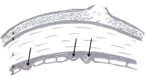
Although the cause of Fuchs’ dystrophy is still being studied, there are characteristic findings associated with it: small outgrowths on Descemet’s membrane called “guttae” or “guttata”, thickening of Descemet’s membrane, and defects in the endothelial cells (Figure 1).

fuchs dystrophy 1
Figure 1: Fuchs’ dystrophy can affect all layers of the cornea. Layers of the cornea from anterior to posterior, or frontside to backside, include (A) epithelial cells where blisters and bullae may form in late-stage disease, (B) Bowman’s layer where scarring can occur in late-stage disease, (C) stroma where corneal swelling occurs early in disease, (D) Descemet’s membrane where guttae form (arrows) and thickening occurs, and (E) endothelial cells that decrease in number and change shape and size with disease progression.

Descemet’s membrane is a thin corneal layer between the endothelial cell and the stromal layers of the cornea. Endothelial cells make up the backside of the cornea and function as a barrier and pump for keeping fluid out of the cornea and maintaining corneal clarity. As guttae accumulate on Descemet’s membrane, patients experience progressive loss and change in endothelial cells. Dysfunction of endothelial cells causes corneal swelling, which distorts vision. First, the back of the cornea swells, and eventually, swelling can reach the epithelial cells at the front of the cornea. Swelling can range from mild moisture accumulation, to painful “bullae”, or blisters. In very late-stage disease, significant corneal scar tissue can form and dramatically reduce vision. The progression to late stage Fuchs’ varies from person to person, but usually takes a couple of decades.

What are signs and symptoms?

A patient may be asymptomatic for years despite having guttae. Initial symptoms, including blurry, hazy, or cloudy vision, are typically due to corneal swelling from dysfunction of the endothelial cell layer. Patients may also experience glare or halos around light in the early stages just from the density of guttae. New studies suggest that patients can get glare and higher order aberrations from guttae without any corneal swelling. Symptoms tend to be worse on awakening, but usually improve throughout the day. This is because the closure of eyelids during sleep results in the accumulation of fluid in the cornea. For the same reason, humid weather can also worsen symptoms. As the disease progresses, poor vision may last longer into the day. There may be associated pain if blisters develop.

How is it diagnosed?

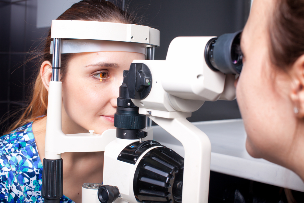
The presence of any of the above signs and symptoms, especially with a family history of Fuchs’, should prompt a consult with an ophthalmologist who will diagnose the disorder and follow its progression with regular checkups. An ophthalmologist will conduct a microscopic slit-lamp examination of the eyes, looking for guttae and Descemet’s membrane thickening (Figure 2).

fuchs dystrophy 2
Figure 2: Slit-lamp examination showing speckling pattern on the backside of the cornea characteristic of guttae in Fuchs’ dystrophy.

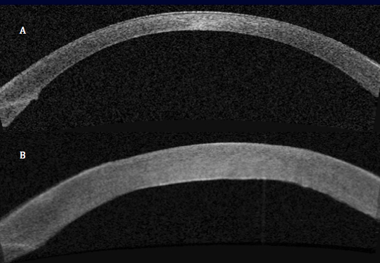
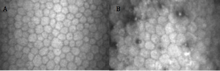
Special tests may be done to measure corneal thickness, a marker of swelling, or count endothelial cells to track disease progression (Figure 3 and 4).

fuchs dystrophy 3
Figure 3: Optical Coherence Tomography (OCT) showing (A) a normal, healthy cornea and (B) corneal swelling typical in Fuchs’ dystrophy.
fuchs dystrophy 4
Figure 4: In-vivo slit-lamp scanning confocal microscopy showing (A) normal endothelial cells and (B) guttae causing endothelial cell loss and change in Fuchs’ dystrophy.

How is it managed?

Management can be medical or surgical depending on symptoms. Patients may have mild or slow progression of disease that can be managed medically including over the counter salt solution drops (5% NaCl) to reduce corneal edema.

When there is late-stage disease, a corneal transplant may be necessary to improve vision. A corneal transplant replaces the patient’s corneal tissue with human donor corneal tissue. Donor corneas are readily available via excellent eye banks throughout the United States. The surgery is outpatient surgery with regular follow-up appointments and suture removal during the subsequent months. The postoperative healing of the cornea and vision stabilization can take up to a year.

Great strides have been made in the last decade in corneal transplantation surgery, giving patients better treatment options. Patients used to be limited to penetrating keratoplasty (PK), a full-thickness replacement of the cornea. We now have newer surgeries known as endothelial keratoplasty (EK), which is a partial-thickness transplant that replaces only the damaged part of the cornea (the endothelial layer). The different types of EK are DSEK (Descemet’s-Stripping Endothelial Keratoplasty) and DMEK (Descemet’s Membrane Endothelial Keratoplasty). The techniques vary by thickness of the transplanted tissue. The type of EK most appropriate is determined by the corneal surgeon and is variable on a case to case basis. Both types of EK surgeries provide comparable long-term visual results. In both surgeries, the patient’s diseased Descemet’s membrane and endothelial cells are stripped from the inner layer of their cornea. The thin lamellar donor graft is then inserted into the eye and positioned onto the back of the patient’s cornea via a gas or air bubble. The patient is then instructed to lie in a face up position for several hours post surgery during which time the bubble supports the graft until the new endothelial cell pumps begin to wake up and naturally adhere to the back side of the recipient cornea. Occasionally, the doctor may replace another air bubble into the eye the next day to allow more time for the graft to adhere. Visual recovery is on the order of 1-2 weeks in DMEK and 2-3 months in DSEK surgery. Rejection risk is still a possibility in EK surgery but has a much lower rate than traditional full thickness PK surgery.

Other surgical considerations depend on the presence of cataracts. Cataract surgery can worsen Fuchs’ dystrophy because of damage to the endothelial cell layer. For this reason, patients with cataracts and Fuchs’ requiring surgical intervention are often recommended to undergo cataract surgery before or at the same time as corneal transplantation to ensure the best outcome for the transplant.

Patients should work with an ophthalmologist to determine the best management plan. Ultimately, vast improvements in treatment options have given many Fuchs’ dystrophy patients the exciting opportunity to regain vision with improved healing times and reduced infection and rejection of the graft.

Citations: Figure 2 and 4 are from Zhang J, Patel DV. The pathophysiology of Fuchs’ endothelial dystrophy—a review of molecular and cellular insights. Exp Eye Res. 2015 Jan

6/4/15

priscilla-thumbnailPriscilla Q. Vu, MS
Medical Student
University of California, Irvine School of Medicine

–
–
–

Farid 3.6.14Marjan Farid, MD
Director of Cornea, Cataract, and Refractive Surgery
Vice-Chair of Ophthalmic Faculty
Director of the Cornea Fellowship Program
Associate Professor of Ophthalmology
Gavin Herbert Eye Institute, University of California, Irvine

9 Ways To Relieve Cataract Surgery Stress

It’s an extremely rare person who would not feel nervous before surgery of any kind, even if it’s an outpatient procedure that will only take a few minutes. In the case of cataract surgery, the fear can be even worse than the procedure itself.


9 Ways to Relieve Cataract Surgery Stress
People who are under intense stress can suffer a range of symptoms, including irregular or racing heartbeat, nausea, upset stomach, difficulty breathing, and an inability to sleep. It can even affect your mind, causing you to forget important details about the operation, like advice on how to get ready or what to do after you come home from surgery.

Here are 9 ways to relieve cataract surgery stress:

1.  Just Think About It – Let’s start with the first, and most difficult, suggestion—change your own mind about how you feel. Admittedly, it takes a great deal of discipline that’s hard to muster in the face of great anxiety, but try to remind yourself how your sight will be saved after a relatively short, quick, and easy procedure. What you are about to go through will prevent you from going blind.


2.  Learn Everything You Can – For many, it helps to learn as much as possible about the surgery before it happens. Knowing exactly what’s going to happen and how others have dealt with what you’re going through can be a great relief. Knowledge may be all you need to relieve your anxiety.


3.  Talk to Your Surgeon – It almost always helps to just talk to someone, and who better than your surgeon? Who else knows every detail of the procedure you’re about to undergo? It’s fairly likely that your surgeon has performed many successful surgeries of this kind before, and he or she may have some stories of encouragement for you, as well as important and comforting knowledge of his or her own personal experiences.


4.  Imagine the End Result – It may help if you keep focused on what happens after, as if it’s already done and you can go home, the procedure over. This takes a great deal of imagination, rather than the discipline of thinking rationally about it, but if you have that level of imagination, it’s certainly worth trying.


5.  Alternate Methods – This encompasses a whole range of stress-reducing tactics that are not usually under the medical umbrella. Nevertheless, they have done a great many people a lot of good. Yoga, hypnosis, massage, acupuncture, acupressure, and other treatments have allowed those suffering preoperative stress to sleep better at the least.


6.  Herbal Supplements – A form of alternative treatment involving traditional ingredients to produce a more restful state. The herbs are often just infused into tea and drunk. While these supplements are generally called “all natural,” you should always consult your doctor before taking them, since they can have an effect on other medications you may be taking, including the anesthetic you are given before surgery.


7.  Have Some Fun – Do something fun to take your mind off of what’s about to happen. Whatever that specific thing may be is up to you, since an individual’s idea of fun differs from person to person. Whatever you generally do to take your mind off of things and unwind after a hard day may be just the thing to help you out before undergoing cataract surgery.


8.  Treat Yourself – In the same vein has doing something fun, do something that usually relaxes you. If you like going for long walks, do that the day before the surgery. Or listen to music that fills you with peace (or joy). Take a long bath. If it makes you feel relaxed or calm, it will help you deal with your anxiety.


9.  Distract Yourself – Once you get to the hospital, you’ll probably be waiting around, even if you get there right on time, giving plenty of time for stress to ramp up. You’ll do better if you keep yourself entertained, but the hospital waiting room is probably one of the more boring places on earth. Fix that by bringing something along to entertain yourself, like a book or some magazines, or even stream your favorite movie or podcast. You’ll probably want to bring more than one thing to do, in case the wait is long.


Stress adds complication to the body’s systems, and can therefore cause some complication in the upcoming surgery. Do what you can, whether you kick back with friends or take some herbal supplements, to help yourself get into the best mind space possible. Think about the positive outcomes, and you’ll do well.


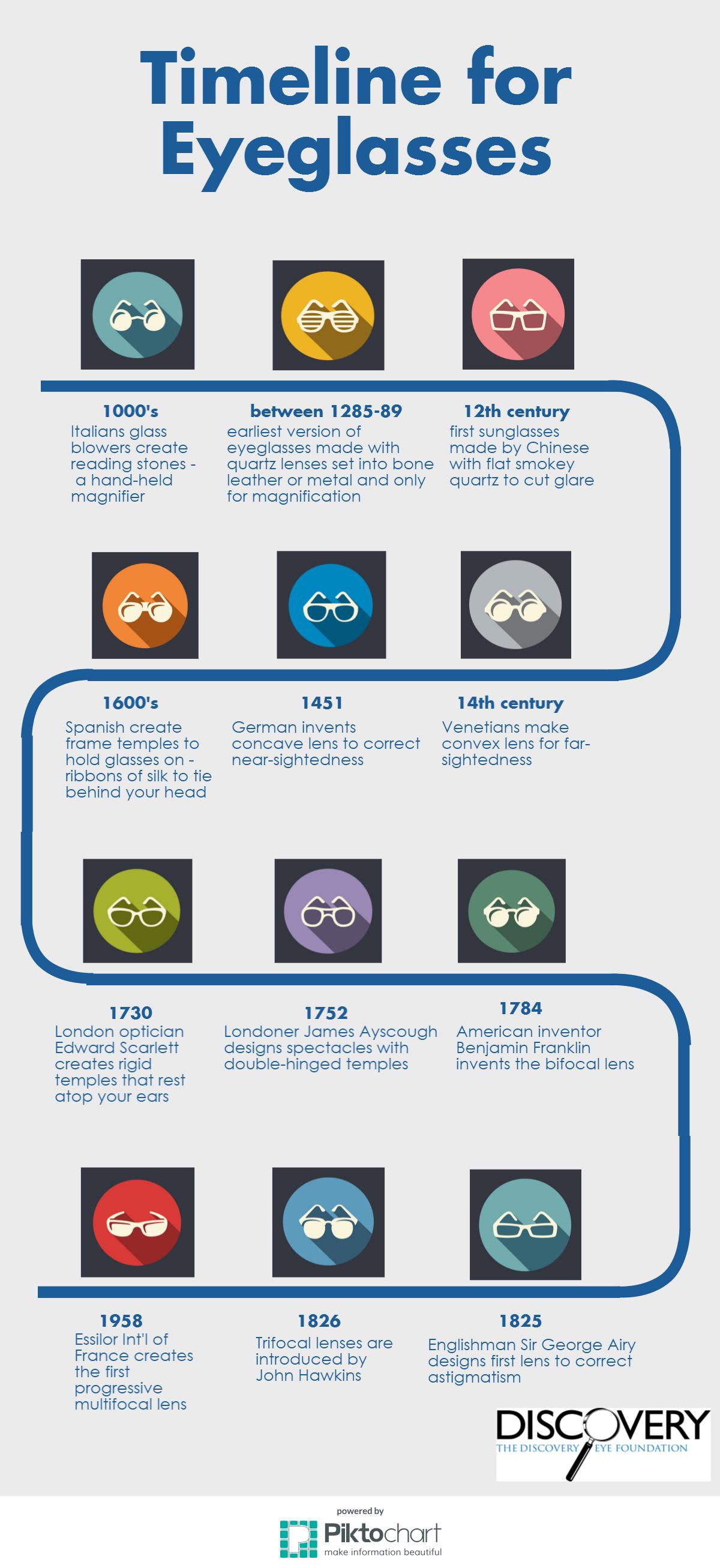
Eyeglasses Timeline

Eyeglasses are something we all take for granted, but they haven’t always existed. More than 700 year ago you had to learn to live with poor vision. Now more than 6 in 10 people in the US wear either glasses or contact lenses, with 60% of them being far-sighted. Here is an eyeglasses timeline to see how eyeglasses have evolved.

Eyeglasses timeline

5/12/15

Susan DeRemerSusan DeRemer, CFRE
Vice President of Development
Discovery Eye Foundation

Itchy Eyes? It Must Be Allergy Season

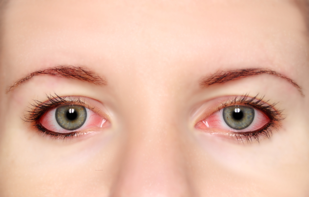
Spring is in the air, which also means it’s the season for allergies (i.e. Itchy, watery, red and generally unhappy eyes). People with keratoconus need to be particularly careful around allergy season, because rubbing your eyes can exacerbate both your allergies and keratoconus. woman with itchy eyesAmong the general keratoconus population, we see a significant incidence of allergic eye disease, which causes itchy eyes. In addition to this, there is a high correlation of keratoconus patients who rub their eyes. If you are one of these, STOP. When you rub your itchy eyes, it damages the mast cells within the eye tissue, causing histamine to release from these cells. More histamine around your eyes will increase itching and your urge to rub, which in turn can cause keratoconus to get worse. So even if rubbing makes your itchy eyes feel good, stop: the potential long term damage outweighs the short-term relief.

Whether your eyes are watery, itchy, or red around this time, you’ll find the best relief beyond your neighborhood drug store’s allergy aisle. While there are over-the-counter medications that help allergies, they may not be the best option for you. Oral medications for allergies tend to dry out the body in general (which is why they make a runny nose stop running), which includes the eyes. When it comes to eye drop options for allergy relief, the results are often unpredictable and short-lived.

Some surprisingly simple changes to your home can help. Keeping the windows and doors closed to keep the allergens out is helpful. Take your shoes off before coming into the house, consider taking a shower before bedtime, or frequently wash your pillowcases, which may remove enough of the allergen to improve your symptoms. If you have a pet that goes outside, consider bathing them twice a week to remove allergens they track in. For direct, immediate relief, place a cold wash cloth on the surface of your eyes to calm the symptoms.

The next step to relief includes personalized recommendations from your eye care provider (ECP). A common medication your ECP may prescribe is an antihistamine-mast cell stabilizer, such as Pataday or Lastacaft. These combination drugs use an antihistamine for immediate comfort and a mast cell stabilizer to carry out the benefits long term. Often these medications are so effective that they may be the only ones you need. When your allergies are severe and this combination drug is not enough, your ECP may add a topical steroid ointment to complete your treatment.

Be extremely diligent in rubbing and rinsing your lenses with fresh solution prior to storing them overnight. This will help to remove the allergens that have accumulated on them during the day.

Even if your annual eye exam doesn’t happen around allergy season, remember to visit your eye care provider if you have allergy symptoms. It may require a combination of prescription medications and environmental changes tailored to your specific symptoms to prepare your eyes now and for allergy seasons to come.

5/7/15


David Kading, OD - itchy eyesDavid L. Kading OD, FAAO
Specialty Eyecare Group
Offices in Kirkland, WA and Seatte, WA

_
_
_

Charissa Young - itchy eyesCharissa Young
Optometric Extern
Eye Care Group of Southern Oregon, PC

May Is Healthy Vision Month

May Is Healthy Vision Month

It’s Healthy Vision Month! Make Your Vision Last a Lifetime.

When it comes to our health, we often visit our doctor or nurse regularly to make sure our bodies are healthy. But what about our eyes? They’re not always top of mind, but they’re just as important.

During Healthy Vision Month, held each May, the National Eye Institute (NEI) reminds you to make your eye health a priority and encourages you to take important steps to protect your sight.
healthy vision month
Get a dilated eye exam. Getting a dilated eye exam is the only way to detect eye diseases early, because with many, there are no warning signs. Talk to your eye care professional about how often you should have one. If you want to see what your eye care professional sees during a dilated eye exam, check out NEI’s new eye exam animation!
Healthy-Lifestyle-FB-Cover-Photo_2015
Live a healthy lifestyle. Eating healthy foods, maintaining a healthy weight, managing chronic conditions, and not smoking can lower your risk of eye disease.
healthy vision month
Know your family history. Talk to your family members about their eye health history. It’s important to know if anyone has been diagnosed with an eye disease, since many are hereditary. This will help to determine if you are at higher risk for developing an eye disease yourself.
Protective-Eyewear-FB-Cover-Photo_2015
Use protective eyewear. Protect your eyes when doing chores around the house, playing sports, or on the job to prevent eye injuries from happening. This includes wearing safety glasses, goggles, safety shields, and eye guards that are made of polycarbonate.
Wear-Sunglasses-FB-Cover-Photo_2015
Wear sunglasses. When purchasing sunglasses, look for ones that block out 99 to 100% of both UVA and UVB radiation, so you can keep your eyes healthy. Prolonged exposure to sunlight can increase your risk for getting an eye disease. A wide-brimmed hat offers great protection, too!

These steps can help you keep your eyes healthy and prevent vision loss and blindness from eye disease.

To learn more about Healthy Vision Month and find additional eye health information, visit www.nei.nih.gov/hvm.

5/5/15

NEI LogoNational Eye Institute
National Institute of Health

Famous People with Vision Loss – Part II

Here is part two of our look at famous people with vision loss. The focus this time is the creative minds of artists, actors and musicians.

Artists

Georgia O’Keeffe (1887-1986) – Best known for her paintings of enlarged flowers, New York skyscrapers, and New Mexico landscapes, she was recognized as the “Mother of American modernism”. In 1972, O’Keeffe’s eyesight was compromised by age-related macular degeneration (AMD), leading to the loss of central vision and leaving her with only peripheral vision. She stopped oil painting without assistance in 1972, but continued working in pencil and charcoal until 1984.

Claude Monet (1840-1926) – A French impressionist painter who had cataracts that blurred his vision and caused a severe loss of color perception. After complaining about his cataracts for ten years, he had cataract surgery in 1923. Those paintings before the surgery have more of a reddish tone, while those painted before his cataracts and after the surgery show more blue tones.

Edgar Degas (1834-1917) – A French artist known for his paintings, sculptures, prints, and drawings. He had a chronic and progressive retinal disease that made him sensitive to light and caused him to lose his central vision. Frustrated by these limitations he switched to oil pastels because less precision was needed. He finally resorted to sculpting to be able to utilize his sense of touch and feeling in order to continue to create art.

Actors

Johnny Depp (1963-) – Known for his many movie roles, Depp doesn’t let his vision loss stop his career. Besides correcting his blurred vision, the tinted lenses of the glasses he wears also help with Meares-Irien ‘visual stress’ syndrome, which causes dizziness, discomfort and eye pain. He is also nearly blind in his left eye.

Issac Lidsky (1979-) – A child actor known for his part in the Saved by the Bell: The New Class TV series, he was diagnosed with retinitis pigmentosa (RP) at 13 and was completely blind by 25 years of age. He changed the course of his career, becoming a lawyer. He was a law clerk for Justices Sandra Day O’Connor and Ruth Bader Ginsburg in 2008-09, making him the the first blind US Supreme Court clerk.

Jon Weller (1975-) – A TV actor who has performed on many shows, he is best known for his recurring role of toxicologist Henry Andrews on CSI: Crime Scene Investigation. He has been diagnosed with RP.

Dame Judith Dench (1934-) – A actress has had a long career in theater and film, receiving many awards including ten BAFTAs, six Olivier Awards, two Screen Actors Guild Awards, two Golden Globes, an Academy Award, and a Tony Award. She is well-known for the part of M in the James Bond series of films. In 2012 She announced that she has age-related macular degeneration (AMD), but has no intentions of slowing down. Scripts are now provided to her in a larger font.

Musicians

Ray Charles (1930-2004) – He was born with congenital glaucoma which resulted in blindness by the age of 7. This American singer, songwriter, musician and composer, received multiple Grammys, was inducted into the Rock and Roll Hall of Fame and a Kennedy Centers Honoree.

Stevie Wonder (1950-) – He is a singer-songwriter, multi-instrumentalist and record producer who signed with Motown Records at the age of 12. He has won 25 Grammys and received the Grammy Lifetime Achievement Award. He is blind due to retinopathy of prematurity due to excessive oxygen in his incubator when he was born.

Andrea Bocelli (1958-) – An Italian classical tenor, he was born with congenital glaucoma, but lost his vision at the age of 12 following a soccer accident in which he was hit in the head. He started playing piano when he was six, also learning the saxophone and flute. Bocelli has recorded fourteen solo studio albums, of both pop and classical music, three greatest hits albums, and nine complete operas, selling over 75 million records worldwide, making him one of the best-selling music artists of all time.

Ella Fitzgerald (1917-1996) – An American jazz singer often referred to as the First Lady of Song. She also appeared on TV and in movies. She won 14 Grammy Awards, National Medal of Arts and the Presidential Medal of Freedom. She was diagnosed later in life with diabetic retinopathy due to her advanced diabetes, which also cost her both legs.

Do you know of any creative famous people You could add to this list? Please share them below in the comments.

4/30/15

Susan DeRemerSusan DeRemer, CFRE
Vice President of Development
Discovery Eye Foundation

Famous People with Vision Loss – Part I

With all the recent news about Rosanne Barr’s vision loss due to age-related macular degeneration (AMD) and glaucoma, I thought it would be interesting to see other famous people with vision loss who didn’t let it stop their impact on the world around them. Today’s post will focus on writers, politicians, business and military leaders, scientists. Thursday we will explore artists, actors and musicians.

Authors

Harper Lee (1926-) – Best known for her 1960 Pulitzer Prize winning book To Kill a Mockingbird, she has been diagnosed with AMD. Her second novel, Go Set a Watchman, which was actually written before To Kill a Mockingbird, will be published this July.

John Milton (1608-1674) – English poet who wrote the poem Paradise Lost, among others, and became blind at the age of 43.

Alice Walker (1944-) – American author and activist who wrote The Color Purple which won the Pulitzer Prize and the National Book Award in 1983. She was blinded in one eye as a child when shot with a BB gun.

Joseph Pulitzer (1847-1911) – American newspaper publisher who established the prestigious journalism award, the Pulitzer Prize. He became blind at the age of 42 due to a retinal detachment.

James Thurber (1894-1961) – American humorist who switched his attention from sports to writing when his brother shot him in the eye with an arrow while recreating the legend of William Tell shooting the apple off his son’s head.

James Joyce (1882-1941) – Irish novelist and poet who had numerous eye surgeries for various conditions starting with iritis.

Stephen King (1947-) – American author of contemporary horror, supernatural fiction, suspense, science fiction, and fantasy. He has been diagnosed with AMD.

Leaders

Horatio Nelson (1758-1805) – British naval admiral lost an eye as a young seaman. He was said to have used this to his advantage by raising his telescope to his blind and then claim not to see the flags of surrender being raised by enemy ships.

Thomas Gore (1870-1949) – Blinded as a child, he became the first senator from Oklahoma and the first blind member of the US Senate.

Steve Wynn (1942-) – A well-known business leader having helped build up Las Vegas, and the owner of The Wynn and The Encore resorts, he was diagnosed with retinitis pigmentosa in 1971 and declared legally blind in 2010.

David Alexander Paterson (1954-) – He was the first African American Governor of New York and the second legally blind governor of any state, after Bob Riley of Arkansas. Paterson became blind at the age of three months when an ear infection spread to his optic nerve.

Willie Brown (1934-) – He spent over 30 years in the California State Assembly and served as the first African American Mayor of San Francisco for eight years. He has retinitis pigmentosa.

Hellen Keller (1880-1968) – She was an American activist, lecturer and author. She was the first deaf/blind person to graduate from college. At the age of 19 months she came down with infection that left her deaf and blind. She inspired the Lions Clubs International to become “knight of the blind,” leading them to focus their community service efforts toward vision-related causes.

Science & Medicine

Dr. Jacob Bolotin (1888-1961) – Was the first congenitally blind person to receive a medical license. This Chicago physician’s specialties were diseases of the heart and lungs.

John Glenn (1921-) – He was the first man to orbit the earth in 1963 on the Friendship 7 mission. He suffers from glaucoma.

Joseph Plateau (1801-1883) – Belgian physicist who invented an early stroboscopic device, the phenakistiscope, in 1836 that allowed still images to create an animated effect. It eventually led to the development of cinema. He performed an experiment in which he gazed directly into the sun for 25 seconds, leading to his eventual blindness.

Join us next Tuesday for part two.

4/28/15

Susan DeRemerSusan DeRemer, CFRE
Vice President of Development
Discovery Eye Foundation

Your Comprehensive Eye Exam

Your Comprehensive Eye Exam

Being able to see clearly is important to all of us. But it is also something we have a tendency to take for granted until we notice changes in our vision. The point of the yearly comprehensive exam is to monitor your eyes before any problems arise, and address any concerns that could affect your vision later. Here is what to expect at your next comprehensive eye exam.

An eye exam involves an external examination of your eyes followed by a series of tests designed to evaluate your vision and check for eye diseases. Each test evaluates a different aspect of your vision and includes specific tests for visual acuity, pupil function, muscle function, visual fields, eye pressure and viewing the back of the eye through a dilated pupil.
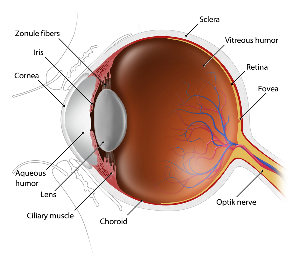
cross section of eye - eye exam
External Exam
The external examination consists of inspecting the eyelids, surrounding tissues and the eyeball including the sclera (white part of the eye), iris and cornea.

Visual Acuity & Fields
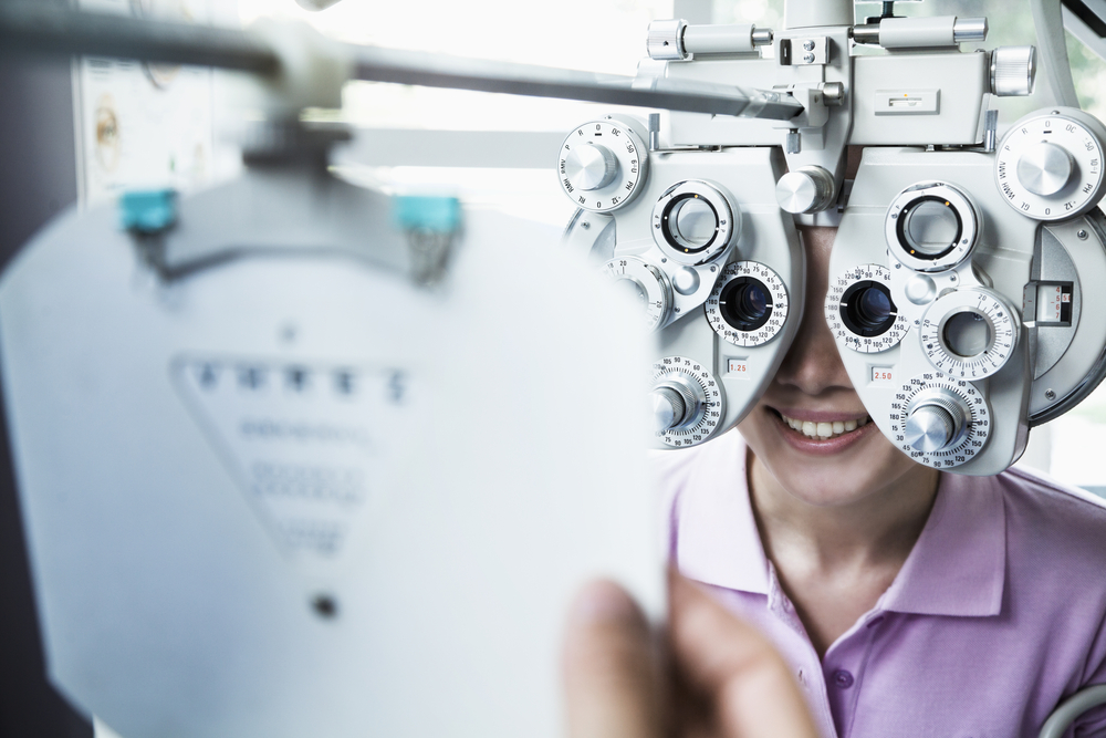
Visual acuity is your eye’s ability to detect fine details and see an in-focus image at a certain distance. A Snellen chart and a phoropter are used. The standard definition of normal visual acuity is 20/20. The term 20/20 comes from even sized objects that can be seen by a “person of normal vision” atvisual acuity eye exam the specified distance. For example, if a person can see at a distance of 20 feet an object that normally can be seen at 20 feet, then they have 20/20 vision. If they can see at 20 feet what a normal person can see at 40 feet, then they have 20/40 vision. For the visual acuity test each eye is tested separately to gauge your side or peripheral vision.

Pupil Functionpupil function eye exam
An examination of the pupil begins with inspecting your pupils for equal size, regular shape, reaction to light, and direct and consensual reaction (meaning the pupil of one eye constricts when the other eye is exposed to light).

Eye Muscle Function
Eye movement is assessed two ways. First by having you move your eye quickly to a target at the far right, left, top and bottom. Then by slow tracking which uses the ‘follow my finger’ test, which tests all the muscles that move your eye.

tonometer eye examEye Pressure Measurement
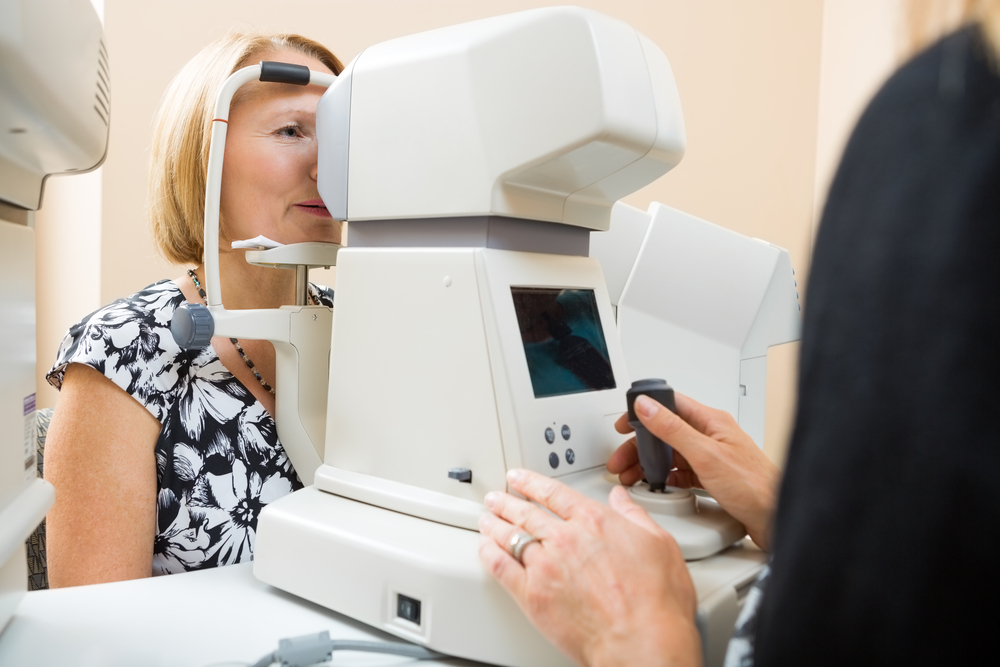
Intraocular pressure, or IOP, is measured using a tonometer to determine the fluid pressure inside your eye. This test provides information regarding your potential for glaucoma.

Viewing the Back of the Eyedilated eye - eye exam
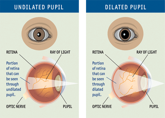
Increasing the size of your pupil with eye drops (known as dilating your eyes) allows the doctor to have a larger view of the back of your eye, including the retina, as demonstrated by this diagram. This is very important for diagnosis and tracking of macular degeneration and diabetic retinopathy.

Eye Structureslit lamp eye exam
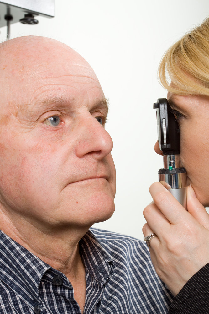
A special, high-powered microscope, called a slit-lamp, is used to view the structures of your eye clearly and in detail, enabling early diagnosis of a variety of eye conditions such as cataracts, presbyopia and corneal injury.

Taking care of your eyes, especially as you become older, is very important. Changes in vision may be gradual, or fast, but in both cases, early diagnosis is key to successful treatment and retaining your vision. It is suggested that individuals 40 and over have a comprehensive eye exam every one to two years. Exceptions would be if you have diabetes, in which case you should see your eye doctor yearly; or there is a family history of eye diseases such as glaucoma, macular degeneration, or corneal diseases, which may require more frequent visits to your eye doctor. If you have any degree of sudden vision loss, eye pain, or significant irritation, contact your eye doctor immediately.

4/23/15

Susan DeRemerSusan DeRemer, CFRE
Vice President of Development
Discovery Eye Foundation

Do I Need Vision Insurance?

With the rising costs of health insurance, many people are looking for ways to reduce their costs. Since not all insurance packages include vision insurance, many people wonder, do I need vision insurance?
vision insurance

Standard Vision Insurance

Vision insurance is a type of health insurance that entitles you to specific eye care benefits such as routine eye exams and other procedures, as well as a specified dollar mount or discount for the purchase of eyeglasses and contact lenses. It only supplements regular health insurance and is designed to help reduce your costs for routine preventative eye care and eyewear.

You can get vision insurance as part of a group, such as your employer, an association, etc., through a government program such as Medicare or Medicaid, or as an individual. It is often a benefit linked to your regular HMO (health maintenance organization) or PPO (preferred provider organization) health insurance.

There are two primary vision insurance plans available:

  • Vision Benefits Package – provides free eye care services and eyewear within a fixed dollar amount for which you pay an annual premium or membership fee and a small co-pay. It may also include a deductible.
  • Discount Vision Plan – provides eye care and eyewear at a discounted rate after you pay an annual premium or membership fee.

Both insurance plans generally include:

  • Annual eye exams
  • Eyeglass frames (usually once every 24 months)
  • Eyeglass lenses (usually once every 24 months)
  • Contact lenses (usually once every 24 months)
  • Discounted rates for LASIK and PRK

Here is where you can check for a list of some vision insurance providers.

Medicare and Medicaid

Different kinds of vision care are included in the US government programs, Medicare and Medicaid. These programs are for qualifying American age 65 and older, individuals with specific disabilities and people with low income.

The Types of Medicare For Vision:

    • Medicare Part A (Hospital Insurance) –Medical eye problems that require a hospital emergency room attention, but routine eye exams are NOT covered.
    • Medicare Part B (Medical Insurance) – Visits to an eye doctor that are related to an eye disease, but routine eye exams are NOT covered.
    • Medicare Part D (Prescription Drug Coverage) – Will help pay for prescription medications for eye diseases.

If you have Medicare Parts A & B you are generally eligible for the following vision coverage, however, there is a deductible before Medicare will start to pay, at which point you will still be paying a percentage of the remaining costs.

  • Cataract surgery – covers many of the cost including a standard intraocular lens (IOL). If you chose a premium IOL to correct your eyesight and reduce your need for glasses, you must pay for this added cost out-of-pocket.
  • Eyewear after cataract surgery – one pair of standard eyeglasses OR contact lenses.
  • Glaucoma screening – an annual screening for people at high risk for glaucoma, including people with diabetes or a family history, and African-Americans whom are 50 or older.
  • Ocular prostheses – costs related to the replacement and maintenance of an artificial eye.

There is also Medicare Supplement Insurance (Medigap) which is sold by private insurance companies to supplement only Medicare Parts A & B. It is intended to cover your share of the costs of Medicare-covered services including coinsurance, co-payments and deductibles. For more details about Medicare plans and coverage check their website or call 800-633-4227.

Medicaid is the US health program that gives medical benefits to low-income people who may have no or inadequate medical insurance. A person eligible for Medicaid may be asked to make a co-payment at the time medical service is provided. Vision benefits for children under the age of 21 include eye exams, eyeglass frames and lenses. Each state determines how often these services are provided and some states offer similar vision services to adults. To learn more about Medicaid eligibility requirements and vision benefits call your state’s Medicaid agency or visit their website.
vision dial - vision insurance

Defined Contribution Health Plans

A way to lower your vison care costs is to take part in a defined contribution health plan (DCHP). You are given a menu of health care benefits to choose from where a portion of the fees you receive for health coverage come from money that is deducted from our paycheck before federal, state and social security taxes are calculated. Four types of DCHP are:

Cafeteria Plans – your employer takes a portion of your salary and deposits it into a non-taxable account for health care spending. The amount taken depends on the number and costs of the benefits you select.

Flexible Spending Accounts (FSA) – your employer takes a predetermined portion of your pre-tax salary and deposits it into health care account for you to pay medical expenses. But generally preventative care such as routine eye exams and are not reimbursable. Nor are eyeglasses and contact lenses reimbursable. You would need to verify with your employer. If you do not use all the money at the end of a 12 month period, the money goes back to your employer.

Health Reimbursement Arrangement (HRA) – this is similar to an FSA except you can use it for preventative care like eye exams and you do not lose the money if it isn’t spent within a certain time period as it can be carried from year to year.

Health Savings Account (HSA) – it can be employer-sponsored of you can set up one independently; however you must purchase a high-deductible health insurance plan to open an HSA and you cannot exceed the annual deductible of your health insurance plan. You cannot be enrolled in Medicare of be a depended on someone else’s tax return. You can use it for preventive care such as eye exams. You can learn more about HSAs by visiting the US Treasury’s website.

There are a variety of options when it comes to vision insurance. You just need to determine your needs and ask providers the correct questions.

4/14/15


Susan DeRemerSusan DeRemer, CFRE
Vice President of Development
Discovery Eye Foundation