Treatment Options For Children

8/19/14

Here is part two in Buddy Russell’s series; this one focusing on contact lenses as a treatment option for children.

We Are Not Born With Good Vision

The human visual system at birth is poorly developed, but rapidly becomes the remarkable combination of nerve tissue, muscles and optics that provide us with the sense of vision. Those babies born with “perfect” eyes have only the opportunity to develop normal vision. The information processed by the eyes is sent directly to the brain and is interpreted as vision.Toddler looking through glasses - treatment options for children During the first few weeks, the child sees shapes, lines and space between objects. The child’s visible world is most usable within 8-14 inches of his/her eyes. During this time, the eyes may appear to wander. After about a month or so, the normal child’s eyes will appear more coordinated and they start to show more interest in looking at objects. It is usually in the third month that a child who has normal eyes can fix and follow on a near object. The growth of the eye is a dynamic process, influenced by genetics and the environment.
Early detection of any eye problem is key to treating the disorder. The prevalence of vision problems in children is higher than you might think. For example:

  • 1 in 10 children are at risk from undiagnosed vision problems
  • 1 in 25 will develop strabismus
  • 1 in 30 will be affected by amblyopia
  • 1 in 33 will show significant refractive error
  • 1 in 100 will exhibit evidence of eye disease
  • 1 in 20,000 children have retinoblastoma

As a result of his granddaughter and her eye problem, former President Jimmy Carter initiated a program in 2002 called InfantSEE. This program allows children to have an eye exam at a very young age at no charge to the family. Participating eye doctors provide a more thorough exam than the busy pediatrician. As a result, there is a greater opportunity to detect and treat eye disorders that may otherwise go undetected.

“Have to” Contact Lenses

Fitting pediatric patients is not usually about routine visits and patients who want to wear contact lenses. It is about critical and often urgent situations and patients who have to wear contact lenses. The more common medical indications for contact lenses can be categorized into three groups; anisometropia, irregular corneal astigmatism and “large” refractive errors.

Anisometropia

One of the more common conditions potentially leading to a permanent loss of vision in a young patient is anisometropia. This difference in the refractive errors of the two eyes can lead to suppression of the less clear image. As a result of the non-focused eye, the brain of a young patient simply turns off the blurred eye. Early detection is key to successful treatment. Following the diagnosis of this problem being present, simply correcting the refractive error may be enough. However, it has been reported that as little as one diopter difference between the two eyes corrected with spectacles and the resultant anisokonia, can lead to foveal suppression impacting stereopsis and depth perception. The use of a contact lens or contact lenses alters the effective image size due to the vertex distance being zero compared to either the magnification or minification of the image size due to the vertex distance with spectacles. One of the most severe examples of this condition would be a child with a unilateral congenital cataract and managed with spectacles postoperatively.

Irregular Corneal Astigmatism

Whether acquired or congenital, the presence of irregular corneal astigmatism of the anterior curve of the cornea is best managed with a contact lens. This condition is to be considered urgent if the patient is of a young age. The eye may forever loose the opportunity to be corrected as the resultant amblyopia develops over a short period of time. By neutralizing the corneal irregularities with a contact lens, the eye of a young child will hopefully gain enough vision improvement to avoid the potential permanent loss.
Obviously, patching the better eye may also be necessary if the treated eye’s vision is not as correctable as the unaffected eye. The length of time the child is to be patched is to be determined by the pediatric ophthalmologist or optometrist, as this area of treatment is sometimes controversial. The factors that are considered include the level of vision obtained, age of the child and the condition of the other eye.

Large Refractive Errors

The optics of spectacle correction in high powers have inherent properties that include distortion, prismatic effect and minification / magnification. For instance, the decrease in image size when one views an object through high minus spectacles may result in less vision. This decrease in image size may impact the opportunity to fully develop normal vision in a young child. The smaller image size that is due to the vertex distance of spectacles may be better managed with a contact lens that has a vertex distance of zero thus providing a larger image. This larger image size often increases best-corrected vision.

“Fitting” the Caregiver

Arguably, the most important factor with young children having a good outcome is the parents / caregivers. The technical challenges that exist in these cases are secondary to the ability the fitter must possess to effectively explain and train the person or persons that will take care of the child outside of the office. They must be your partner in the child’s treatment. They must understand the urgency of the situation, they must understand the seriousness of the problem, they must be trained to properly apply, remove and care for the lens / lenses, they must also follow any and all instructions concerning the child. Many of these parents struggle with feelings of nervousness, guilt and sadness. My strategy is to be sensitive to their feelings but not let them feel sorry for themselves too long as the clock is ticking. I provide verbal instructions, written instructions, videos, my email address and a 24-hour phone number. I welcome the caregiver to ask any question at any time. I do my best to let them know that I do care and that I want them and their child to be successful. I am tough on them. There is no good excuse not to do as I have instructed them to do.

When the child and the parent / caregiver are convinced that I am confident in my ability and they know that I do care, the partnership develops as we walk the path together. I want the child to know that they are coming to see me. I want them to know I will reward their cooperation with all phases of the visit. This positive reinforcement may be in the form of a piece of candy, a small toy or just a sticker when the child allows me to see their eye, measure their cornea or intraocular pressure or they just tell me what they can see. Kids love to please us just like they love to please their parents. Reward them for it. Whether you consider this approach bribery or positive reinforcement, it works.

Buddy Russell - pediatric contact lensesBuddy Russell, FCLSA, COMT
Associate, Specialty Contact Lens Service
Emory University Eye Center

Breathing, Patience and Keratoconus

7/1/14

Besides hearing from eye care professionals and Discovery Eye Foundation staff, we also think it is important to hear from people with sight threatening eye diseases such as age-related macular degeneration, keratoconus, retinitis pigmentosa, etc. They can share their experiences with others that are newly diagnosed with eye disease, while providing insights to family members, friends and caregivers. What follows is the first of these occasional posts focusing on the experiences and insights gained from vision loss.

“The expression on my optometrist’s face was as if he was giving me a cancer sentence,” Jeanette Hasseman remembers. “I had never heard of keratoconus, but he told me it was an incurable vision disability. He said he was aware of some evolving technology, but he wanted to give me some time to absorb and research the disease.
Jeanette Hasseman - keratoconus
“When I went back three months later, he told me a about a cross-linking study that was going on not-too-far from here, but I hadn’t fully absorbed all the information I was reading yet,” she recalls. I did not understand — even though it was plainly stated in the information I was looking at — that once you lose some vision, you don’t get it back.

“I kept thinking, ‘OK, this certainly can be fixed,” so instead of getting cross-linking in July, I waited until December. I regret not acting on my doctor’s suggestion of getting in the study in July, because I lost a tremendous amount of vision by December.”

Hasseman was tested and qualified for the clinical trial in December and had epi-on cross-linking in both eyes in January 2013. “The topography scans show the KC progression has been halted,” she says. “I rejoice in that.”

While her vision seemed better shortly after the procedures, she was diagnosed one week after crosslinking with a “gouge” (4 mm corneal abrasion) in her left cornea that resulted in severe hazing. While the severity of the corneal hazing has decreased in the past year, she is waiting to find out if it is permanent. If so, she may need to have a corneal transplant at some point to restore any visual acuity.

A registered nurse, Hasseman finished her BSN degree two years ago at age 54 — just months before she was diagnosed with KC. The native Ohioan has since stopped working as a nurse, and more recently, she had to give up teaching and doing tatting — lace-making — a hobby she had enjoyed for decades.

Hasseman found the National Keratoconus Foundation (NKCF) when she first Googled “kerotoconus.” “KC-Link has been a great blessing,” she says. “I’ve downloaded information; I signed up for KC-Link and asked questions of Catherine [Warren, director of NKCF] and the moderating doctors. I found great support for my own spirit, as well as information on the latest technologies.

“When people ask questions on KC-Link, if I can relate, I answer. Just the other day, someone who was just diagnosed asked, ‘Well, what should I do?’ I wrote: ‘First thing is: Breathe. Second thing is: Ask for information from your eye doctor and ask who is the best corneal specialist in your area who is really good for keratoconus. Most of all: Be patient.’

“It’s really hard to lose your vision. Even in your own heart, you can feel isolated. KC-Link gives you a body of other people who totally understand what you’re thinking, what you’re feeling, what frustrates you. There is so much advice you can get on KC-Link regarding contacts, how to keep eyes moist, how to handle low-light driving, what type of e-reader works best for people with keratoconus — you cannot exhaust the information that is shared; you cannot exhaust the different emotions that are shared.”

Jeanette HassemanJeanette Hasseman
Keratoconus Advocate

Corneal Donor’s Age Not Critical for Transplant Success

6/17/14

In the United States, more than 40,000 corneal transplants are performed each year with a high success rate in comparison to other types of organ transplants. According to the Eye Bank Association of America (EBAA), keratoconus was the leading cause of anterior lamellar keratoplasty (DALK/ALK partial thickness transplant) and was the fourth most common indication for penetrating keratoplasty surgery in 2012 (their last reporting period).
corneal transplant-original size
Advances in technology have led to increasingly successful outcomes for all who need corneal transplants. New long-term research of corneal transplant patients have shown that the age of corneal donors is no longer as important as once thought by eye health providers. According to a study funded by the National Institutes of Health, ten years after a transplant, a cornea from a 71-year-old donor is likely to remain as healthy as a cornea from a donor half that age.

The Cornea Donor Study (see www.ClinicalTrials.gov), funded by NIH’s National Eye Institute (NEI), was designed to compare graft survival rates for corneas from two donor age groups, aged 12-65 and aged 66-75. It was coordinated by the Jaeb Center for Health Research in Tampa, Fla., and involved 80 clinical sites across the United States. The study enrolled 1,090 people eligible for transplants, ages 40-80. Donor corneas were provided by 43 eye banks, and met the quality standards of the Eye Bank Association of America. The study found that 10-year success rates remained steady at 75 percent for corneal transplants from donors 34-71 years old. In the United States, three-fourths of cornea donors are within this age range, and one-third of donors are at the upper end of the range, from 61-70 years old.

Prior to this study, many surgeons would not accept corneas from donors over 65. Since the supply of young donor corneas is limited, these study results are encouraging for those who face a corneal transplant . The high level of success rates using corneas from older donors (over age 60) greatly increases the pool of donated corneas and corneal tissue available for transplant. In 2012, corneal donors under age 31 comprised less than 10 percent of the U.S. donor pool. “Our study supports continued expansion of the corneal donor pool beyond age 65,” said study co-chair Edward J. Holland, M.D., professor of ophthalmology at the University of Cincinnati and director of the Cornea Service at the Cincinnati Eye Institute. “We found that transplant success rates were similar across a broad range of donor ages.”

“Overall, the findings clearly demonstrate that most corneal transplants have remarkable longevity regardless of donor age,” said Mark Mannis, M.D., chair of ophthalmology at the University of California, Davis, and co-chair of the study. “The majority of patients continued to do well after 10 years, even those who received corneas from the oldest donors.”

SOURCE: National Eye Institute Press Releases

For information about Eye Bank Association of America

CathyW headshotCathy Warren, RN
Executive Director
National Keratoconus Foundation

20 Facts About the Amazing Eye

6/10/14

We don’t often give our eyes as much thought as we should, that is until something goes wrong and our vision is affected. But when you learn more about eyes, you realize just how amazing they are. Here are a few facts you may enjoy:

1. Eyes began to develop 550 million years ago. The simplest eyes were patches of photoreceptor protein in single-celled animals.

2. Your eyes start to develop two weeks after you are conceived.
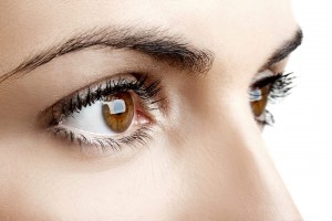
Brown eyes 6.10.14
3. The entire length of all the eyelashes shed by a human in their life is over 98 feet with each eye lash having a life span of about 5 months.

4. To protect our eyes they are positioned in a hollowed eye socket, while eyebrows prevent sweat dripping into your eyes and eyelashes keep dirt out of your eyes.

5. Your eyeballs stay the same size from birth to death, while your nose and ears continue to grow.

6. An eye is composed of more than 2 million working parts.

7. Only 1/6 of the human eyeball is exposed.

8. Corneas are the only tissues that don’t have blood.

9. The human eye weights approximately just under an ounce and is about an inch across.

10. An eye cannot be transplanted. More than 1 million nerve fibers connect each eye to the brain and currently we’re not able to reconstruct those connections.

11. 80% of our memories are determined by what we see.

12. Eyes heal quickly. With proper care, it takes only about 48 hours to repair a minor corneal scratch.

13. There are about 39 million people that are blind around the world.

14. 80% of vision problems worldwide are avoidable or even curable.

15. Humans and dogs are the only species known to seek visual cues from another individual’s eyes, and dogs only do this when interacting with humans.

16. A fingerprint has 40 unique characteristics, but an iris has 256, a reason retina scans are increasingly being used for security purposes.

17. People who are blind can see their dreams if they weren’t born blind.

18. “Red eye” occurs in photos because light from the flash bounces off the back of the eye. The choroid is located behind the retina and is rich in blood vessels, which make it appear red on film.

19. 80% of what we learn is through our eyes.

20. Eyes are the second most complex organ after the brain.


Susan DeRemerSusan DeRemer, CFRE
Vice President of Development
Discovery Eye Foundation

What You Need to Know About Cataracts

6/5/14

Do you feel like your vision is getting worse? Do you feel like colors are not as vibrant as they used to be? Are you having more trouble with glare? If you have any of these symptoms, you may be experiencing the effects of cataracts.

Cataracts are a normal aging process of the crystalline lens in the eye. Well you may ask — what is the crystalline lens? It is easiest to think of the eye as a camera. The eye has a lens (actually two) – the cornea (the front window of the eye) and the crystalline lens (inside the eye). It also has an aperture (the colored iris), and film (the retina). All these structures work together to focus light and allow us to see – just like a camera. When we are young (less than 40), the crystalline lens is flexible. This is why we are able to see distance and then near without the need for reading glasses. The crystalline lens is able to change its shape depending on where one is looking.

Figure 1 – Slit-lamp photo of a visually significant cataract.

As we age, the crystalline lens becomes less flexible, thereby causing one’s near vision to be more blurry. This necessitates the need for reading glasses. As the crystalline lens become less flexible with age, the lens also starts to become more yellow and can also become cloudy. When the yellowing and/or clouding become visually significant, we refer to this as a cataract (figure 1).

Are cataracts dangerous? The simple answer is no. In the vast majority of cases, a cataract can be monitored until it becomes visually significant (drop in vision, glare, decreased contrast, vision related difficulties with day to day activities, etc). However, there are a few instances in which cataract removal is a medical necessity. Routine examinations by your eyecare provider can help you determine if you are at risk for these less common instances.

Figure 2 – Intraoperative photo during cataract surgery (prior to cataract removal).

What can I expect during cataract surgery? Do you have to replace the crystalline lens with anything? Cataract surgery involves removing the clouded crystalline lens (figure 2) and replacing it with an artificial lens known as an intraocular lens (IOL) (figure 3). Surgery generally takes 10-15 minutes under a mild sedative, and you don’t have to stop any of your current medications. Anesthesia is achieved with drops and you will only feel mild pressure during the surgery. IOLs come in different styles – Standard IOLs grossly correct your vision and you can fine tune your vision (distance and near) with glasses post-operatively;

Figure 3 – Intraoperative photo during cataract surgery (after implantation of an IOL).

Toric (astigmatism correcting) IOLs allow for increased spectacle independence, but glasses will still be needed for distance or near; Accommodating IOLs “flex” within the eye to decrease your dependence on distance and near glasses; Multifocal IOLs allow spectacle independence for distance and near. I always counsel patients that there is no perfect IOL and you have to determine which IOL is best for your particular situation. Your doctor can help you decide which IOL is best for you. Generally, cataract surgery is extremely safe. Your doctor will discuss particular risks specific to your eye.

How do I know if cataract surgery is right for me? The best way to know if you have a cataract and if it time to consider surgery is to consult with your local ophthalmologist. If you have experienced a recent drop in vision, that is not correctable with glasses, cataract surgery may be able to restore your vision!

Garg feb 2014 thumbSumit “Sam“ Garg, MD
Medical Director
Vice Chair of Clinical Ophthalmology
Assistant Professor of Ophthalmology
Gavin Herbert Eye Institute – UC, Irvine

Little Bottle, Big Relief

6/3/14

What you need to know about eye drops.

Have you been staring at a computer all day and your eyes are tired? Have allergies been making your eyes watery and itchy? Are your contact lenses irritating your eyes? If you have experienced any of these conditions, you have probably turned to eye drops for relief.

While eye drops are an easy and effective means of treating a number of eye issues, there are many different eye drops available, both over the counter (OTC) and by prescription. It is wise to know what your underlying condition is before trying to get relief.
eye drops 6.3.14
If your eyes are red and you may want to try a decongestant eye drop, which will shrink the tiny blood vessels in the “whites” of your eyes (sclera), but they also cause dryness so may not be a good choice if you wear contact lenses.
For lens wearers you are better off with a re-wetting drop to lubricate the eye and lens making you more comfortable. Another problem with the decongestant eye drops is over use – which can cause irritation and an increased tolerance that could lead to more redness.

If you suffer from allergies and antihistamine eye drop would be the best choice for relief from itchy, watery, red and swollen eyes. They work by reducing histamine in the eye tissue.

Lubricating eye drops, also known as artificial tears, are for short-term relief caused by temporary situations such as eye strain form computer use, being tired or being outdoors in windy and/or sunny conditions. If the condition is chronic, a prescription eye drop will be the best choice.

It is important to remember that if any of the above symptoms worsen or continue for an extended period of time, it is time to see your eye doctor to determine the underlying cause of your issue and to rule out eye disease. Postponing a visit could also lead to an eye infection.

Prescription drops are used to treat a wide variety of eye diseases such as glaucoma, dry eye and the symptoms of ocular herpes. They are also used to help with healing from cataract surgery, corneal transplants, glaucoma surgery and even Lasik. it is extremely important to use them as often as your ophthalmologist recommends to improve healing and prevent infection.

Because of the ease of applying eye drops researchers are working toward using them to treat other eye diseases. Ocular herpes symptoms are sometimes treated with antiviral and steroid drops. But this only is targeted at the symptoms and not the underlying cause, the herpes simplex virus. Lbachir BenMohamed, PhD and Steven Wechsler, PhD at the University of California, Irvine, Gavin Herbert Eye Instittue have been working to determine what reactivate the herpes simplex virus and develop an eye drop that would either stop the reactivation of the virus or kill it.

Using eye drops to treat age-related macular degeneration (AMD) is also being explored. Researchers at the Institute of Ophthalmology at University College London are working with nanoparticles to deliver anti-VEGF drugs such as Lucentis and Avasitn to the back of the eye via drops. “The study shows that Avastin can be transported across the cells of the cornea into the back of the eye, where is stops blood vessels from leaking and forming new blood vessels, the basis for wet AMD.” While researchers in the Department of Ophthalmology, Tufts University School of Medicine in Boston “reported in their “proof of concept” study that topical application of a compound called PPADS inhibits damage to the tissues in the eye that impacts the individual’s ability to see color and fine detail, as well as reduces the growth of extraneous blood vessels in the back of the eye related to AMD.” It would work in both dry and wet AMD reduce the need for direct injections.

Eye drops, when properly applied, can provide temporary relief from symptoms of eye discomfort. But if the symptoms worsen or continue for an extended period of time, consult your eye doctor. To make sure you apply the eye drops correctly check out the article in our February 2013 newsletter for 12 easy steps to get the drops into your eyes and avoid infection.

One final note – keep your eye drops out of reach of children. Eye drops come in small bottles that are the perfect size for small hands and don’t have the same security tops found on other medications. The FDA has warned that ingredients found in some eye drops that relieve redness have caused abnormal heart rate, decreased breathing, sleepiness, vomiting and even comas in children five and younger that have ingested them. If you child has swallowed eye drops, call the Poison Help Line 800-222-1222.

Susan DeRemerSusan DeRemer
Vice President of Development
Discovery Eye Foundation

New Hope for Corneal Scarring

5/22/14

There are several etiologies for limbal stem cell deficiency of the front of the eye. These include chemical and thermal burns, Steven-Johnson syndrome (which is an autoimmune severe allergic reaction that causes a burn from within), congenital aniridia, and a few other insults such as contact lens over-wear. All of these cause severe ocular surface scarring and problems with the cornea. Many eyes with these diseases have problems with corneal healing. They do not have the stem cells to support ocular surface health. The scarring can be so severe in many cases that severe corneal blindness can result.

Limbal stem cells from the human cornea, with a protein known as p63 stained yellow. Cell nuclei (which hold the DNA) are stained red.  From eurostemcell.org
Limbal stem cells from the human cornea, with a protein known as p63 stained yellow. Cell nuclei (which hold the DNA) are stained red. From eurostemcell.org

In these cases, a simple corneal transplant will quickly fail and not result in any visual improvement. The reason for this is that the stem cells of the ocular surface have been damaged or burned out.

Visual rehabilitation for these eyes usually requires a limbal-corneal stem cell transplantation. The stem cells can be taken from the other healthy eye of the same patient, a living related donor, and or cadaveric tissue. In most cases systemic immunosuppression medications need to be taken for 1 to 3 years following surgery in order to minimize risk of rejection. Management of these patients is done in conjunction with an immunologist or a transplant specialist who can co-manage and monitor for systemic toxicity while the patient is on the these immunosuppressive medications. As most of these eyes also have concomitant glaucoma and scarring of the eyelids to the globe, co-management with a glaucoma specialist and an oculoplastic specialist is also required.

For patients who cannot be on systemic immunosuppression for other health reasons such as diabetes or cancer, they may require an artificial corneal transplantation. The artificial corneal transplantation is reserved as a last step for visual rehabilitation in these eyes. The only artificial cornea that has shown potential, is the Boston keratoprosthesis. Even this artificial cornea carries a high risk for infection and glaucoma. Very close monitoring of eyes that have an artificial cornea is required to monitor for infection and glaucoma progression. However these eyes do not require systemic immunosuppression.

Eye with Boston keratoprosthesis


The management of eyes with severe ocular surface disease is a difficult one for the cornea specialist. A subspecialist in severe ocular surface disease and limbal stem cell transplantation is required to manage these very sick eyes. At the Gavin Herbert Eye Institute, we have developed a team approach for the management of severe ocular surface disease patients and have successfully treated and are managing many patients who have otherwise no place to go.

Farid 3.6.14Marjan Farid, MD
Director of Cornea, Cataract, and Refractive Surgery
Vice-Chair of Ophthalmic Faculty
Director of the Cornea Fellowship Program
Associate Professor of Ophthalmology
Gavin Herbert Eye Institute, University of California, Irvine

New Technology for Evaluating Contact Lenses

5/1/14

Successful management with contact lenses can sometimes be a frustrating process for those with keratoconus. The fitting and evaluation process involves numerous visits to the optometrist, out of pocket expenses as well as medical insurance co-pays. Luckily, the contact lens industry has responded to the need to have better contact lens materials for patients with keratoconus. It used to be that the only contact lenses available to manage keratoconus were gas-permeable (GP) lenses. Now, more and more patients are being fit with newer generation hybrid lenses (GP lens core with a skirt of soft lens material to aid in fit and comfort) and scleral lenses (large diameter GP lenses that do not rest on the cornea, only the outlying sclera). These newer designs are intended to vault over the central cornea and do not rest on the cornea at all. They have resulted in much more comfortable and wearable strategies for full-time use. As a result of the newer lens designs, the game has changed when it comes to the science of fitting and evaluating the lenses.

For years, optometrists have used corneal topography to guide their decision making on fitting keratoconic eyes. A corneal topographer is an instrument that maps the shape of the cornea, and gives information much like that of a topographical map for hiking. Corneal topography is still an absolutely mandatory part of evaluating the shape of the cone, the simulated corneal curvature, and monitoring for progression of the disease, and is not a standard part of a typical eye examination. However, doctors have a new tool at their disposal for fitting contact lenses on patients with keratoconus.

Optical Coherence Tomography (OCT) was once reserved for use in the back of the eye, or retina. OCT uses visible light passed through the clear structures of the eye to generate a cross-sectional image of the layers of the retina, much like an image generated by an MRI. Advances in OCT technology has improved the resolution to image the eye on the micrometer scale (one-thousandth of a millimeter). OCT technology is now commercially available not only for the retina, but the structures of the front part of the eye. The obvious application is to aid the doctor in the fitting and evaluating complex contact lenses that vault the cornea.

Figure 1.  OCT image of a scleral lens fit on a keratoconic patient.  The cornea is the opaque white band located at the bottom of this picture, the tear film reservoir is the middle clear band and the contact lens is the top band.  Using an electronic caliper tool, the precise amount of vault can be measured, leaving no doubt as to the precision of the fit.
Figure 1. OCT image of a scleral lens fit on a keratoconic patient. The cornea is the opaque white band located at the bottom of this picture, the tear film reservoir is the middle clear band and the contact lens is the top band. Using an electronic caliper tool, the precise amount of vault can be measured, leaving no doubt as to the precision of the fit.

OCT allows the optometrist to view a cross-sectional image of the contact lens on the eye in real time and to monitor the health of the cornea in the presence of the contact lens. This view is valuable for judging the vault of new designs of contact lenses over the cornea and judging where the lenses land on the eye. It is the most specific way to determine if the fit is acceptable and to troubleshoot if lenses are not fitting appropriately.

Figure 2.  OCT image of the periphery of a scleral lens on a patient with pellucid marginal degeneration.  The lens contacts the cornea over an area of 0.87mm long.  These types of measurements help guide decision making in modifying the lens fit and were impossible before the advent of this technology.
Figure 2. OCT image of the periphery of a scleral lens on a patient with pellucid marginal degeneration. The lens contacts the cornea over an area of 0.87mm long. These types of measurements help guide decision making in modifying the lens fit and were impossible before the advent of this technology.
Figure 3.  Hybrid lens on a highly irregular eye after corneal transplant.  The point of contact of the soft skirt with the cornea is visible to the right of the image.
Figure 3. Hybrid lens on a highly irregular eye after corneal transplant. The point of contact of the soft skirt with the cornea is visible to the right of the image.

Optometrists now have a much more powerful tool for evaluating and managing even the most challenging contact lens fits. It remains to be seen whether this technology has the ability to reduce the number of visits required for successful fit. But, the precision afforded by this technology does have the ability to improve patient outcomes.

Sonsino HeadshotJeffrey Sonsino, OD, FAAO
The Contact Lens Center at Optique Diplomate
Cornea, Contact Lens, and Refractive Therapies

Dry Eye and Tear Dysfunction

4/24/14

With an aging population and increasing awareness of the condition, dry eye has become one of the hottest topics in eye care today. There have been numerous advances in our understanding of the condition as well as several breakthroughs in treatment.
eye drops in eye
Lets start with the most fundamental question. Why do we have tears? Tears serve many functions, but perhaps the most important is that they create a perfectly smooth optical surface at the front of the eye to allow sharp focusing. The sharper your vision the more quickly you can spot and run from a predator or find a supply of food. Hence, a stable tear film is critical for survival. As a result, the body has evolved many ways to help keep the tears healthy and intact.

When I describe the tears to a patient I describe it as a structure. Tears have a foundation that anchors them to the eye’s surface, an elastic protective support section not that different than the walls in a building and even a roof, just like a house has. Like a house, problems with any structural part can adversely impact the entire building.

Over the past few years the most important breakthroughs in understanding dry eye have focused on the outer portion of the tear structure – what you might think of as the roof. When a roof develops holes and it rains, water gets in which can destroy the entire structure of the house. With the tears, holes in the “roof” can let moisture evaporate causing the structure to collapse and become unstable.

The outer layer of the tears consist of lipid or oil produced by the meibomian glands – glands that run radially through the lids and express small amounts of complex oil with each blink. When these glands work properly, the oils they produce coat the tears, preventing evaporation and stabilizing and lubricating. When the glands become blocked – which can occur for a number of reasons including decreased blinking due to excessive computer use, the lack of oil can lead to a down spiral of damage to the eye’s surface with symptoms that can range from annoying to life altering.

Lets talk for a moment about symptoms. We know that dry eye is progressive and, if left untreated will worsen over time, so the sooner we know about it, the sooner treatment can begin. I am often surprised that even with increased awareness, most patients don’t recognize that they suffer from dry eye until the condition is fairly advanced. One reason is that many people think that the symptoms of dry eye are a normal and expected part of aging. For the most part they are not. If you experience, burning, irritation, unstable vision, grittiness or any change in your vision or eye comfort, you should bring this to the attention of your eye care practitioner.

One of the most common signs of dry eye is excessive tearing. Patients often complain that their eyes are too wet and I have to explain that the excessive tearing is the eye’s way of trying to correct for dryness and prevent further damage. Unfortunately excessive tearing further destabilizes the tears and makes matters worse.

Lipiview System
Lipiview System

Perhaps the greatest advance today is the recognition that meibomian gland dysfunction causes or contributes to nearly 90% of all dry eye. Many eye care providers are adding high tech tools like meibography which can show the actual state of the meibomian glands like an X-Ray and LipiView which measure the actual thickness of the lipid layer of the tears. As a result diagnostic ability has improved.

Lipiflow Device
Lipiflow Device

Recently introduced eyedrops like Alcon’s Systane Balance and Allergan’s Refresh Optive Advanced Formula drops can help restore absent or deficient oils in the tears. And new treatments including moist heat goggles like Tranquileyes by EyeEco can help improve comfort. LipiFlow, a technology pioneered by TearScience, offers a breakthrough treatment that can clear blocked and reset meibomian glands. It can produce amazing improvement.

There have also been significant advances for contact lens wearers suffering from dry eye. Scleral lenses are especially helpful for patients with advanced dry eye who also must wear contact lenses to see, such as those who have keratoconus. The large scleral lens serves as a barrier that protects the eye and provides sharp stable vision. In fact, scleral lens designs are used as a treatment for severe dry eye and ocular surface disease.

Finally, if you have been diagnosed with dry eye previously and are still suffering despite treatment, this may be a good time to return to your eye care provider to discuss your condition and explore the possibility of new treatment options.

AArthur B. Epstein, OD, FAAO
co-founder of Phoenix Eye Care
and the Dry Eye Center of Arizona
Fellow of the American Academy of Optometry
American Board of Certification in Medical Optometry
Chief Medical Editor of Optometric Physician™

The Evolving Contact Lens

4/22/14

Contact lenses give a person the ability to see without glasses. If you have keratoconus, they are essential for seeing as regular glasses don’t work with an irregularly shaped cornea. But lately these relatively simple lenses have created a whole new world where they can dispense eye medication, measure blood glucose levels and even help the blind see.

Courtesy Google
Courtesy Google

Monitoring Blood Sugar
You have heard about Google Glasses, but Google is looking beyond the smartphones of eye wear to monitoring health. They are currently working on a lens with tiny wireless chips and glucose sensors that are sandwiched between two lenses. They would monitor glucose levels once a second and use tiny LED lights, also inside the lenses, to flash when the levels are too high or low. And how big are these electronics? They are no larger than a speck of glitter, with a wireless antenna that is thinner than a human hair. While they are still in development – Google has run clinical research studies and is in discussions with the FDA – it could make blood sugar monitor far less invasive than pricking your finger several times a day.

Drug Delivery for Glaucoma
Getting glaucoma patients to regularly use their eye drops to regulate the pressure in their eyes has always been a problem. They forget, don’t want to be bothered, or have a hard time getting the drops into their eyes. This could change with two research projects exploring the use of contact lenses to deliver medication over a prolonged period of time.

Researchers at Massachusetts Eye and Ear/Harvard Medical School Department of Ophthalmology, Boston Children’s Hospital, and the Massachusetts Institute of Technology who are working on a lens designed with a clear central area and a drug-polymer film made with the glaucoma drug latanoprost, around the edge to control the drug release. These lenses can be made with no refractive power or the ability to correct the refractive error in nearsighted or farsighted eyes.

Another team from University of California, Los Angeles have combined glaucoma medication timolol maleate with nanodiamonds and embedded them into contact lenses. When the drugs interact with the patient’s tears, the drugs are released into the eye. While the nanodiamonds strengthen the lens, there is no difference in water content so they would be comfortable to wear and allow oxygen levels to reach the eye.

Seeing in the Dark
Researchers out of the University of Michigan have developed an infrared sensor that could eventually be used in the production of night vision contact lenses. Thanks to graphene, a tightly-packed layer of carbon atoms, scientists were able to create a super-thin sensor that can be stacked on a contact lens or integrated with a cell phone.

Stem Cells for Cornea Damage
Researchers in Australia are working on a way to treat corneal damage with stem cell infused contact lenses. Stem cells were taken from the subject’s good eye and then plated them onto contact lenses (if there is a defect in both eyes, stem cells are taken from a different part of the eye). After wearing for about two weeks the subjects reported a significant increase in sight.
Braille-Tracile-Contacts
Helping the Blind See
And what good are contact lenses if you are blind? At Bar Ilan University in Israel researchers are creating special lenses that translate images into sensations felt on the eye. It works by taking an image with a smartphone or camera, it is then processed and sent to the contact lens. The custom-made lens is fitted with a series of electrodes that use small electric impulses to relay shapes onto the cornea, similar to braille. After some practice, test subjects were able to identify specific objects.

In expanding the uses of contact lenses, these projects seem to be just the beginning, all reported in the first four months of this year. Researchers and developers are working together to find more and better ways help with vision and medical issues, using contact lenses.

Susan DeRemerSusan DeRemer, CFRE
Vice President of Development
Discovery Eye Foundation