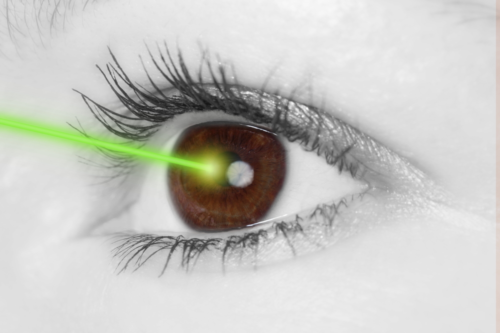
Treatments for Dry Eye Disease

This is the third and final installation of the comprehensive series we have presented on dry eye disease. Dr. Wade first discussed the symptoms you might experience if you have dry eye and the Dr. Garg explained the process of diagnosing they type of dry eye disease you might have. In this article Dr. Farid reviews treatment options based on your diagnosis of dry eye disease.treatments for dry eye

Treatments for Dry Eye Disease

As our understanding of dry eye disease expands, so do treatment options. We now know that dry eye disease is a multifactorial disease. There is no one cause so there is no one magic cure. Treatments aim to improve tear composition, reduce eye surface inflammation, and target eyelid margin disease. Here, we will review many treatment options, but the treatment combination or “cocktail” that is appropriate for you will depend on your specific type of dry eye disease. This is usually determined after some testing by your eye care provider.

Environmental, Dietary, and Medication Adjustments
Before going into specific dry eye treatments, there are modifiable causes and preventative methods to improve dry eyes. Simple changes in the environment, diet, and medications can be easy ways to improve symptoms.

Environmental Changes
As expected, a dry environment will worsen dry eyes. Humidifiers and moisture goggles have been shown to help alleviate these symptoms. Furthermore, situations that cause decreased blinking, such as prolonged use of computer screens, can worsen dry eyes. Patients should take frequent breaks from computer screens and reading, allowing their eyes to rest and resume normal blinking. When in windy, smoky, or dusty situations, sunglasses can act as a barrier to the eyes, reducing dry eye symptoms. Avoiding wind, fans, or any source of air blowing into the eyes may also help.

Dietary Changes
Drinking adequate water keeps patients hydrated and reduces exacerbations of dry eye symptoms. Omega-3 fatty acids, particularly eicosapentaenoic acid (EPA) and docosahexaenoic acid (DHA), have been shown in many clinical studies to improve dry eyes. It is believed that these fatty acids help inhibit inflammatory mediators. Essential fatty acids cannot be synthesized and must be ingested through diet. Foods rich in omega-3 fatty acids include fish, other seafood, and flaxseed oil. Supplements are also available.

Adjusting Medications
Many medications are associated with dry eyes, and patients may benefit from adjusting doses or finding alternative treatments. You can work with your physician to weigh the costs and benefits about modifying your current medications if they are suspected to worsen dry eyes. Some of these medications include: hypertensive drugs, antihistamines, decongestants, antidepressants, acne medications, birth control, and hormone replacement therapy. Eye drops with preservatives, such as glaucoma medications, can also worsen dry eyes.

Lubricating Treatments
For patients with decreased tear production, supplementation of tears or reduction of tear drainage will improve symptoms.

Artificial tears, gels, and ointments
Artificial tears, gels, and ointments are readily available over the counter. Artificial tears are eye drops that are used throughout the day as needed, up to eight times for dry eyes. You must make sure to get “lubricating” drops. There are multiple available brands that are excellent and equivalent in providing artificial moisture. Avoid “redness relief” brands as they are focused on reducing appearance of the vessels in the eyes as opposed to actual dry eye relief. Gels and ointments are thicker and very effective but can blur vision. Non-commercial comparison testing between brands has not been done. Because the ingredients, preservatives, and consistency vary from brand to brand, it is recommended for patients to choose the best option that works well for them based on trial and error.

Preservatives are used in many eye drops to help them to last longer. Unfortunately, they can be irritating to the eyes, especially when used often (more than 4 drops/day) or with other drops containing preservatives (ie. glaucoma medications). Patients who frequently use eye drops are recommended to use preservative-free (PF) drops. Unfortunately, preservative-free drops are more expensive because they come in “single use” containers. You may be able to extend the life of each single use container by spreading out its contents over multiple uses throughout the day.

Lipid-containing lubricants, such as those with castor oil, attempt to mimic the oils found in the tears, reducing evaporative dry eyes, but more research is necessary to study their efficacy.

Punctal Plugs
Your ophthalmologist can place small plugs at a clinic visit into the punctum, a small hole at the upper and inner lids that drain tears from the eyes. One plug can be added initially, and if more tear retention is required, another plug can be added. There are absorbable and non-absorbable types. Absorbable types are made of collagen and last 1 week to 6 months. Once placed, they are not visible or removable. Non-absorbable plugs are usually silicone, and permanent. They are easily placed by your ophthalmologists and remain visible in follow-up exams. Some patients feel the plugs, and there may be mechanical rubbing, especially when the plug is not the proper size. Some patients may also experience excessive tearing with plugs. If problematic, patients can wait for the plugs to dissolve if absorbable or they can be removed with forceps if non-absorbable.

Autologous Serum
Serum is extracted from a patient’s blood and turned into an eye drop. The growth factors, vitamins, and antibodies present in serum are the same as those in natural tears. Evidence is showing significant promise in alleviating symptoms and signs of chronic dry eye disease. A good collaboration between a phlebotomy lab and compounding pharmacy is necessary to make the products. The products must be kept refrigerated or cold between uses. They can be frozen for long-term storage. Each blood draw provides a supply of drops that can last 3-6 months.

Hydroxypropyl Cellulose Ophthalmic Inserts (Lacrisert)
An insert is available for people who find regular artificial tear use to be difficult. A physician can order the inserts through a pharmacy. The patient places the insert in the inferior fornix of the eye, the area between the lower lid and sclera of the eye. It slowly dissolves over 24 hours, giving constant lubrication.

Anti-inflammatory Treatments
Inflammation is now being recognized as a major underlying cause of chronic and worsening dry eye disease. Many patients who have been suffering from chronic dry eyes, particularly those with autoimmune diseases like Sjogren’s Syndrome, will do well on treatments that reduce the amount of inflammation in the tear film and ocular surface. Your physician can order these medications for you if needed.

Cyclosporine A (Restasis)
The FDA approved Restasis for dry eyes in 2002. This is an immunosuppressive and anti-inflammatory eye drop medication. Relief is not instant, and may take 6-8 weeks of sustained use for improvement in dry eye symptoms. Less than 20% of patients may experience a burning sensation with the drops, but the safety and tolerability profile is otherwise excellent.

Antibiotics
Doxycycline and minocycline are used for inflammatory ocular surface and eyelid disease. The antibiotics have a dual effect: they act as anti-inflammatories and anti-microbials. As an anti-microbial, these medications can improve meibomian gland function. They can decrease lid bacterial flora, reducing a cause of meibomian lipid breakdown. A low-dose (doxycycline 20 mg BID) regimen for 1-2 months has been shown to be effective. Side effects are usual mild but include stomach upset, yeast infections, and photosensitivity.

Steroids
Steroids act as anti-inflammatories. Because steroids are associated with complications in long-term use, they are mainly used in short pulses either at the initiation of treatment or as rescue during exacerbations. A short 4-6 week course is generally well tolerated as a “rescue treatment,” or an urgent treatment, to relieve intolerable symptoms quickly before resorting to other treatment options.

Meibomian Gland Treatments
Meibomian glands produce oils that are crucial in preventing our tears from evaporating too quickly. Treatments that target the glands can help patients with meibomian gland dysfunction or lid margin disease.

Warm compresses and lid scrubs
Warm compresses provide heat that warms the oils in the glands, unclogging the glands and improving oil flow. Warm washcloths, small rice bags heated in the microwave, or commercial hydrogel pads are all effective, and no studies have been done to compare the different methods. The heat. The compress should be placed over the eyes for 5-10 minute. Gentle circular or rolling massage of the eyelids can help express the oils from the glands.

Lid scrubs are useful particularly in cases of blepharitis, or mild inflammation of the lids, that cause them to become crusted. There are excellent over the counter commercial lid soap formulations that work well to clean the lid margins. Alternatively, baby shampoo and a washcloth gently applied to the lids can work as well.

Thermal pulsation (Lipiflow)
Lipiflow is an FDA approved in-office treatment for meibomian gland dysfunction and dry eyes. During the 12-minute procedure, a device is placed over the eye and eyelids that provides localized warmth and pressure on the lids (Figure 1).

treatments for dry eye
Image 1 – Lipiflow treatment

The procedure is 100% safe and very effective at clearing out the trapped oil glands and allowing smooth flow to be re-established. After approximately one month, the consistency of the oils in the tear flow will have improved remarkably with associated improvement in dry eye symptoms. With one procedure, the effects last between 12-24 months.

Intense Pulsed Light (IPL)
Originally approved for acne and rosacea dermatologic disease, IPL uses bursts of light to minimize blood vessel size. It can be used off-label for ocular rosacea and meibomian gland dysfunction, but results have not been reported.

Summary
There are many treatment options for dry eye disease. Generally, conservative over-the-counter treatments should be tried first. Commonly, environmental changes, dietary changes, artificial tears, and warm compresses will improve the majority of dry eye symptoms to tolerable levels. However, when these options are exhausted, there are many additional options for patients. Work closely with a trusted health professional to determine the optimal treatment combination as each patient is different.

8/25/15


treatments for dry eyeMarjan Farid, MD
Director of Cornea, Cataract, and Refractive Surgery
Vice-Chair of Ophthalmic Faculty
Director of the Cornea Fellowship Program
Associate Professor of Ophthalmology
Gavin Herbert Eye Institute, University of California, Irvine


treatments for dry eyePriscilla Q. Vu, MS
Medical Student
University of California, Irvine School of Medicine

Dry Eye Diagnosis

As covered earlier by Dr. Wade, the symptoms of dry eye disease (DED) can be variable. Simply put, dry eyes can be separated into two categories: aqueous tear deficient (ATD) or dysfunctional tear syndrome (DTS). More commonly there is a combination of the two that I like to refer to as ocular surface disease (OSD). Lucky for you, as clinicians, we have several tools that will allow us determine what type of DED you have.

Steps In A Dry Eye Diagnosis

First is a review of your symptomatology. This is crucial to determining if you 1) have DED, and 2) what type you have. This determination can drive our treatment plan that is individual to you. In addition, we utilize various questionnaires that can help us hone in on your OSD.

Second is the ocular examination. We use a microscope (slit-lamp) to carefully examine the surface of the eye. When we look at your tear film we are looking to see the amount and health of your tears, how well they are working, and what effect they are having on the ocular surface (conjunctiva and cornea). Not only do we look at your tears, but we pay special attention to your eyelids. In your eyelids, there are oil producing glands called Meibomian Glands. These glands are responsible for creating a key component to the tear film: lipid. Human tears are very complex, but simply put, tears have 3 main components – water, mucus, and oil. I like to describe tears like salad dressing. In order to have tasty salad dressing, there needs to be a balance of oil, vinegar, and spices. Human tears are very much similar. In order for your tears to work properly, There needs to be the proper balance of the aqueous component (water), lipid component (from the Meibomian glands), and mucus component (image 1).

dry eye diagnosis
Image 1 – A relatively healthy meibiomian gland examination

If there is an imbalance in your tears, this will reflect in their function, and ultimately cause signs and symptoms of ocular surface disease. To highlight the appearance and function of the tears on the ocular surface, clinicians often use special stains that can aid us in determining the amount and function of your tears. Two of the most common stains are fluorescein and lissamine green. Each of these stains has particular characteristics that help determine the severity and extent of your ocular surface disease. For example, if you have significant staining near the bottom part of your cornea, your eyes maybe slightly open when you sleep, and therefore you may benefit from using an ointment at nighttime. Alternatively, if your tears appear to break up very quickly on your ocular surface, there is likely an imbalance in the tear composition that may benefit from institution of warm compresses along with tear replacement in the form of artificial tears.

Third is the use of ancillary testing to help confirm our clinical diagnosis. We are fortunate to have access to several commercially available OSD diagnostics at the Gavin Herbert Eye Institute. A brief description of if you have these diagnostics follows.

    1) Schrimer Testing – This is a very simple and common method of determining whether a patient has hey aqueous tear deficiency. Essentially, the eye is numbed and a sterile piece of special paper is placed in the lower outer corner of the eye. After a specified amount of time, the amount of tears is recorded, and if under a threshold value (generally 10 millimeters at five minutes) there is a high suspicion of aqueous tear deficiency. Treatments for this subtype of OSD will be covered in the next blog.
    2) Tear osmolarity – The most available tear osmolarity system is from TearLab. With this test, we look at the integrity of the tears by determining the osmolarity – essentially the ultrastucture of the tears. If the tear osmolarity is high (hyperosmolar), we know that the tears are not functioning properly. With proper institution of treatment, the osmolarity can normalize indicating a healthier tear film. This test is very noninvasive, requiring only a tear sample of 50 nanoliters – less than the volume of a single tear!
    3) InflammaDry – Inflammation has long been accepted as a hallmark of dry eye disease/ocular surface disease. As such, many of our treatment modalities have focused on treating ocular surface inflammation (discussed in the next installment of this blog). Prior to having access to the InflammaDry test, we would have to assume that there was inflammation involved in an individual’s OSD. Now, however, we can test the ocular surface for inflammatory markers and have an answer within just a few minutes. This test not only allows us to custom tailor treatments to an individual, but also we are able to see if our treatments are working. Again, this test is minimally invasive requiring the small sample of tears for testing.
      4) LipiView II – This test allows us to Image of the structure and function of the meibomian glands in vivo. The images obtained allow for several things. First, we are able to determine the extent of meibomian gland dysfunction. Second we are able to determine the extent of meibomain gland drop out (image 2).
dry eye diagnosis
Image 2 – Significant dropout of meibomian glands
    And third we are able to educate our patients so they can see the importance of treatment of their MGD. Again, this information can help us custom tailor treatment options for the individual patient.

In conclusion, as you can see diagnosis of ocular surface disease can be quite intricate. We are fortunate to be in and age where there has been significant improvements in our tools to help us better diagnose our patients and use this information to individualize treatment options. Stay tuned for the next installment of this blog focusing on treatments for ocular surface disease.

8/20/15


Sam Garg, MDSumit “Sam“ Garg, MD
Medical Director and Vice Chair of Clinical Ophthalmology
Assistant Professor of Ophthalmology
Gavin Herbert Eye Institute – UC Irvine

Symptoms of Dry Eye Disease

Introduction

If you could be a fly on the exam room wall of your local ophthalmologist, you would hear patient after patient report symptoms of dry eye disease.

Some patients come in already knowing they have dry eyes. However, the variability of symptoms that can occur from dry eye disease is so wide many patients don’t even use the word “dry.” We will cover some of these symptoms in this article.

symptoms of dry eye
Redness often associated with dry eye

Underlying Factors

Dry eye disease has many underlying factors including an inadequate production of tears, rapid evaporation of tears, poor eyelid function and an imbalance in the tear composition of water, oil, and mucus. Dry eye disease can be associated with systemic conditions such as Sjogrens disease, Sarcoid disease and sleep apnea among many others. It is more common in females especially after hormonal changes such as menopause.

Exacerbating Influences

Many medications can exacerbate dry eye disease. Over the counter antihistamines are one example.

Environmental factors may also worsen dry eye symptoms. These include dry climates, windy weather conditions, smoky environments and the dry air found in airplanes.

Modern life includes hours and hours of focusing our eyes on everything from cell phones to computer screens to television. Prolonged focusing reduces the blink rate resulting in more tear evaporation and worsening of dry eye symptoms. Increased evaporation can also occur with exposure to heating, air conditioning, fans and rolling down the car windows while driving.

Symptoms Fluctuate

It is very common for dry eye symptoms (especially blurred vision) to wax and wane throughout the day. Symptoms can even change from blink-to-blink. Dry eye disease which is predominately due to insufficient tears tends to worsen throughout the day with symptoms worse at night. Dry eye disease that is more associated with blepharitis can be worse in the morning. Blepharitis is associated with burning and itching of the eyes.

Visual Symptoms

The front surface of the eye is the most powerful focusing surface of the eye. Thus, a dry ocular surface will produce visual symptoms. These symptoms can include:

Blurred vision: A decrease or fluctuation in visual acuity. This is manifested in the inability to see fine detail. Objects at both near and far may appear out of focus.

Sensitivity to light: Sensitivity to light is termed photophobia. It occurs because a dry ocular surface has more irregularities than a health surface. These irregularities scatter light entering the eye. This scattered light can cause significant discomfort. The inability to tolerate light may lead to squinting and headaches.

Difficulty with nighttime driving: During low light conditions, such as at night, the pupil enlarges and allows more light into the eye. When the ocular surface is dry, the incoming light becomes unfocused and scattered. Many of these abnormalities are filtered out by the small size of the pupil during the day. However, at night, the larger pupil size allows more light abnormalities to pass through to the retina. This results in nighttime glare and halos. Glare is a decreased tolerance of bright lights. Halos present as circles or auras around a bright source of light. Glare and halos from the headlights of oncoming traffic are especially troublesome.

Physical Symptoms

The front surface of the eye is richly supplied with nerve endings. As such, a dry ocular surface can result in significant symptoms of discomfort. In addition to feeling dry, these symptoms include:

Foreign body sensation: Patients may feel as if there is something present in the eye.

Redness of the eye: Enlarged blood vessels on the ocular surface cause the eye to look red.

Ocular and periocular pain: Pain from dry eye can be mild or severe. Pain from dry eye can be felt on the ocular surface. Pain can also be felt in structures around the eye such as the eyelids or scalp.

Periocular irritation: Stinging, burning, or itching sensations of the ocular surface and eyelids.

symptoms of dry eye
Watery eyes can be a symptom of dry eye

Watery eyes: Patients typically raise an eyebrow or two when I explain how the tearing they are experiencing is from dryness. “How can my eyes be dry if they are watering all of the time?” Although this may seem counter-intuitive, when the ocular surface is very dry it will overproduce the watery component of the tears as a protective mechanism.

Eye fatigue: A tired sensation of the eyes and heaviness of the eyelids.

Decreased tolerance of sustained visual focusing: As noted earlier, any activity that requires prolonged visual attention will decrease the blink rate and increase tear evaporation.

Discomfort while wearing contact lenses: Individuals may experience pain and irritation in the eyes while inserting or wearing contact lenses.

Inability to cry: Tears associated with emotional discomfort or watching a sad movie may be decreased in some types of dry eye disease.

Stringy discharge from the eye: A dry ocular surface can result in the overproduction of a sticky, mucus discharge.

Conclusion

There are many symptoms of dry eye disease. Some symptoms affect vision and others affect ocular comfort. If symptoms persist, an evaluation by your eye care provider can help clarify the cause and offer information on treatment options.

It is important to remember the symptoms of dry eye disease can overlap with the symptoms of other ocular conditions. One example is cataracts which, like dry eye disease, can also cause blurred vision and nighttime glare. The next post in this series will review how dry eye disease (and its sub-types) are diagnosed.

As a final note, while the name “dry eye disease” may sound innocuous, the symptoms of dry eye disease can be very severe in many patients. If you suffer from dry eye disease, you are not alone. Today there are many treatment options which can be very helpful. Significant research is underway to continue improving our ability to treat dry eye disease.


Matthew Wade, MD - toric intraocular lensesMatthew Wade, MD
Assistant Professor of Ophthalmology
Gavin Herbert Eye Institute


Minal Reddy was also a contributor to the is article.

Which Eye Care Specialist Do You Need?

It’s time to get your eyes checked – do you go to an ophthalmologist, optometrist or optician? Your optometrist sees the beginnings of age-related macular degeneration, but is sending you to see and ophthalmologist, why?

One of the most confusing things about taking care of your eyes can be differentiating between an ophthalmologist, optometrist and optician. Each eye care specialist has a very important part to play in the health of your eyes and here is a quick synopsis of what each does so you can choose the best one for your vision issues and treatment.

Ophthalmologist
These specialists are fully trained medical doctors that have completed the eight years of training beyond a bachelor’s degree. Their training has included a full spectrum of eye care, from prescribing glasses and contact lenses and giving eye injections, to carrying out intricate eye surgeries. Many doctors may also be involved eye research to better understand vision, improve eye disease treatments or potentially find a cure. They are easily identified by the MD following their name.

Optometrist
These medical professionals have completed a four-year program at an accredited school of optometry. They have been trained to prescribe and fit glasses and contact lenses, as well as diagnose and treat various eye diseases. They provide treatments through topical therapeutic agents and oral drugs, and are licensed to perform certain types of laser surgery, such as Lasik. They are easily identified by the OD following their name.

Optician
These eye care professionals are not licensed to perform eye exams, medical tests or treat patients. Their purpose is to take the prescription from the ophthalmologist or optometrist and work with you to determine which glasses or contact lenses work best for you. If you suffer from an eye disease like keratoconus, these specialists can make the difference between a relatively normal life, or one that is dictated short periods of vision because of contact lens pain. These eye care professionals may hold and associate optician degree or have apprenticed fore required number of hours.

While each one of these eye specialists has their own area of expertise, they can form a team whose only concerns are your eye health and the ability to see as clearly as possible.

8/11/15


Susan DeRemerSusan DeRemer, CFRE
Vice President of Development
Discovery Eye Foundation

Toric Intraocular Lenses after Cataract Surgery

Toric Intraocular Lenses for Post-Transplant Astigmatism

Corneal transplants can be very successful at replacing diseased or damaged corneas. However, vision after a corneal transplant is often limited by high amounts of astigmatism. Treating this astigmatism is often difficult. Typically the amount of astigmatism is higher than can be corrected with glasses. Rigid contact lenses are often required. LASIK, PRK and astigmatic incisions in the cornea (astigmatic keratotomy) have all been tried with varying success.

This month, doctors at the Gavin Herbert Eye Institute at the University of California, Irvine, published a paper describing the use of commercially available, FDA approved toric (astigmatism correcting) intraocular lenses (IOL) during cataract surgery in patients with previous corneal transplant surgery.

Good candidates for this procedure are those who have had all transplant sutures removed and had corneal astigmatism that was stable, and for the most part symmetric and regular. (Image 1A shows topography that is both regular and symmetric. Image 1B is regular but not symmetric and image 1C is irregular.)

toric intraocular lenses

The study showed improvement in uncorrected vision (post-treatment average 20/40) and vision corrected with glasses only (post-treatment average 20/25). The images below, 1D and 1F, illustrate how toric intraocular lenses are positioned along the axis of corneal astigmatism.

toric intraocular lenses

While any intraocular surgery after corneal transplant can decrease the life expectancy of the graft, no complications or graft failures were seen during the course of the study. Not all types of astigmatism can be treated with this procedure.

This study highlights an effective treatment for regular symmetric corneal astigmatism after corneal transplant in patients needing cataract surgery.

7/24/15

Matthew Wade, MD - toric intraocular lensesMatthew Wade, MD
Assistant Professor of Ophthalmology
Gavin Herbert Eye Institute
University of California, Irvine

Eye Issues For Every Age Recap

Vision is something we take for granted, but when we start to have trouble seeing it is easy to panic. This blog has covered a variety of eye issues for every age, from children through older adults. Here are a few articles from leading doctors and specialists that you may have missed and might be of interest.
Artistic eye 6
Bill Takeshita, OD, FAAO – Visual Aids and Techniques When Traveling

Michelle Moore, CHHC – The Best Nutrition for Older Adults

Arthur B. Epstein, OD, FAAO – Understanding and Treating Corneal Scratches and Abrasions

The National Eye Health Education Program (NEHEP) – Low Vision Awareness
Maintaining Healthy Vision

Sandra Young, OD – GMO and the Nutritional Content of Food

S. Barry Eiden, OD, FAAO – Selecting Your Best Vision Correction Options

Suber S. Huang, MD, MBA – It’s All About ME – What to Know About Macular Edema

Jun Lin, MD, PhD and James Tsai, MD, MBA – The Optic Nerve And Its Visual Link To The Brain

Ronald N. Gaster, MD FACS – Do You Have a Pterygium?

Anthony B. Nesburn, MD, FACS – Three Generations of Saving Vision

Chantal Boisvert, OD, MD – Vision and Special Needs Children

Judith Delgado – Driving and Age-Related Macular Degeneration

David L. Kading OD, FAAO and Charissa Young – Itchy Eyes? It Must Be Allergy Season

Lauren Hauptman – Traveling With Low Or No Vision  /  Must Love Dogs, Traveling with Guide Dogs  /  Coping With Retinitis Pigmentosa

Kate Steit – Living Well With Low Vision Online Courses

Bezalel Schendowich, OD – What Are Scleral Contact Lenses?

In addition here are few other topics you might find of interest, including some infographics and delicious recipes.

Pupils Respond to More Than Light

Watery, Red, Itchy Eyes

10 Tips for Healthy Eyes (infographic)

The Need For Medical Research Funding

Protective Eyewear for Home, Garden & Sports

7 Spring Fruits and Vegetables (with some great recipes)

6 Ways Women Can Stop Vision Loss

6 Signs of Eye Disease (infographic)

Do I Need Vision Insurance?

How to Help a Blind or Visually Impaired Person with Mobility

Your Comprehensive Eye Exam (infographic)

Famous People with Vision Loss – Part I

Famous People with Vision Loss – Part II

Development of Eyeglasses Timeline (infographic)

What eye topics do you want to learn about? Please let us know in the comments section below.

7/21/15


Susan DeRemerSusan DeRemer, CFRE
Vice President of Development
Discovery Eye Foundation

Vision Recap Of Previous Articles of Interest

Besides the comments that we get, one of the best parts of putting together this blog is the wonderful group of guests who share their expertise and personal stories. I want to thank all of the eye care professionals and friends that have contributed to make this blog a success.
Vision Recap
Here is a quick vision recap of some of the articles we had in the past that you may have missed.

Jullia A. Rosdahl, MD, PhDCoffee and Glaucoma and Taking Control of Glaucoma

David Liao, MD, PhDWhat Are A Macular Pucker and Macular Hole?

Kooshay MalekBeing A Blind Artist

Dan Roberts15 Things Doctors Might Like Us To Know

Jennifer VilleneuveLiving With KC Isn’t Easy

Daniel D. Esmaili, MDPosterior Vitreous Detachment

Donna ColeLiving With Dry Age-Related Macular Degeneration

Pouya N. Dayani, MDDiabetes And The Potential For Diabetic Retinopathy

Robin Heinz BratslavskyAdjustments Can Help With Depression

Judith DelgadoDrugs to Treat Dry AMD and Inflammation

Kate StreitHadley’s Online Education for the Blind and Visually Impaired

Catherine Warren, RN
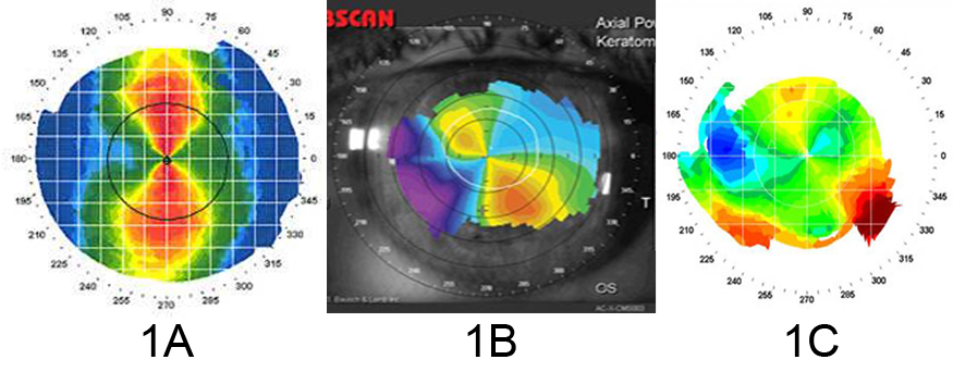
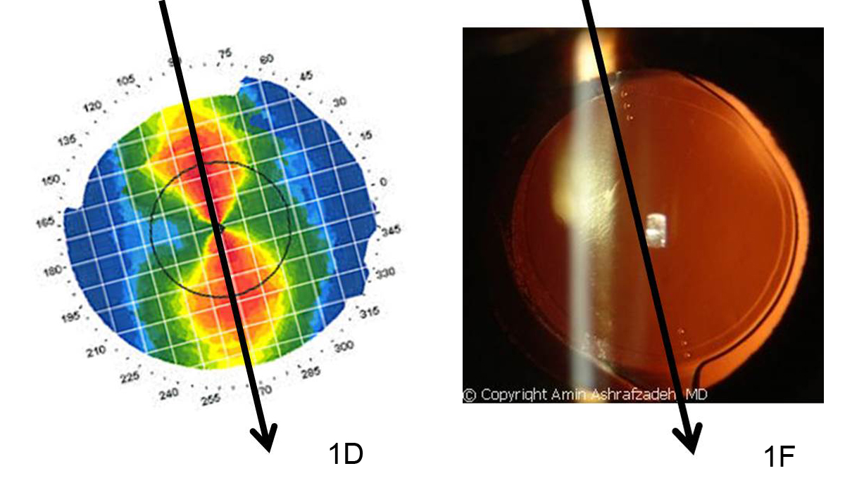
Can Keratoconus Progression Be Predicted?

Richard H. Roe, MD, MHSUveitis Explained

Sumit (Sam) Garg, MDCataract Surgery and Keratoconus

Howard J. Kaplan, MDSpotlight Text – A New Way to Read

Gerry TrickleImagination and KC

In addition to the topics above, here are few more articles that cover a variety of vision issues:

If you have any topics that you would like to read about, please let us know in the comments section below.

6/23/15


Susan DeRemerSusan DeRemer, CFRE
Vice President of Development
Discovery Eye Foundation

Fuchs’ Dystrophy: Current Insights

What is Fuchs’ Dystrophy?

Corneal dystrophies are a debilitating group of progressive diseases that can ultimately deprive a person of sight. The cornea, which forms the front of the eye, is a window for vision, and dystrophies due to intrinsic defects in the corneal tissue cause this window to become opaque and hazy. Fuchs’ dystrophy, also known as Fuchs’ corneal endothelial dystrophy (FCED), is amongst the most commonly diagnosed corneal dystrophies requiring corneal transplantation. The ophthalmologist Ernest Fuchs first described the disease in 1910.

Who gets it?

The disease is rare, and it is difficult to predict who will get it. We know that it affects women more than men (3:1 ratio), older adults (older than 50 years of age), and those with a family history. There are forms in which there could be up to a 50% chance of transmission to children of parents with Fuchs’ dystrophy. Most cases, however, occur sporadically.

What causes it and how does it progress?

Although the cause of Fuchs’ dystrophy is still being studied, there are characteristic findings associated with it: small outgrowths on Descemet’s membrane called “guttae” or “guttata”, thickening of Descemet’s membrane, and defects in the endothelial cells (Figure 1).

fuchs dystrophy 1
Figure 1: Fuchs’ dystrophy can affect all layers of the cornea. Layers of the cornea from anterior to posterior, or frontside to backside, include (A) epithelial cells where blisters and bullae may form in late-stage disease, (B) Bowman’s layer where scarring can occur in late-stage disease, (C) stroma where corneal swelling occurs early in disease, (D) Descemet’s membrane where guttae form (arrows) and thickening occurs, and (E) endothelial cells that decrease in number and change shape and size with disease progression.

Descemet’s membrane is a thin corneal layer between the endothelial cell and the stromal layers of the cornea. Endothelial cells make up the backside of the cornea and function as a barrier and pump for keeping fluid out of the cornea and maintaining corneal clarity. As guttae accumulate on Descemet’s membrane, patients experience progressive loss and change in endothelial cells. Dysfunction of endothelial cells causes corneal swelling, which distorts vision. First, the back of the cornea swells, and eventually, swelling can reach the epithelial cells at the front of the cornea. Swelling can range from mild moisture accumulation, to painful “bullae”, or blisters. In very late-stage disease, significant corneal scar tissue can form and dramatically reduce vision. The progression to late stage Fuchs’ varies from person to person, but usually takes a couple of decades.

What are signs and symptoms?

A patient may be asymptomatic for years despite having guttae. Initial symptoms, including blurry, hazy, or cloudy vision, are typically due to corneal swelling from dysfunction of the endothelial cell layer. Patients may also experience glare or halos around light in the early stages just from the density of guttae. New studies suggest that patients can get glare and higher order aberrations from guttae without any corneal swelling. Symptoms tend to be worse on awakening, but usually improve throughout the day. This is because the closure of eyelids during sleep results in the accumulation of fluid in the cornea. For the same reason, humid weather can also worsen symptoms. As the disease progresses, poor vision may last longer into the day. There may be associated pain if blisters develop.

How is it diagnosed?

The presence of any of the above signs and symptoms, especially with a family history of Fuchs’, should prompt a consult with an ophthalmologist who will diagnose the disorder and follow its progression with regular checkups. An ophthalmologist will conduct a microscopic slit-lamp examination of the eyes, looking for guttae and Descemet’s membrane thickening (Figure 2).

fuchs dystrophy 2
Figure 2: Slit-lamp examination showing speckling pattern on the backside of the cornea characteristic of guttae in Fuchs’ dystrophy.

Special tests may be done to measure corneal thickness, a marker of swelling, or count endothelial cells to track disease progression (Figure 3 and 4).

fuchs dystrophy 3
Figure 3: Optical Coherence Tomography (OCT) showing (A) a normal, healthy cornea and (B) corneal swelling typical in Fuchs’ dystrophy.
fuchs dystrophy 4
Figure 4: In-vivo slit-lamp scanning confocal microscopy showing (A) normal endothelial cells and (B) guttae causing endothelial cell loss and change in Fuchs’ dystrophy.

How is it managed?

Management can be medical or surgical depending on symptoms. Patients may have mild or slow progression of disease that can be managed medically including over the counter salt solution drops (5% NaCl) to reduce corneal edema.

When there is late-stage disease, a corneal transplant may be necessary to improve vision. A corneal transplant replaces the patient’s corneal tissue with human donor corneal tissue. Donor corneas are readily available via excellent eye banks throughout the United States. The surgery is outpatient surgery with regular follow-up appointments and suture removal during the subsequent months. The postoperative healing of the cornea and vision stabilization can take up to a year.

Great strides have been made in the last decade in corneal transplantation surgery, giving patients better treatment options. Patients used to be limited to penetrating keratoplasty (PK), a full-thickness replacement of the cornea. We now have newer surgeries known as endothelial keratoplasty (EK), which is a partial-thickness transplant that replaces only the damaged part of the cornea (the endothelial layer). The different types of EK are DSEK (Descemet’s-Stripping Endothelial Keratoplasty) and DMEK (Descemet’s Membrane Endothelial Keratoplasty). The techniques vary by thickness of the transplanted tissue. The type of EK most appropriate is determined by the corneal surgeon and is variable on a case to case basis. Both types of EK surgeries provide comparable long-term visual results. In both surgeries, the patient’s diseased Descemet’s membrane and endothelial cells are stripped from the inner layer of their cornea. The thin lamellar donor graft is then inserted into the eye and positioned onto the back of the patient’s cornea via a gas or air bubble. The patient is then instructed to lie in a face up position for several hours post surgery during which time the bubble supports the graft until the new endothelial cell pumps begin to wake up and naturally adhere to the back side of the recipient cornea. Occasionally, the doctor may replace another air bubble into the eye the next day to allow more time for the graft to adhere. Visual recovery is on the order of 1-2 weeks in DMEK and 2-3 months in DSEK surgery. Rejection risk is still a possibility in EK surgery but has a much lower rate than traditional full thickness PK surgery.

Other surgical considerations depend on the presence of cataracts. Cataract surgery can worsen Fuchs’ dystrophy because of damage to the endothelial cell layer. For this reason, patients with cataracts and Fuchs’ requiring surgical intervention are often recommended to undergo cataract surgery before or at the same time as corneal transplantation to ensure the best outcome for the transplant.

Patients should work with an ophthalmologist to determine the best management plan. Ultimately, vast improvements in treatment options have given many Fuchs’ dystrophy patients the exciting opportunity to regain vision with improved healing times and reduced infection and rejection of the graft.

Citations: Figure 2 and 4 are from Zhang J, Patel DV. The pathophysiology of Fuchs’ endothelial dystrophy—a review of molecular and cellular insights. Exp Eye Res. 2015 Jan

6/4/15

priscilla-thumbnailPriscilla Q. Vu, MS
Medical Student
University of California, Irvine School of Medicine


Farid 3.6.14Marjan Farid, MD
Director of Cornea, Cataract, and Refractive Surgery
Vice-Chair of Ophthalmic Faculty
Director of the Cornea Fellowship Program
Associate Professor of Ophthalmology
Gavin Herbert Eye Institute, University of California, Irvine

9 Ways To Relieve Cataract Surgery Stress

It’s an extremely rare person who would not feel nervous before surgery of any kind, even if it’s an outpatient procedure that will only take a few minutes. In the case of cataract surgery, the fear can be even worse than the procedure itself.


9 Ways to Relieve Cataract Surgery Stress
People who are under intense stress can suffer a range of symptoms, including irregular or racing heartbeat, nausea, upset stomach, difficulty breathing, and an inability to sleep. It can even affect your mind, causing you to forget important details about the operation, like advice on how to get ready or what to do after you come home from surgery.

Here are 9 ways to relieve cataract surgery stress:

1.  Just Think About It – Let’s start with the first, and most difficult, suggestion—change your own mind about how you feel. Admittedly, it takes a great deal of discipline that’s hard to muster in the face of great anxiety, but try to remind yourself how your sight will be saved after a relatively short, quick, and easy procedure. What you are about to go through will prevent you from going blind.


2.  Learn Everything You Can – For many, it helps to learn as much as possible about the surgery before it happens. Knowing exactly what’s going to happen and how others have dealt with what you’re going through can be a great relief. Knowledge may be all you need to relieve your anxiety.


3.  Talk to Your Surgeon – It almost always helps to just talk to someone, and who better than your surgeon? Who else knows every detail of the procedure you’re about to undergo? It’s fairly likely that your surgeon has performed many successful surgeries of this kind before, and he or she may have some stories of encouragement for you, as well as important and comforting knowledge of his or her own personal experiences.


4.  Imagine the End Result – It may help if you keep focused on what happens after, as if it’s already done and you can go home, the procedure over. This takes a great deal of imagination, rather than the discipline of thinking rationally about it, but if you have that level of imagination, it’s certainly worth trying.


5.  Alternate Methods – This encompasses a whole range of stress-reducing tactics that are not usually under the medical umbrella. Nevertheless, they have done a great many people a lot of good. Yoga, hypnosis, massage, acupuncture, acupressure, and other treatments have allowed those suffering preoperative stress to sleep better at the least.


6.  Herbal Supplements – A form of alternative treatment involving traditional ingredients to produce a more restful state. The herbs are often just infused into tea and drunk. While these supplements are generally called “all natural,” you should always consult your doctor before taking them, since they can have an effect on other medications you may be taking, including the anesthetic you are given before surgery.


7.  Have Some Fun – Do something fun to take your mind off of what’s about to happen. Whatever that specific thing may be is up to you, since an individual’s idea of fun differs from person to person. Whatever you generally do to take your mind off of things and unwind after a hard day may be just the thing to help you out before undergoing cataract surgery.


8.  Treat Yourself – In the same vein has doing something fun, do something that usually relaxes you. If you like going for long walks, do that the day before the surgery. Or listen to music that fills you with peace (or joy). Take a long bath. If it makes you feel relaxed or calm, it will help you deal with your anxiety.


9.  Distract Yourself – Once you get to the hospital, you’ll probably be waiting around, even if you get there right on time, giving plenty of time for stress to ramp up. You’ll do better if you keep yourself entertained, but the hospital waiting room is probably one of the more boring places on earth. Fix that by bringing something along to entertain yourself, like a book or some magazines, or even stream your favorite movie or podcast. You’ll probably want to bring more than one thing to do, in case the wait is long.


Stress adds complication to the body’s systems, and can therefore cause some complication in the upcoming surgery. Do what you can, whether you kick back with friends or take some herbal supplements, to help yourself get into the best mind space possible. Think about the positive outcomes, and you’ll do well.