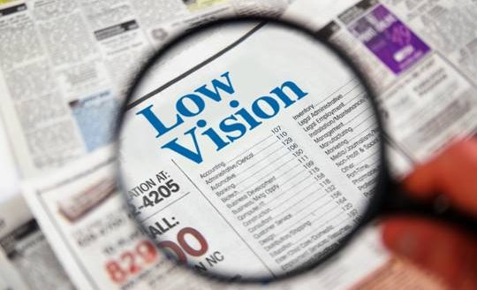
 Healthy Aging Month is an annual health observance designed to focus national attention on the positive aspects of growing older. Aging is a process that brings many changes. Vision loss and blindness, however, do not have to be one of them. There are several simple steps you can take to help keep your eyes healthy for the rest of your life.
Eye diseases often have no early symptoms, but can be detected during a comprehensive dilated eye exam. A comprehensive dilated eye exam is different from the basic eye exam or screening you have for glasses or contacts. By dilating the pupils and examining the back of the eyes, your eye care professional can detect eye diseases in their early stages, before vision loss occurs. By performing a comprehensive eye exam, your eye care professional can check for early signs of –
Here are some other tips to help maintain healthy vision and body now and as you age:
- Eat a healthy, balanced diet. Loading up on fruits and vegetables can help keep your eyes healthy and disease free.
- Maintain a healthy weight. Being overweight increases your risk for heart disease and diabetes. Complications from diabetes, such as diabetic retinopathy or glaucoma, can eventually lead to vision loss.
- Don’t smoke. Smoking increases your risk for age-related macular degeneration, cataract, and other systemic diseases, including cancer. Wear protective eyewear when outdoors. Protecting your eyes from the sun’s ultraviolet rays when you are outdoors is important for your eye health. Choose sunglasses that block 99 to 100 percent of both UV-A and UV-B radiation.
Even if you are not experiencing vision problems, visiting an eye care professional regularly for a comprehensive dilated eye exam is the most important thing you can do to reduce your risk of vision loss as you age.
Download “Everyone’s vision can change with age”
A handout with explanation on how vision can change with age.
 When you think of cancer, most of us do not think about the eye or vision. Though rare, cancer can start inside or outside of the eye. If cancer starts inside the eyeball it’s called intraocular and if it starts outside the eye (eyelid or in the eye socket) then it’s called extraocular tumor. It can occur in both children and adults. Most major eye centers have specialists who are trained in the diagnosis and treatment of eye cancers.
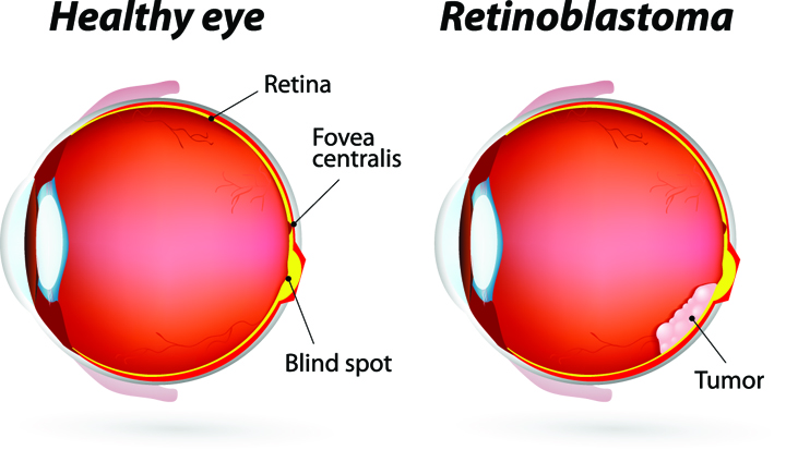 At the later stage of this cancer, the only one way to survive is to remove the eyeball (enucleation). Like many of other types of cancer, retinoblastoma has a genetic component so genetic testing needs to be done. The tumor begins with the RB1 gene mutation that stimulates retinal cells to develop into a tumor called a retinoblastoma. The RB1 mutation can be inherited from the parents, but in some cases it is sporadic and not inherited. There are various treatments such as surgery, chemotherapy, radiotherapy etc. to cure retinoblastoma cancer. Rarely it can spread beyond the eye.
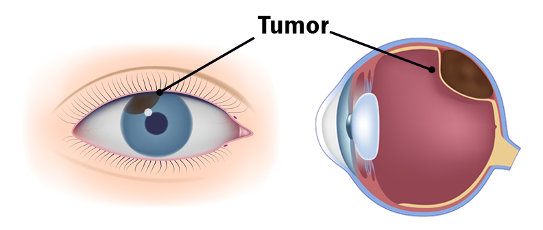
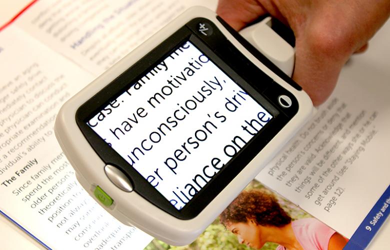 Portable magnifiers and lighted magnifiers- offer magnified reading on the go. Perfect for menus, shopping lists, label reading, and more, portable magnifiers can fit in your pocket, purse, or be worn on the belt for quick, easy use.
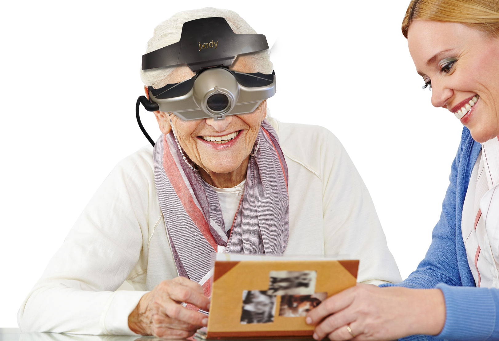 Wearable magnifiers – wearable technology is the future for those with low vision who live an active lifestyle. Wearable options make it possible to see and take part in everyday tasks, such as reading and recognizing faces.
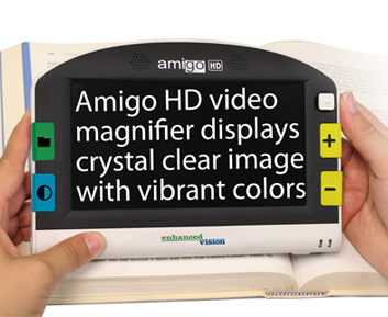 Transportable magnification screens– are perfect for close up viewing as well as distance viewing. These great viewers offer great flexibility, from watching TV to using the mirror image feature for self-viewing. There are APPS for smart phones that can be used to magnify reading material.
 Tom Sullivan

 The other day my daughter Blythe asked me which Christmas I consider to be my favorite. I had to think a minute, because as a family, the Sullivan’s have had some great ones. I was about to say the first time you and your brother Tom were old enough to really get into Santa, being absolutely sure that the fat man brought your presents right down the chimney. I was about to say that, and then I remembered.
  Colorado, when our children were teenagers and our friend, the marvelous Betty White, joined us for a Christmas Eve sleigh ride none of us will ever forget. The night was perfect. It had snowed earlier that day, and the air had a feeling of Christmas that you could almost taste. Oh, sure, it was cold, but we were bundled up under tons of blankets as two beautiful Clydesdale horses with bells jingling took us through the woods to a magical barn where dinner would be served and carols sung.
 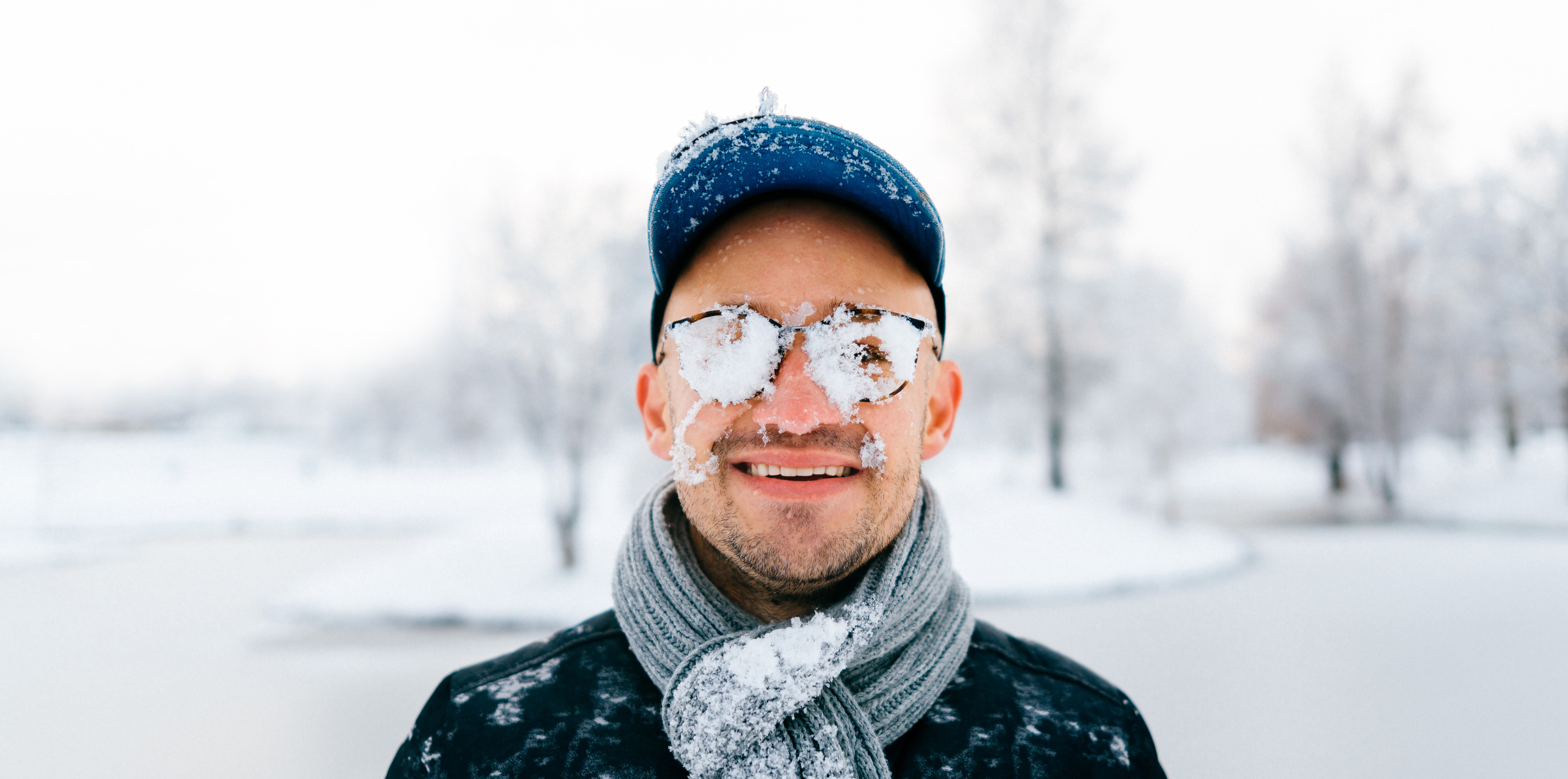 Harsh weather conditions can reduce the natural moisture in your eyes and the irritation usually results in a burning or itching sensation that often leads to rubbing or scratching your eyes which can worsen the symptoms. Sometimes it feels like there is a foreign object in your eye and for some, dry eyes can even cause excessive tearing, as your eyes try to overcompensate for their lack of protective tears. Prolonged, untreated dry eyes can lead to blurred vision as well. Between the harsh winter winds outside and the dry heat radiating inside, our eyes are very quickly irritated and dried in the winter months. The result is itchy, dry eyes that may cause pain, blurred vision, a burning sensation, or even watery vision as our eyes try to compensate for the dryness.
 For 8 years I served as a member of the Academy of Ophthalmology’s Foundation Board. In that time we conducted a number of studies in all areas of vision preservation. The one that I believe was most meaningful occurred when we asked thousands of people to express what frightened them most in life. Frankly, I was really surprised at the results of the study. I was sure that people would say maybe stage four cancer, or ALS, or some other terminal disease would be the thing that would frighten them the most. I would have imagined that they might talk about the loss of a loved one or even the fear of a natural disaster. The results of the study were very clear. 62% of all the participants said that the loss of vision was the single most frightening possibility they would ever have to face.
 Tom Sullivan
 I can only imagine my wife’s beautiful face. Oh sure, I’ve touched it and kissed it many times. I’ve felt the lines with the tips of my fingers, tracing our lives together, and I’ve heard her smile. I understand that’s not really seeing it. It’s not seeing her eyes as they sparkle with something funny I said; or, when she looks at me with love reserved only for those who are truly in love.
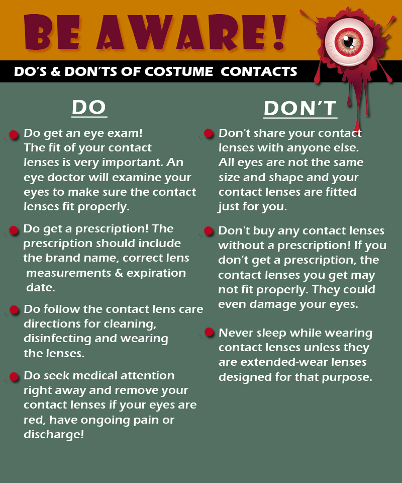
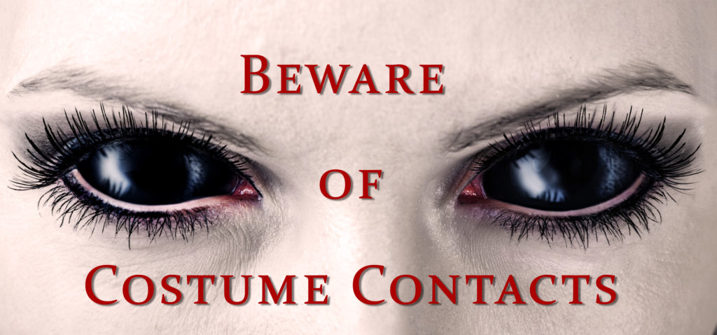 Costume Contact Lenses such as cat eyes or zombie may make your Halloween costume a bit more frightful although wearing those lenses without a prescription can be more terrifying, as it could result in vision loss or even blindness.