The “A B C’s” of Contact Lens Fitting for Keratoconus

4/18/14


Keratoconus is a disease of the cornea in which the cornea loses its natural round shape and becomes distorted with cone-like bulging, progressive thinning, and associated reduction in vision quality. It is the front surface of the cornea that is the most significant focusing element of the eye. The irregularity of the front surface of the cornea in keratoconus is primarily responsible for the poor vision obtained with glasses since light that is focused through glasses lenses once again passes through air prior to reaching the corneal surface where again it is distorted. Contact lenses utilized for vision correction in keratoconus serve to “mask” the irregularity of the corneal surface thus creating a new smooth and regular front surface to the optical system.

Today contact lens correction for keratoconus is NOT synonymous with rigid corneal lenses. There are numerous options available to the contact lens specialist that can provide significant vision improvement for patients with keratoconus. It is the responsibility of the contact lens specialist to determine which design of contact lens is most appropriate for the individual patient. Let’s review the various options available today for the contact lens management of keratoconus.

Corneal rigid gas permeable contact lenses have been the mainstay for contact lens correction of keratoconus for many decades.

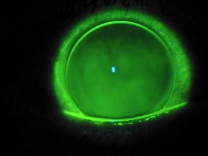
Large diameter corneal gas permeable lens fit with clearance of the cone apex. - contact lens
Large diameter corneal gas permeable lens fit with clearance of the cone apex.

These are made of oxygen permeable rigid materials and are typically of a size that is smaller than the overall corneal diameter and they are fit to center as well as possible over the optical center of the cornea. Due to their rigid nature they do a wonderful job in masking the corneal irregularity and provide excellent vision. However, certain limitations have required us to develop other contact lens options for keratoconus. The limitations include; difficult initial adaptation and ongoing comfort issues for some patients, difficulty in centering over the optics of the eye in cases of more advanced and decentered cones, and occasional cases where lenses are not stable on the cornea and will dislocate. Most importantly, corneal rigid lenses that are fit “flat” and bear on the apex of the cone may induce progressive corneal scaring over time. Current fitting philosophy for these lenses attempts to fit with clearance over the cone apex.

Soft contact lenses have a significant role in the treatment of keratoconus. Mild and early cases can be managed with traditional non-specialty soft contact lenses as long as the degree of corneal distortion is not too severe.

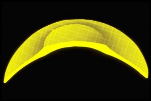
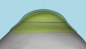
Custom specialty soft lens for keratoconus with thick central optical zone and thin periphery.  Courtesy of Visionary Optics. - contact lens
Custom specialty soft lens for keratoconus with thick central optical zone and thin periphery. Courtesy of Visionary Optics.

Typically soft astigmatism correcting contact lenses are required. More advanced cases can be treated with specialty keratoconic designed soft lenses. These lenses typically have the central optic zone thickened in order to mask the corneal irregularity. However, the periphery of these lenses is thinned and provides excellent comfort and eye health response, especially when designed with highly oxygen permeable materials. We have been able to manage many cases of moderate and somewhat severe keratoconus with these specialty lenses. The advantage primarily is in lens wearing comfort and positional stability on the corneal surface. The limitation is typically if these lenses can provide adequate vision correction for the individual.

Piggyback or tandem lens systems utilize a combination of rigid corneal gas permeable lenses and soft lenses. Typically we utilize a thin, highly oxygen permeable soft disposable lens with insignificant power and then fit an appropriate gas permeable rigid lens on top of the soft disposable lens. The rigid lens provides the vision correction and the soft lens provides superior comfort compared to the rigid lens being fit directly on the eye surface and also occasionally helps with centration of the rigid lens over the optics.

Recessed Pillow Lens System (RPLS). Courtesy of EyeVis Eye and Vision Technologies and Fusion Technologies.- contact lens
Recessed Pillow Lens System (RPLS). Courtesy of EyeVis Eye and Vision Technologies and Fusion Technologies.

A variant of this system is called a “recessed pillow lens system” (RPLS) where a custom soft lens is created with a partial thickness cut out or recess that can hold a rigid lens in place. This can be used when a typical piggyback/tandem system cannot allow the rigid lens to center properly.

Hybrid contact lenses are available in specialty designs to address keratoconus. A hybrid lens is constructed with a rigid gas permeable lens center and a soft lens “skirt”. Again, the rigid lens provides the vision correction and the soft skirt provides excellent lens centration and improved comfort. Limitations of hybrid lenses include the inability to fit on more advanced and decentered cones and their potential to tighten up on the eye making removal more challenging and potential for eye irritation. However, new designs and fitting methods have addressed these issues in most cases.

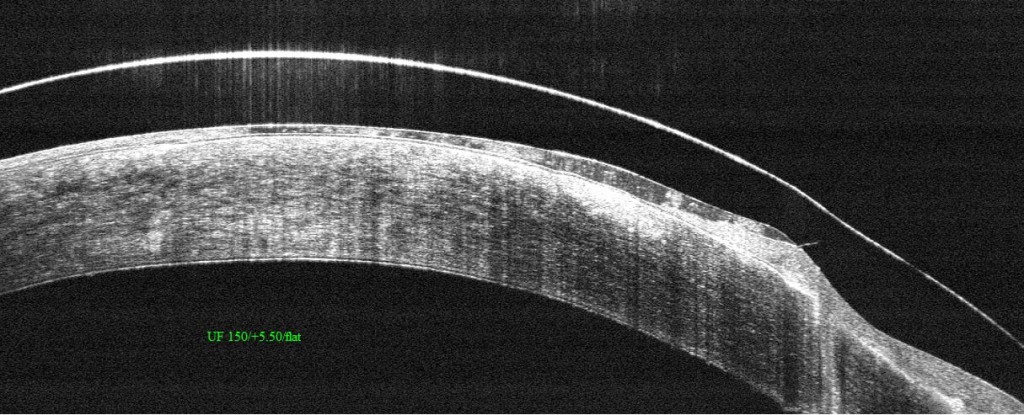
Finally, scleral lenses are an excellent option for keratoconus. These are large diameter rigid gas permeable lenses. They vault the entire cornea and do not touch the corneal surface at all.

Fully customized scleral lens design based on an “Eye Print” impression. Courtesy of EyePrint Prosthetics.- contact lens
Fully customized scleral lens design based on an “Eye Print” impression. Courtesy of EyePrint Prosthetics.

A tear layer is created below the scleral lens and the lens comes to rest gently on the white of the eye (the scleral surface). This results in very good lens comfort and it also protects the corneal surface from the negative effects of contact lens bearing. The rigid material provides excellent optics. Limitations of scleral lenses include the complexity of fitting, difficulty in insertion and removal on occasion and entrapment of mucous and debris below the lens requiring removal and rinsing of the lens for some patients. Overall scleral lens fitting is taking a larger role for keratoconus over the past few years. We are now developing totally customized scleral lens systems based on taking scans or impressions of the ocular surface to design a totally customized lens made uniquely for the individual eye that can address even the most distorted ocular surface.

Today we have multiple contact lens options to address the vision needs of our keratoconic patients. It is the responsibility of the contact lens practitioner to prescribe the most appropriate design for the individual patient.

Barry Eiden OD, FAAOS. Barry Eiden, OD, FAAO
North Suburban Vision Consultants, Ltd., President and Medical Director
www.nsvc.com
National Keratoconus Institute, President and Medical Director
www.nkci.org
EyeVis Eye and Vision Research Institute
sbeiden@nsvc.com

Wavefront Sensing Applied to Custom Contact Lens Research in Keratoconus

4/10/14

During a trip to the optometrist or ophthalmologist, a patient will encounter the process of subjective refraction.  This technique involves the clinician asking the patient to make a series of judgments (which is better, one or two?) about the clarity of their vision when looking through a series of lenses.  The choices that the patient makes guide the clinician in identifying an optical prescription which is typically made up of sphere, and potentially, cylinder lenses.

Why is it that glasses don’t always work for patients with keratoconus?

In many instances, individuals with keratoconus do not achieve excellent visual performance with spectacles or traditional soft contact lenses.  One cause for the failure of these corrections is that the changes in corneal shape that accompany keratoconus induce refractive errors which traditional spectacles simply cannot correct.  So, even when sphere and cylinder in the keratoconic eye are well-corrected, these “other refractive errors” or “other aberrations” remain uncorrected and can lead to a blurred retinal image and blurred vision.  Collectively these other aberrations can be referred to as higher order aberration, while the aberrations that are typically corrected with spectacles and soft contact lenses are referred to as lower order aberration.

What kinds of higher order aberrations are present in keratoconus:

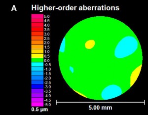
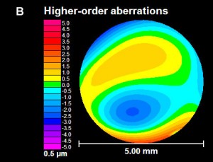
Pantanelli et al. have stated that the level of higher order aberration present in an eye with keratoconus is, on average, approximately 5.5 times higher than the level experienced in a control group.  In an effort to visualize higher-order aberration data, they are commonly represented graphically as shown in the figures below.  Examples of higher order aberration measured in one normal eye are shown in figure A, while an example of higher order aberration from one keratoconic eye are shown in figure B.  The circular nature of the map denotes the boundary of the measurement, which is defined by the round pupil of the eye.  A majority of the higher order aberration map in figure A is green (denoting a relative absence of higher order aberration).  However, the map in figure B displays a much larger variation in color, indicating the presence of higher order aberration  in this individual keratoconic eye in a greater quantity than the normal eye shown in figure A.

Figure A - normal-keratoconus
Figure A – normal
Figure B - keratoconus
Figure B – keratoconus

A wavefront aberration map of the “other aberrations” or higher order aberrations of two eyes. Figure A is an example of data for a normal eye and figure B is an example of data for an eye with keratoconus.

If refraction is not capable of quantifying higher order aberrations, how are they measured?

One method for obtaining the information regarding higher order aberration shown above is with a wavefront sensor.  The wavefront sensor objectively (without patient feedback) collects information on the optical performance of the eye that can be used to calculate the amount of both lower and higher order aberration present.

Laboratory-based research related to custom contact lenses:

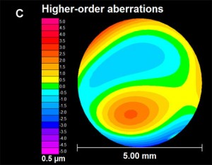
Several investigators in the laboratory (e.g. Katsoulos et al., Sabesan et al., Chen et al., Marsack et al.) have reported on work that attempts to further reduce higher order aberration by targeting the eye-specific higher order aberration seen in a given keratoconic eye.  The general philosophy behind these customized lenses is that the aberration pattern measured with the wavefront sensor is a more complete optical prescription for implementation of a custom contact lens.  Figure C demonstrates, in principle, the optical properties of a contact lens designed to correct the higher-order aberration in figure B.  Where the map of the eye (figure B) is red, the map of the correction (figure C) is blue, and vice versa.  When the lens is worn, the net effect as light propagates through the lens-eye system is the cancellation of the higher order aberration in a targeted manner.

Figure C -keratoconus correction

In principle, this figure pictorially represents the higher order optical properties of a contact lens designed to fully correct the higher-order aberration of the eye represented in figure B.

What is next:

Investigators continue to push the technology behind custom contact lenses for keratoconus towards clinical relevance.  However, like every novel intervention strategy, we must manage our expectations.  Complexity in measuring keratoconic eyes, a need for specialized equipment and expertise to design and manufacture the lenses, the infrastructure needed to coordinate the clinical exam and manufacture efforts and cost associated with the process are a subset of the barriers that must be removed if this type of correction is to become more mainstream.  For this reason, it is my opinion that if/when these corrections become commonly available in the clinic, they will likely add to, and not replace, existing forms of corrections that patients and clinicians now utilize to correct vision.

jmarsack-bio-picJason Marsack, PhD
Research Assistant Professor
University of Houston, College of Optometry.
Dr. Marsack’s work focuses on the relationship between visual performance
and optical aberration in individuals with highly aberrated eyes.

End of the Day Syndrome

4/2/14

“Dr. S., my eyes are red and burning at the end of my work day.”

“Patient, what sort of work do you do?  Tell me something about your work conditions.”

“I am a computer graphics artist.  I sit and stare at my twenty-seven inch HD screen for hours on end gently adjusting the composition of each pixel.  My studio is air-conditioned but not humidified, so after some hours of work, I feel dry as a bone.”

“One more question…can you cry tears?  Say, when you peel and slice an onion?”

Rule of 20 - blinking

The need to blink

Blinking is a complex function of the eyelids that when completed results in a clean, refreshed, re-wetted corneal surface.  The tears that are washed across the outside of the eye with each blink bring oxygen and other nutrients to the outer cell layer aiding in the rebuilding and revitalizing of the surface tissue.

Blinking is characterized by a full sweep of the upper lid over the eye to meet the lower lid.  The completion of this motion is performed gently without squeezing.  And, to be effective full eye closure needs to be repeated fairly often.  Blink rates vary according to investigators but most sources report an average of between six and ten full blinks per minute under normal viewing circumstances.

The anti-blink problem of our generation

In olden times – say the years between 1750-1950 – the most aggravating problem to the ocular surface was a good book or intense study.  The reader would concern himself with the text at hand and slowly his eyes would dry until a “rest break” was necessary.

Environmental or vocational changes to our lifestyle over the generations have promoted reduced blink rates.  Most recently in this negatively developmental progression is the effect of the television screen, the CRT, the LED screen, the handheld and pocket computer on the blink rate.  It appears that as attention level increases, blinking suffers.  First the eyes close less, then incompletely, and finally rarely only when surface dryness drives the individual to desperate measures.  He must blink or (so he feels) his eyes will pop out of their sockets.

Adding insult to injury increasingly over the decades is air conditioning – both heating and cooling – when not humidified.  Staring at console screens in dry environs speeds the desiccation of the cornea and results in discomfort.

The surface of the eye is a biological system.  Living systems require some degree of moisture.  If the cells of the eye – or any biological surface — are permitted to dry out, they will die.  Dead corneal cells fall off the cornea and float in the tears on the surface of the eye until washed away with a blink.  Until the surface is cleaned the dead cells are considered by the eye to be foreign bodies with the consequent irritation and induced reflex to blink.

When cells die and fall off, the underlying nerve endings send pain signals to the nervous system.  The sensation can be felt as pain, burning, or mere irritation or itching depending upon the severity of cell loss.

How to handle environmentally induced dry eye

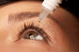
After the ocular surface is dry most treatments will seem to make matters worse:  to cause burning and stinging, perhaps, even more than the dry eye itself.  Any tear substitute, any amount of blinking will be irritating at first. But, that is really all that can be done at this stage:  wetting and blinking.

Prevention

As in many conditions, the best treatment, in fact a cure, for recurrent environmentally induced dry eye is prevention.  For the eye that has a naturally flowing tear supply, the act of blinking is the surest prevention to stinging and burning after a day’s work at the computer.  Additionally, many sources recommend using the ‘rule of 20’:  after each twenty minutes of work, look up from the text or away from the screen; blink and refocus on the page twenty times.  This repetitive exercise simultaneously re-wets the eye and relaxes the focusing mechanism of the eye.

The result is relaxed and comfortable eyes that can continue to provide important and high quality information for longer hours of work.

Bezalel-SchendowichBezalel Schendowich, OD
Chairperson and Education Coordinator, JOS
Fellow, IACLE
Member, Medical Advisory Board NKCF
Sha’are Zedek Medical Center, Jerusalem, ISRAEL

Help for Computer Users

Working long hours in front of the computer requires a fairly unchanging body, head and eye position which can cause discomfort.  Correct working position, periodic stretch breaks, frequent eye blinking, artificial tears for lubrication are all very important.  However, it’s not always easy to remember this when you are engrossed in work. Here are a few fun, free and easy-to-install “break reminders” to help:

WorkSafe Sam - break reminder
WorkSafe Sam
WorkSafe Sam is a desktop tool that provides stretching tips to help reduce eye and muscle strain for office workers (clicking on this link will open a file on your computer because this is a zip file).

Workrave is break reminder program that alerts you to take “micro-pauses” and stretch breaks.

Take Your Break is another break reminder designed to prevent or minimize repetitive strain injury, computer eye strain and other computer related health problems.  It has a friendly interface and a tray icon status indicator.  It runs quietly in the background, monitoring your activity and reminding you to take regular breaks.

And remember to blink.  Blinking cleans the ocular surface of debris and flushes fresh tears over the ocular surface. Each blink brings nutrients to the eye surface structures keeping them healthy. The flow of tears is responsible for wetting the lower third of the cornea. This is very important in KC, since this area is generally below the bulge of the cone and in many cases irritated by wobbly RGP lenses.  Maybe your job requires hours of work at a computer. Maybe you like to spend your free time surfing the internet. Whatever the reason, your body is probably feeling the effects of spending too much time staring at a computer monitor, which could result in Computer Vision Syndrome (CVS).  The most common symptoms are: eye strain, dry or irritated eyes,redness in eyes,difficulty in refocusing eye,neck pain,double vision,blurred vision, fatigue, and headaches.

Please join us on Thursday when Dr. Bezalel Schendowich will be providing a detailed insight into the importance of blinking, going beyond computer usage.

CathyW headshotCathy Warren, RN
Executive Director
National Keratoconus Foundation

Corneal Transplant Surgery Options

In this day and age of advancing technology, corneal transplants have changed from a long arduous ordeal to a more simple and precise procedure that offers faster visual recovery.  Instead of replacing the entire cornea for any and all corneal diseases, we now perform disease targeted partial corneal transplants.  If the disease involves the back layer of the cornea, we perform endothelial keratoplasty and replace only the diseased inner layer of the cornea.  Conversely, if the problematic portions are the front layers of the cornea, we perform anterior lamellar keratoplasty.  The co-morbidity and risk of rejection from partial corneal transplants are significantly less than the traditional full thickness transplants.

With endothelial keratoplasty, a small incision, about 4-5 mm is made and a sheet of donor endothelial cells are placed into the anterior chamber of the eye.  A large air bubble is then used to float this sheet up so that it opposes the posterior or back portion of the cornea.  The patient is asked to position face up for 24 hours.  Over this period of time, the cells will “stick” on their own and thus no sutures are required to keep the graft in place.,/span>

Figure 1 - corneal transplant
Figure 1

Anterior lamellar keratoplasty is done for superficial scars and opacities of the cornea or for keratoconus, a genetic degeneration of the cornea that is seen in younger individuals.  In this case, the native endothelial cells of the patient are healthy and therefore are left intact while the remainder of the cornea is transplanted.  This significantly lowers the risk of rejection, which is traditionally a much higher risk in young patients.  Multiple sutures are required to maintain this graft in place however, with the advent of femtosecond laser technology, the wound configuration is made in such a way as to promote rapid healing and visual recovery. (Figure 1)  Sutures are removed at an earlier time than with traditional surgery and the eye is able to undergo visual rehabilitation with glasses or contact lenses in 3-6 months’ time.

Corneal transplantation does not require waiting on a list for a donor to become available like it once did.  There are now multiple excellent eye banks across America that harvest, screen, and distribute donor tissue to surgeons.  This way, tissue is readily available and patients only need to schedule a time based on their own and their surgeon’s time schedule.  Post operatively, patients are asked to return to regular activity with the exception of no heavy lifting or bending for a period of 2 months.  Antibiotic and anti-rejection drops are started immediately after surgery and continued for several months after.  No oral medications aside from the patient’s regular medications are required.

Farid 3.6.14Marjan Farid, MD
Director of Cornea, Cataract, and Refractive Surgery
Vice-Chair of Ophthalmic Faculty
Director of the Cornea Fellowship Program
Associate Professor of Ophthalmology
Gavin Herbert Eye Institute, University of California, Irvine