New Technology for Evaluating Contact Lenses

5/1/14

Successful management with contact lenses can sometimes be a frustrating process for those with keratoconus. The fitting and evaluation process involves numerous visits to the optometrist, out of pocket expenses as well as medical insurance co-pays. Luckily, the contact lens industry has responded to the need to have better contact lens materials for patients with keratoconus. It used to be that the only contact lenses available to manage keratoconus were gas-permeable (GP) lenses. Now, more and more patients are being fit with newer generation hybrid lenses (GP lens core with a skirt of soft lens material to aid in fit and comfort) and scleral lenses (large diameter GP lenses that do not rest on the cornea, only the outlying sclera). These newer designs are intended to vault over the central cornea and do not rest on the cornea at all. They have resulted in much more comfortable and wearable strategies for full-time use. As a result of the newer lens designs, the game has changed when it comes to the science of fitting and evaluating the lenses.

For years, optometrists have used corneal topography to guide their decision making on fitting keratoconic eyes. A corneal topographer is an instrument that maps the shape of the cornea, and gives information much like that of a topographical map for hiking. Corneal topography is still an absolutely mandatory part of evaluating the shape of the cone, the simulated corneal curvature, and monitoring for progression of the disease, and is not a standard part of a typical eye examination. However, doctors have a new tool at their disposal for fitting contact lenses on patients with keratoconus.

Optical Coherence Tomography (OCT) was once reserved for use in the back of the eye, or retina. OCT uses visible light passed through the clear structures of the eye to generate a cross-sectional image of the layers of the retina, much like an image generated by an MRI. Advances in OCT technology has improved the resolution to image the eye on the micrometer scale (one-thousandth of a millimeter). OCT technology is now commercially available not only for the retina, but the structures of the front part of the eye. The obvious application is to aid the doctor in the fitting and evaluating complex contact lenses that vault the cornea.

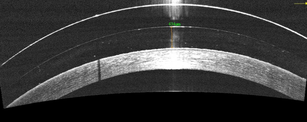
Figure 1.  OCT image of a scleral lens fit on a keratoconic patient.  The cornea is the opaque white band located at the bottom of this picture, the tear film reservoir is the middle clear band and the contact lens is the top band.  Using an electronic caliper tool, the precise amount of vault can be measured, leaving no doubt as to the precision of the fit.
Figure 1. OCT image of a scleral lens fit on a keratoconic patient. The cornea is the opaque white band located at the bottom of this picture, the tear film reservoir is the middle clear band and the contact lens is the top band. Using an electronic caliper tool, the precise amount of vault can be measured, leaving no doubt as to the precision of the fit.

OCT allows the optometrist to view a cross-sectional image of the contact lens on the eye in real time and to monitor the health of the cornea in the presence of the contact lens. This view is valuable for judging the vault of new designs of contact lenses over the cornea and judging where the lenses land on the eye. It is the most specific way to determine if the fit is acceptable and to troubleshoot if lenses are not fitting appropriately.

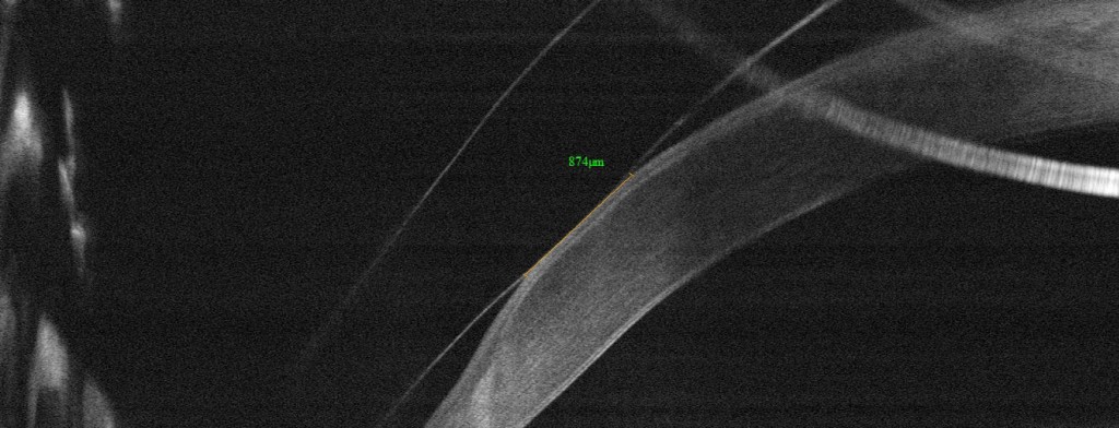
Figure 2.  OCT image of the periphery of a scleral lens on a patient with pellucid marginal degeneration.  The lens contacts the cornea over an area of 0.87mm long.  These types of measurements help guide decision making in modifying the lens fit and were impossible before the advent of this technology.
Figure 2. OCT image of the periphery of a scleral lens on a patient with pellucid marginal degeneration. The lens contacts the cornea over an area of 0.87mm long. These types of measurements help guide decision making in modifying the lens fit and were impossible before the advent of this technology.
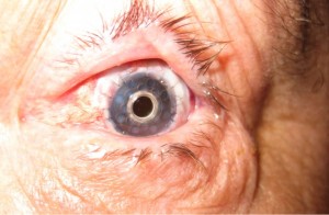
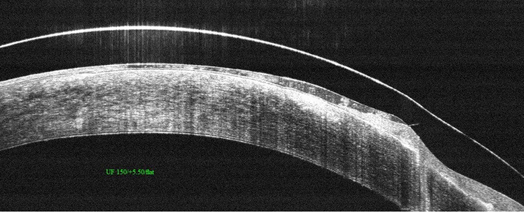
Figure 3.  Hybrid lens on a highly irregular eye after corneal transplant.  The point of contact of the soft skirt with the cornea is visible to the right of the image.
Figure 3. Hybrid lens on a highly irregular eye after corneal transplant. The point of contact of the soft skirt with the cornea is visible to the right of the image.

Optometrists now have a much more powerful tool for evaluating and managing even the most challenging contact lens fits. It remains to be seen whether this technology has the ability to reduce the number of visits required for successful fit. But, the precision afforded by this technology does have the ability to improve patient outcomes.

Sonsino HeadshotJeffrey Sonsino, OD, FAAO
The Contact Lens Center at Optique Diplomate
Cornea, Contact Lens, and Refractive Therapies

Dry Eye and Tear Dysfunction

4/24/14

With an aging population and increasing awareness of the condition, dry eye has become one of the hottest topics in eye care today. There have been numerous advances in our understanding of the condition as well as several breakthroughs in treatment.
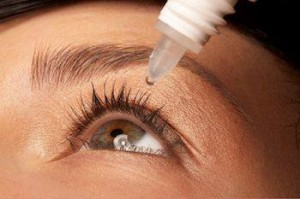
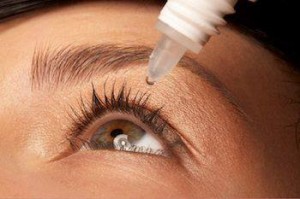
eye drops in eye
Lets start with the most fundamental question. Why do we have tears? Tears serve many functions, but perhaps the most important is that they create a perfectly smooth optical surface at the front of the eye to allow sharp focusing. The sharper your vision the more quickly you can spot and run from a predator or find a supply of food. Hence, a stable tear film is critical for survival. As a result, the body has evolved many ways to help keep the tears healthy and intact.

When I describe the tears to a patient I describe it as a structure. Tears have a foundation that anchors them to the eye’s surface, an elastic protective support section not that different than the walls in a building and even a roof, just like a house has. Like a house, problems with any structural part can adversely impact the entire building.

Over the past few years the most important breakthroughs in understanding dry eye have focused on the outer portion of the tear structure – what you might think of as the roof. When a roof develops holes and it rains, water gets in which can destroy the entire structure of the house. With the tears, holes in the “roof” can let moisture evaporate causing the structure to collapse and become unstable.

The outer layer of the tears consist of lipid or oil produced by the meibomian glands – glands that run radially through the lids and express small amounts of complex oil with each blink. When these glands work properly, the oils they produce coat the tears, preventing evaporation and stabilizing and lubricating. When the glands become blocked – which can occur for a number of reasons including decreased blinking due to excessive computer use, the lack of oil can lead to a down spiral of damage to the eye’s surface with symptoms that can range from annoying to life altering.

Lets talk for a moment about symptoms. We know that dry eye is progressive and, if left untreated will worsen over time, so the sooner we know about it, the sooner treatment can begin. I am often surprised that even with increased awareness, most patients don’t recognize that they suffer from dry eye until the condition is fairly advanced. One reason is that many people think that the symptoms of dry eye are a normal and expected part of aging. For the most part they are not. If you experience, burning, irritation, unstable vision, grittiness or any change in your vision or eye comfort, you should bring this to the attention of your eye care practitioner.

One of the most common signs of dry eye is excessive tearing. Patients often complain that their eyes are too wet and I have to explain that the excessive tearing is the eye’s way of trying to correct for dryness and prevent further damage. Unfortunately excessive tearing further destabilizes the tears and makes matters worse.

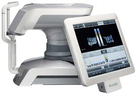
Lipiview System
Lipiview System

Perhaps the greatest advance today is the recognition that meibomian gland dysfunction causes or contributes to nearly 90% of all dry eye. Many eye care providers are adding high tech tools like meibography which can show the actual state of the meibomian glands like an X-Ray and LipiView which measure the actual thickness of the lipid layer of the tears. As a result diagnostic ability has improved.

Lipiflow Device
Lipiflow Device

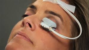
Recently introduced eyedrops like Alcon’s Systane Balance and Allergan’s Refresh Optive Advanced Formula drops can help restore absent or deficient oils in the tears. And new treatments including moist heat goggles like Tranquileyes by EyeEco can help improve comfort. LipiFlow, a technology pioneered by TearScience, offers a breakthrough treatment that can clear blocked and reset meibomian glands. It can produce amazing improvement.

There have also been significant advances for contact lens wearers suffering from dry eye. Scleral lenses are especially helpful for patients with advanced dry eye who also must wear contact lenses to see, such as those who have keratoconus. The large scleral lens serves as a barrier that protects the eye and provides sharp stable vision. In fact, scleral lens designs are used as a treatment for severe dry eye and ocular surface disease.

Finally, if you have been diagnosed with dry eye previously and are still suffering despite treatment, this may be a good time to return to your eye care provider to discuss your condition and explore the possibility of new treatment options.

AArthur B. Epstein, OD, FAAO
co-founder of Phoenix Eye Care
and the Dry Eye Center of Arizona
Fellow of the American Academy of Optometry
American Board of Certification in Medical Optometry
Chief Medical Editor of Optometric Physician™

The Evolving Contact Lens

4/22/14

Contact lenses give a person the ability to see without glasses. If you have keratoconus, they are essential for seeing as regular glasses don’t work with an irregularly shaped cornea. But lately these relatively simple lenses have created a whole new world where they can dispense eye medication, measure blood glucose levels and even help the blind see.

Courtesy Google
Courtesy Google

Monitoring Blood Sugar
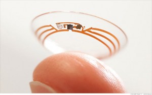
You have heard about Google Glasses, but Google is looking beyond the smartphones of eye wear to monitoring health. They are currently working on a lens with tiny wireless chips and glucose sensors that are sandwiched between two lenses. They would monitor glucose levels once a second and use tiny LED lights, also inside the lenses, to flash when the levels are too high or low. And how big are these electronics? They are no larger than a speck of glitter, with a wireless antenna that is thinner than a human hair. While they are still in development – Google has run clinical research studies and is in discussions with the FDA – it could make blood sugar monitor far less invasive than pricking your finger several times a day.

Drug Delivery for Glaucoma
Getting glaucoma patients to regularly use their eye drops to regulate the pressure in their eyes has always been a problem. They forget, don’t want to be bothered, or have a hard time getting the drops into their eyes. This could change with two research projects exploring the use of contact lenses to deliver medication over a prolonged period of time.

Researchers at Massachusetts Eye and Ear/Harvard Medical School Department of Ophthalmology, Boston Children’s Hospital, and the Massachusetts Institute of Technology who are working on a lens designed with a clear central area and a drug-polymer film made with the glaucoma drug latanoprost, around the edge to control the drug release. These lenses can be made with no refractive power or the ability to correct the refractive error in nearsighted or farsighted eyes.

Another team from University of California, Los Angeles have combined glaucoma medication timolol maleate with nanodiamonds and embedded them into contact lenses. When the drugs interact with the patient’s tears, the drugs are released into the eye. While the nanodiamonds strengthen the lens, there is no difference in water content so they would be comfortable to wear and allow oxygen levels to reach the eye.

Seeing in the Dark
Researchers out of the University of Michigan have developed an infrared sensor that could eventually be used in the production of night vision contact lenses. Thanks to graphene, a tightly-packed layer of carbon atoms, scientists were able to create a super-thin sensor that can be stacked on a contact lens or integrated with a cell phone.

Stem Cells for Cornea Damage
Researchers in Australia are working on a way to treat corneal damage with stem cell infused contact lenses. Stem cells were taken from the subject’s good eye and then plated them onto contact lenses (if there is a defect in both eyes, stem cells are taken from a different part of the eye). After wearing for about two weeks the subjects reported a significant increase in sight.
Braille-Tracile-Contacts
Helping the Blind See
And what good are contact lenses if you are blind? At Bar Ilan University in Israel researchers are creating special lenses that translate images into sensations felt on the eye. It works by taking an image with a smartphone or camera, it is then processed and sent to the contact lens. The custom-made lens is fitted with a series of electrodes that use small electric impulses to relay shapes onto the cornea, similar to braille. After some practice, test subjects were able to identify specific objects.

In expanding the uses of contact lenses, these projects seem to be just the beginning, all reported in the first four months of this year. Researchers and developers are working together to find more and better ways help with vision and medical issues, using contact lenses.

Susan DeRemerSusan DeRemer, CFRE
Vice President of Development
Discovery Eye Foundation