How to Design for Older Adults
Reading the small print can be very challenging as you age. Your eyes lose their elasticity due to a hardening of the lens inside your eye. This condition is called presbyopia and begins to affect many people after the age of 40, continuing to advance as you age. Readers glasses or a single prescription is no longer the best solution. You may find that you need one pair of eyeglasses for reading a book that you hold in your lap, while a different strength may be needed to use a computer at your desk, because it is further away. But it is not just the font size that can affect how easily you can read. Font shape, spacing and color all contribute to readability. Here are some helpful hints if you are producing printed materials for people over 40.

Print Size
Ideal size will vary depending on the font you choose as not all fonts are the same size. A 14 point type size in New Times Roman is smaller than a 14 point Verdana font. Therefore smaller fonts should not be less than 14 points and you may find they are easier to read at 16 points.
Font Type
Decorative fonts are difficult to read and should be used sparingly. For the body of text stick to a regular font that is bolder, with thick lines that are more legible.
Some people prefer a serif font, such as Times New Roman, as they say it is easier to read because of the “tails” at the end of the letters that create an illusionary line, helping to guide the eye along the line. However, others prefer a sans serif font, such as Ariel. It can be easier to read because of the simplicity of the lines. It is a personal choice.
Regardless of the font you select, use both upper and lower case letters in your body text. All capitals letters can be difficult to read. Save them for headlines or to emphasize a word or two.
Avoid using italicized text as the letters appear squeezed together, increasing the reading difficulty.
Presentation Style
Allow for white space as it provides natural places for the eyes to relax and can help you focus on what you are reading.
Align text to the left, as it is easier to read. And don’t wrap text around graphics.
Keep normal spacing between letters, neither expanding nor condensing them which make it more difficult to read the words. Space lines of text at 1.5 instead of single space, to make the lines of text much easier to follow.
Contrast & Color
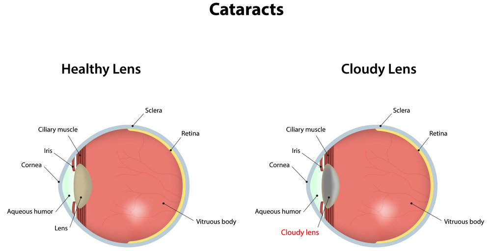
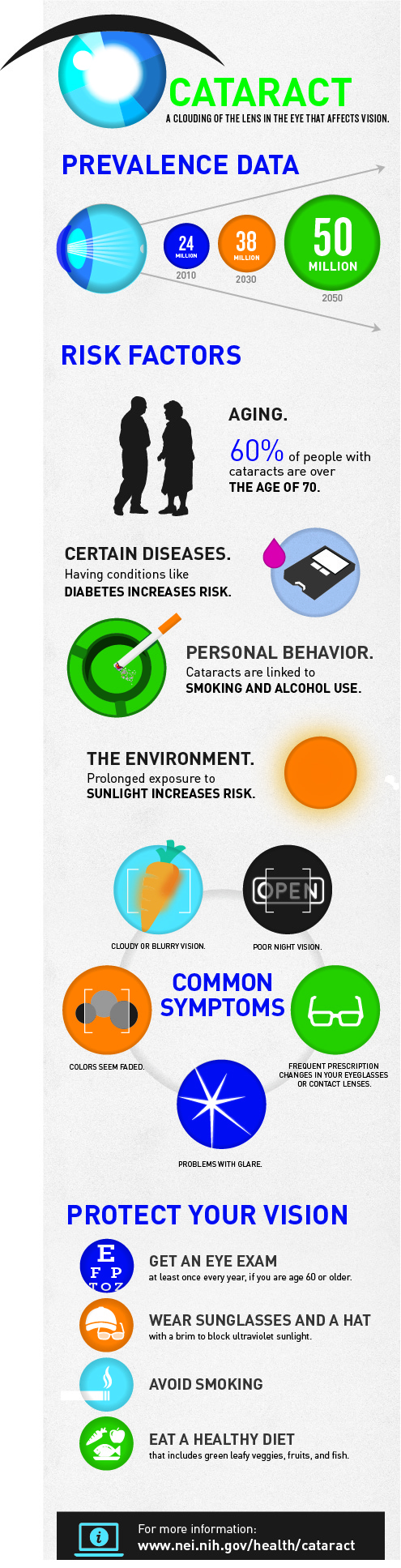
As you get older, yellow, blue and green become increasing difficult to differentiate from each other if they are used in close proximity to each other, especially if you have cataracts. Yellow can almost disappear.
To make it easier for reading, stick with very dark type on a white background. Avoid patterned backgrounds.
Avoid using very glossy paper as it creates glare that can make reading hard. Also make sure your paper is thick enough so print form the other side of the page cannot be seen.

Websites & Blogs
Most of the rules listed above for printed materials also apply to websites and blogs (expect the glossy paper rule). But here are a few additional suggestions for online communications.
Use design templates that are one column (or one and a sidebar) to make it easier read. This is especially true for viewing on mobile devices, even if your web design is mobile responsive.
Allow enough space around clickable items, such as word links and buttons, so they are easy to target and click separately. Make sure the linked text is clearly defined with a color that is easy to differentiate for the surrounding text. Bright royal blue is the most common color used.
Provide a space between paragraphs.
Online a sans serif font is much easier to read, but keep the size at 12 -14 points. Ariel is common font, but Tahoma and Verdana are often used and were specifically designed for online usage. Verdana is a naturally large font, so a 12 point can work well.
Offer a feature where you can easily change the size of the font directly from the screen. An example is the Discovery Eye Foundation site where the control is located at the top right of the page. You can even offer on-screen contrast settings like on the Macular Degeneration Partnership page, at the top center of the page.
Avoid layering shades of the same color, such as dark blue type on a light blue background. Also avoid layering colors that clash such as red type in a purple block. These make reading the text more difficult.
These are just a few of the ways to make text easier to read, both in print and online. Do you have any other tips to share below in the comments?
7/28/15
 Susan DeRemer, CFRE
Vice President of Development
Discovery Eye Foundation