Tear Film Health is Essential for People with Keratoconus

People afflicted with keratoconus (KC) are often obligated to wear contact lenses in order to obtain functional vision. Unfortunately, wearing contact lenses can have detrimental effects on the ocular surface and tear film layers over the course of decades, ultimately reducing lens tolerance. Therefore, any intervention prolonging the comfortable wear time of contact lenses should be aggressively pursued. The tear film covers the surface of the eye, provides lubrication and is the primary defense against foreign bodies and infection. Without a robust and healthy tear film, safe and comfortable contact lens wear is not possible. This article will describe the structure of the tear film and review simple remedies that can keep it healthy throughout life.

Tear Film Layers

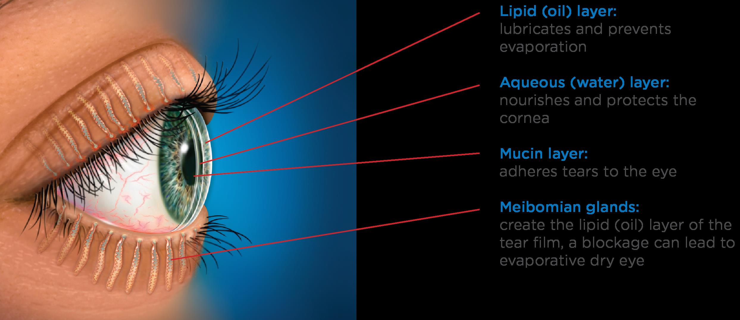
The tear film is a complex, triple layered structure comprised of mucus, water and oil. The surface of the cornea and conjunctiva contain cells specialized to secrete a sticky mucoid substance. These so called goblet cells produce the mucin layer of the tears, which creates a “Velcro” type interface and allows the overlying watery component to stick to the ocular surface without washing away.

The bulk of the tear film is comprised of the watery, or “aqueous” layer which is secreted primarily by the lacrimal gland. This specialized structure is located near the eyebrow. This gland continuously releases small amounts of watery fluid that also contains enzymes and antibodies to help fight infection and wash away contaminants.

The lipid layer is the final, outermost layer of the tears. If the tear film is the first line of defense for the ocular surface, then the lipid layer is the first line of defense for the entire tear film and the ocular surface combined. Because of that role, it is extremely important and helps stabilize the tear film by preventing evaporation. This thin, lipid based layer is released by the meibomian glands, which are modified sebaceous glands that reside in the upper and lower lids. In each lid there are 20-30 glands. These glands open up onto the lid margin and through the action of a complete blink, release the lipid secretion to ocular surface which gets spread with the upward motion of the upper eyelid.

Each one of these layers contributes to the structure of the tear film, and a problem with any one of these structures (goblet cells, lacrimal gland or meibomian glands) will negatively impact the corresponding tear layer.

Tear Film
Image 1 -Layers of the tear film across the ocular surface & Meibomian glands of the eyelids. (Picture courtesy of TearScience™)

Tear Film Issues

Because the tear film is so thin, each individual component is necessary to maintain the integrity of the tears as a whole. When any layer of the tear film is deficient, the tear film becomes unstable and the ocular surface becomes irritated and can progress to developing classic symptoms of dry eye. This includes burning, stinging, redness, tearing, fatigue and contact lens intolerance.

Deficiencies in the mucin layer are uncommon, and are typically the result of chemical or thermal insult, or scarring. An aqueous deficiency, primarily from a lacrimal gland related etiology, is also relatively uncommon, and can arise from autoimmune and inflammatory causes such as Sjögren’s Syndrome. The most common reason for a poor tear film is linked with excessive evaporation of our tears due to a lack of sufficient lipid secretions from non-functioning or obstructed meibomian glands. It is understood that many factors contribute to why these glands stop performing optimally.

One factor has been linked to our habitual working environments. The compressive force exerted by the muscles of our eyelids that control blinking are essential for lipid secretion. However, the use of computers or wearing contact lenses has been shown to negatively impact our blinking habits, both by reducing the number of blinks and making blinks less complete. With an incomplete blink, the upper and lower lids do not make contact. The negative consequences of this are 1) the meibomian glands do not release their lipid contents, 2) the lower part of the eye is chronically exposed to the air, increasing evaporative stress and 3) dead skin cells accumulate on the lid margin which can clog the meibomian gland openings.

When increased evaporation of the tear film occurs chronically, the integrity of the entire ocular system becomes compromised over time and problems to the health of the eye become permanent attributes. This condition is known as Meibomian Gland Dysfunction or MGD and is linked with 86% of all dry eye sufferers.

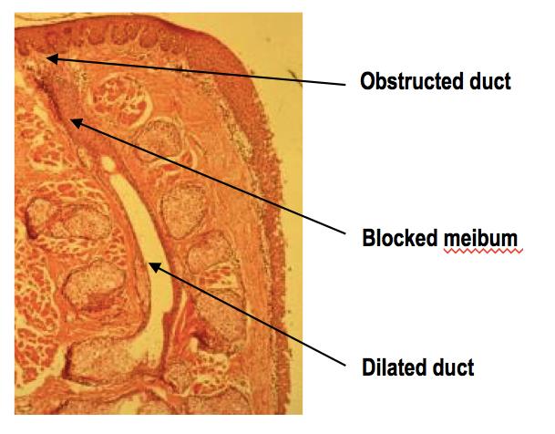
Image 2 - Histology slide of a Meibomian gland with a terminal duct blockage
Image 2 – Histology slide of a Meibomian gland with a terminal duct blockage
Contact lenses have been shown in multiple studies to have a negative impact on the integrity of the tear film. To begin with, placement of a lens onto the eye divides the tears into two sections, referred to as the “post” (behind) and “pre” (in front) lens tear films.

The characteristics of the post lens tear film can differ depending on the type of lens that is worn. For example, soft lenses and scleral lenses have very little turnover of this post-lens tear film. This can cause issues related to the build up of toxic waste and bacterial elements that ultimately aggravate the corneal surface. Conversely, rigid gas permeable lenses are designed to have substantial tear turnover behind the contact lens with every blink.

The pre-lens tear film is also greatly affected by the type of lens material, as well as the interaction between the lid and the contact lens surfaces. Eye doctors know that without a healthy tear film, chances for contact lens intolerance increases. The rate of contact lens intolerance substantially increases as patients enter their fourth decade of life, primarily because of MGD caused by years of poor blinking habits.

Tear Film Care

Fortunately, simple interventions can prevent and/or limit the severity of MGD altogether or help to manage it once it occurs. Just like brushing and flossing one’s teeth can prevent gum disease, attention to complete blinking and lid margin hygiene can improve the tear film and prevent contact lens intolerance problems.

Because partial blinking is strongly linked with developing MGD, it is vitally important that the two lids touch when blinking. It is best to practice this several times throughout the day as well as when you are reading or using the computer.

Akin to flossing the teeth, it is also important to clean the lid margins with a Q-tip soaked in saline solution or a bit of mineral oil by gently brushing the Q-tip across the lid margin 10-20 times each night. It is easiest to get the lower lid.

Finally, performing warm compresses daily can provide heat to the Meibomian glands to soften the hardened oil that can plug the meibomian gland ducts. Warm compresses need to be done continuously for at least 10 minutes with consistent heat in order to attain a temperature that is sufficient to melt the oil that clogs the glands. We recommend folding 5-6 small towels or facecloths into a rectangular shape and wrapped together into a circular bundle, similar to the appearance of a cinnamon roll. The towels should be damp and moist, placed in a microwaveable safe dish with a lid and heated for approximately 1 minute and 50 seconds. After removal, wait a minute or two and then proceed to use the outermost cloth and cover the rest. Replace the first cloth after two minutes and grab the next outer most towel from the bundle, continuing this until all towels are used. In this way, the temperature can be adequately maintained for the full 10 minutes. The high temperatures applied to the lid are transferred to the cornea and very often cause temporary deformation, a phenomenon characterized by transient visual blur immediately following compress application. Therefore, it is vitally important, especially for patients with keratoconus, that pressure never be exerted onto the globe of the eye with a compress or massage administered to the lids of closed eyes after a compress.

It is becoming apparent that MGD is developing in patients at earlier ages. Because of this, the condition has likely been present for decades by the time the patient becomes symptomatic. It may take significant time and effort to rehabilitate not only the glands themselves, but also to reduce the resulting inflammation of the ocular surface.

Meibography is the technique used to image Meibomian glands. In chronic cases of MGD, we see abnormal changes to gland structure, in the form of atrophy or loss of gland tissue and/or dilation of glands where obstructed material causes glands to become widened. In severe cases, the prognosis for recovery is guarded.

The visual clarity that contact lenses provide for patients with keratoconus is incredibly important. But the ability to comfortably wear contact lenses is reliant on our body’s ability to provide a sufficiently thick protective tear film. Taking a small amount of time daily to attend to the lipid producing Meibomian glands by proper blinking habits, exfoliation of the lid margin with a Q-tip and warm compresses will help to extend the number of hours, and ultimately the number of years, that contact lenses can be safely and comfortably worn.

10/20/15

tear filmAmy Nau, OD
Korb and Associates, Boston, MA
Contact lens fitting for keratoconus, other ocular surface disorders and dry eye


tear filmDavid Murakami, MPH, OD, FAAO
Tear Science, Inc.
Researcher, Dry Eye

When Is The Best Time For Cataract Surgery?

As you age, cataracts become a concern prompting the question – when is the best time for cataract surgery?

There are decades worth of old wives tales floating around regarding cataracts that often lead to unnecessary fear and apprehension for many patients. These myths involve concepts such as “ripeness”, having to wear eye patches afterwards, danger in “waiting too long, etc. Just as the techniques of cataract extraction have changed over the decades, so have the indications to proceed to surgery.
best time for cataract surgery - people
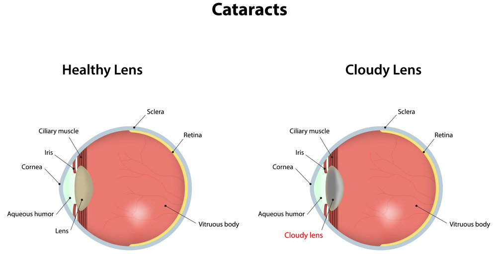
Firstly, cataracts are a normal part of the aging process. Patients should not be alarmed if they are told that they are developing cataracts, even as early as their fifties. As we age, the natural clear lens inside the eye becomes progressively harder, darker, and cloudier. This dark, cloudy lens is what is referred to as a cataract. Cataracts develop at different rates for different people, and even between the two eyes of the same person. It typically takes many years for the lens to become cloudy enough to impact the clarity of vision. There are many different types of cataracts depending of what area of the lens becomes cloudy, but the typical cataract related to normal aging results in a relatively uniform cloudiness with a denser central core, and is referred to as “Nuclear Sclerosis”. Other varieties of cataracts tend to grow more quickly, are relatively uncommon, and often result from certain conditions other than typical aging.
best time for cataract surgery
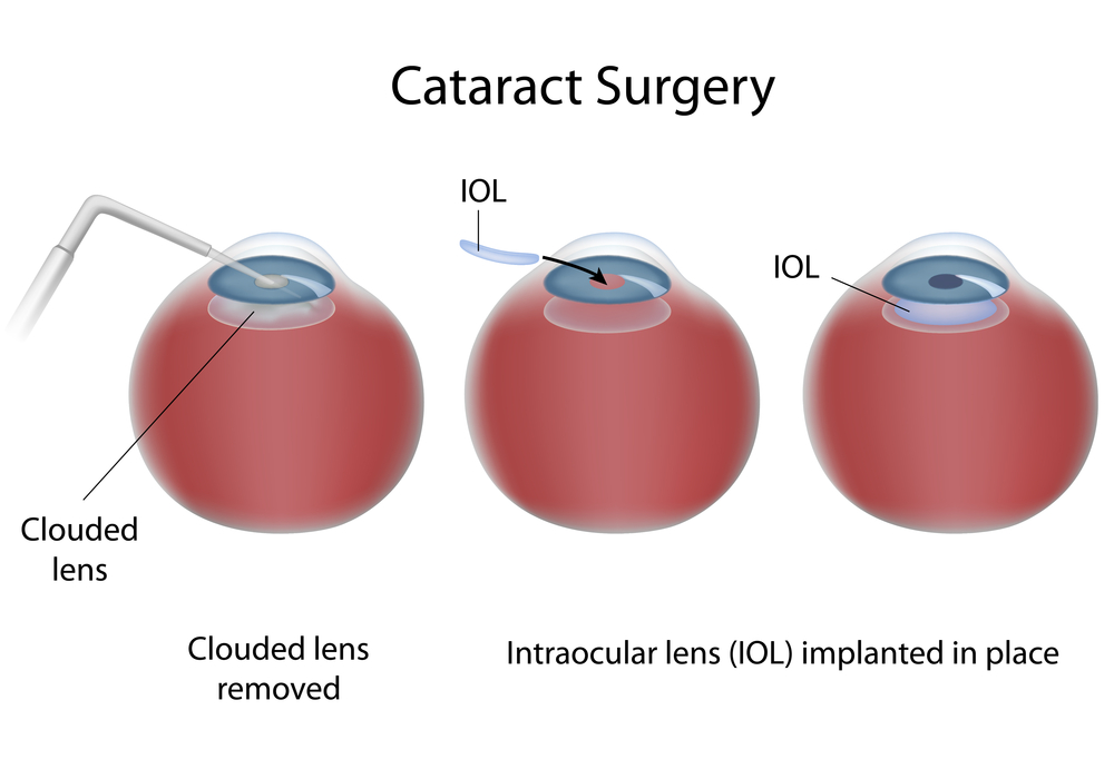
Regardless of what type of cataract the patient has, the treatment is the same: cataract extraction with an implant of an intraocular lens. There have been great advances in lens design over the years, and they now result in excellent, stable, predictable vision for the remainder of the patient’s lifetime and do not typically need to be changed once implanted.

Cataracts result in different symptoms that may be more of less relevant to a specific person’s needs, such as:

  • Glare with bright lights
  • Difficulty with fine print
  • Difficulty following the golf or tennis ball
  • Impairment in night driving
  • Difficulty with seeing street signs
  • Seeing the score or small print on the television
  • Fine visual tasks such as threading a needle, etc.

Although cataract surgery is an incredibly successful procedure with only about a 1-2% risk of complications, it still DOES have some risk. Therefore, cataract surgery should only be undertaken when there is something to gain. In other words, the BENEFITS MUST OUTWEIGH THE RISKS. This means that if your symptoms are mild and are not interfering with your activities of daily living, it is not time to accept the risks of surgery. Once your visual impairment progresses to the point that YOU feel your activities of daily living and enjoyment are impaired, this is the time to proceed to surgery. This threshold is very different between people. Some people feel impaired with vision of 20/25, and others still function within their scope of usual activities until they are 20/100! The best first-step in determining if it is time for your surgery is to get an up-to date refraction. This means a detailed check for new glasses. Often, cataract development will change a person’s glasses prescription, and updating this can improve the visual symptoms for months to years. When a new glasses prescription no longer improves the sight adequately, this is when surgery is indicated.

For the most part, putting off cataract surgery does not impact the final outcome. It will not harm you or your eye to leave the cataract alone until you are ready. There are of course certain exceptions to this rule, such as in Fuchs’ dystrophy, pseudoexfolation, untreated narrow-angle glaucoma, and some others. However, these are relatively rare conditions that your doctor will speak to you about if you have any of these diagnoses.

In summary, the time to proceed to cataract surgery is something that you as the patient determine. YOU assess your lifestyle needs and your vision performance within your scope of activities. When you feel you are impaired in these activities, the benefits will outweigh the risks, and it’s time to take them out. You should not feel any pressure to urgency in this process.

Once you have determined you are ready to have cataract surgery, your surgeon will discuss with you your options for intraocular lens implantation including astigmatism neutralizing lenses, standard distance or near-vision lenses, multiple focal distance lenses, accommodating lenses, and others. The current standard approach for cataract surgery is called “phacoemulsification” and uses ultrasound technology to remove the cataract. There are also laser devices that assist in making the incisions and breaking up the lens, which many surgeons now employ in addition to the phacoemulsification. In general cataract surgery only takes a few minutes, is performed with topical anesthesia, is pain-free, and has a very short recovery time. No pirate-patches are used these days! Most patients are very happy with the results, but this requires adequate discussion with the surgeon prior to the procedure to best assess the needs of the individual patient. A well- informed patient who participates in their care results in the best outcomes!

6/18/15

Sameh Mosaed, MD best time for cataract surgerySameh Mosaed, MD
Director of Glaucoma Services, Gavin Herbert Eye Institute, UC Irvine
Associate Professor, Cataract and Glaucoma Surgery, UC Irvine School of Medicine

Cataract Prevention

The more you know about cataracts, the easier it is to focus on cataract prevention.

What is a cataract?

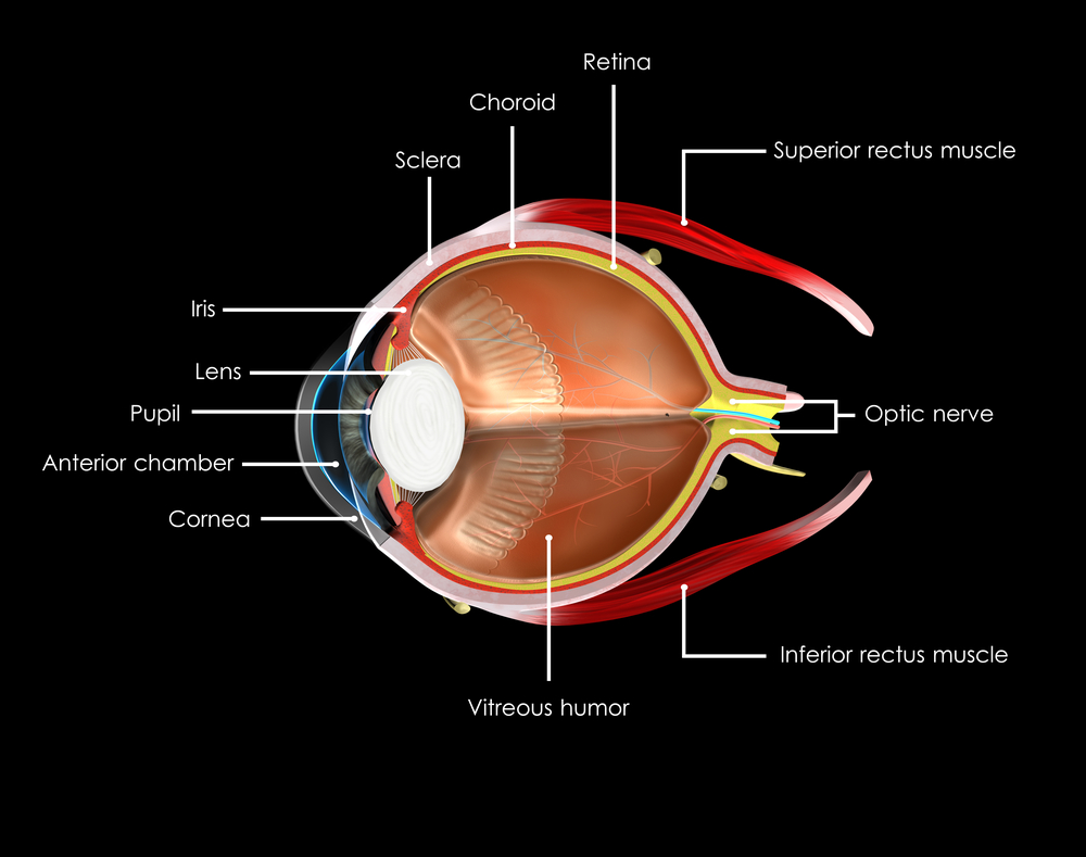
At birth, with rare exceptions, most of us arrive in the world with a clear crystalline lens within each eye. The pathway of our visual images start with light passing through the cornea (the clear front window of the eye), through the pupil (the opening in the center of the iris, or colored portion of the eye) and through crystalline lens which functions to focus light onto the center of the retina (the film of the eye). cataract preventionThe retina, via the optic nerve, will then transmit visual images to the brain. When the crystalline lens becomes opacified (cloudy), this system becomes disrupted, and vision becomes impaired. Opacification of the crystalline lens is called “cataract”, and there are many variations in appearance and type and many causes and can present at any age. The word cataract originates from the Greek word “cataracta”, which means waterfall. The ancient Greeks used this term as they noticed a similarity in the appearance of the white opaque rushing water of a waterfall and the appearance of a white mature cataract.

To understand the different types of cataracts and causes, it is important to understand the anatomy of the lens. Using a metaphor, the lens anatomy can be compared to a Peanut M&M candy™. There is an outer candy coating (the lens capsule), a chocolate layer inside (the lens cortex), and a peanut in the center (the lens nucleus).

The most common cause of a cataract is an age related nuclear clouding which is due to long term accumulation of metabolic and oxidative waste products within the lens and possibly UV-B/Sunlight light exposure. Cortical clouding (within the cortex of the lens), due to similar causes, is also a common cause of an age related cataract.

Cataracts can occur earlier in life with poorly controlled diabetes resulting in cortical and nuclear cataract. Patients who are exposed to steroid medications in any form (orally, topically as eye drops, skin creams etc.) are at an increased risk to develop a posterior subcapsular (PSC) cataract which occurs on the posterior lens capsule. PSC cataracts can have a much more abrupt and earlier onset in life than nuclear or cortical cataract. Smoking has also been known to predispose patients to formation of a PSC cataract. Other less common varieties of cataract can occur with any trauma to the eye or even present at birth as a congenital cataract with a large variety of causes.

What can be done to prevent cataracts?

I often joke with patients that a cataract is such a common occurrence that just like birth, death, and taxes, it is an issue we must all face at some juncture in life (hopefully later than earlier). I am often asked if there are any dietary measures or vitamin supplementation to reduce the formation of a cataract, however this is not as well studied as the use of vitamins in the prevention of macular degeneration. Several scientific epidemiological studies following populations over many decades have shown some merit however that using multivitamins regularly (Vitamin B6 and B12, Vitamin C, beta carotene, antioxidants and possibly lutein and zeaxathin) can reduce the degree of lens opacification over time. As with all medications, you should consult with your physician before deciding to use any vitamin supplementation to clarify if you have any contraindication to using them.

There is conflicting evidence regarding the role of UV-B exposure in sunlight as a causative agent for cataracts. There is some support that using sunglasses on a regular basis to block UV-B light may help to reduce cortical cataract formation. Smoking cessation can also help to reduce the formation of cataract. If a patient is diabetic, strict blood sugar control is also an important measure to reduce the formation of a cataract. If possible, reducing or avoiding the use of steroid medication can reduce the formation of a PSC cataract.

What can be done if a cataract is worsenening and glasses cannot help improve vision significantly?

If you are experiencing gradual painless loss of vision, you should consult with your ophthalmologist as cataract can be a common cause. If you are found to have cataract formation, there is generally a shift in the glasses prescription in the early stage. Having your glasses prescription checked to see if your vision can be improved with glasses is the first step in determining how significant your cataract has become. If glasses are not able to sufficiently improve your vision and your daily activities are affected by the decrease in vision your experience, you may be a candidate to have cataract surgery.

Modern cataract surgery has improved a tremendous degree compared to decades earlier. It is the most common and successful surgery in the world, and is typically performed on an outpatient basis with topical anesthetic and often without any sutures or eye patch. Prior to surgery the pupil is dilated, and once in the operating room, a small self-sealing incision is made on the side of the cornea. The surgeon then makes a circular opening in the anterior lens capsule (the candy coating of the peanut M&M), and uses an ultrasound instrument to emulsify and vacuum out the nucleus (the central peanut), and remove the cortex (the chocolate layer). The inside of the lens capsule is polished and an intraocular lens is folded and introduced into the eye through the corneal incision and seated into the remaining lens capsule to conclude the surgery.

Prior to surgery, measurements are taken to determine the power of lens necessary to achieve the best vision after surgery based on the curvature of the cornea and anterior-posterior length of the eye. Intraocular lenses (IOLs) can potentially have several features depending on a patient’s needs. The most common IOL used is a monofocal lens, which does not typically require an additional out of pocket expense. This lens is chosen to have a point of focus either for distance vision (driving, TV) or near vision (reading), but not both. Typically patients who have the monofocal lens will choose to have distance focus and use reading glasses for near vision. There are multifocal/accommodating IOLs available for patients who are appropriate candidates, to allow the patient a larger range of vision at far, near and intermediate (computer) distance and may allow great independence from glasses. There are still other IOLs which can correct astigmatism (a special type of glasses prescription) at the time of cataract surgery. After discussion of the patient’s needs and preferences, the surgeon can best advise their patient regarding which type of IOL may best suit them.

6/11/15

Anand Bhatt, MD - cataract preventionAnand B. Bhatt, MD
Assistant Professor of Glaucoma and Cataract Surgery, Gavin Herbert Eye Institute
UC Irvine School of Medicine

Fuchs’ Dystrophy: Current Insights

What is Fuchs’ Dystrophy?

Corneal dystrophies are a debilitating group of progressive diseases that can ultimately deprive a person of sight. The cornea, which forms the front of the eye, is a window for vision, and dystrophies due to intrinsic defects in the corneal tissue cause this window to become opaque and hazy. Fuchs’ dystrophy, also known as Fuchs’ corneal endothelial dystrophy (FCED), is amongst the most commonly diagnosed corneal dystrophies requiring corneal transplantation. The ophthalmologist Ernest Fuchs first described the disease in 1910.

Who gets it?

The disease is rare, and it is difficult to predict who will get it. We know that it affects women more than men (3:1 ratio), older adults (older than 50 years of age), and those with a family history. There are forms in which there could be up to a 50% chance of transmission to children of parents with Fuchs’ dystrophy. Most cases, however, occur sporadically.

What causes it and how does it progress?

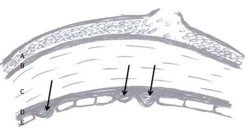
Although the cause of Fuchs’ dystrophy is still being studied, there are characteristic findings associated with it: small outgrowths on Descemet’s membrane called “guttae” or “guttata”, thickening of Descemet’s membrane, and defects in the endothelial cells (Figure 1).

fuchs dystrophy 1
Figure 1: Fuchs’ dystrophy can affect all layers of the cornea. Layers of the cornea from anterior to posterior, or frontside to backside, include (A) epithelial cells where blisters and bullae may form in late-stage disease, (B) Bowman’s layer where scarring can occur in late-stage disease, (C) stroma where corneal swelling occurs early in disease, (D) Descemet’s membrane where guttae form (arrows) and thickening occurs, and (E) endothelial cells that decrease in number and change shape and size with disease progression.

Descemet’s membrane is a thin corneal layer between the endothelial cell and the stromal layers of the cornea. Endothelial cells make up the backside of the cornea and function as a barrier and pump for keeping fluid out of the cornea and maintaining corneal clarity. As guttae accumulate on Descemet’s membrane, patients experience progressive loss and change in endothelial cells. Dysfunction of endothelial cells causes corneal swelling, which distorts vision. First, the back of the cornea swells, and eventually, swelling can reach the epithelial cells at the front of the cornea. Swelling can range from mild moisture accumulation, to painful “bullae”, or blisters. In very late-stage disease, significant corneal scar tissue can form and dramatically reduce vision. The progression to late stage Fuchs’ varies from person to person, but usually takes a couple of decades.

What are signs and symptoms?

A patient may be asymptomatic for years despite having guttae. Initial symptoms, including blurry, hazy, or cloudy vision, are typically due to corneal swelling from dysfunction of the endothelial cell layer. Patients may also experience glare or halos around light in the early stages just from the density of guttae. New studies suggest that patients can get glare and higher order aberrations from guttae without any corneal swelling. Symptoms tend to be worse on awakening, but usually improve throughout the day. This is because the closure of eyelids during sleep results in the accumulation of fluid in the cornea. For the same reason, humid weather can also worsen symptoms. As the disease progresses, poor vision may last longer into the day. There may be associated pain if blisters develop.

How is it diagnosed?

The presence of any of the above signs and symptoms, especially with a family history of Fuchs’, should prompt a consult with an ophthalmologist who will diagnose the disorder and follow its progression with regular checkups. An ophthalmologist will conduct a microscopic slit-lamp examination of the eyes, looking for guttae and Descemet’s membrane thickening (Figure 2).

fuchs dystrophy 2
Figure 2: Slit-lamp examination showing speckling pattern on the backside of the cornea characteristic of guttae in Fuchs’ dystrophy.

Special tests may be done to measure corneal thickness, a marker of swelling, or count endothelial cells to track disease progression (Figure 3 and 4).

fuchs dystrophy 3
Figure 3: Optical Coherence Tomography (OCT) showing (A) a normal, healthy cornea and (B) corneal swelling typical in Fuchs’ dystrophy.
fuchs dystrophy 4
Figure 4: In-vivo slit-lamp scanning confocal microscopy showing (A) normal endothelial cells and (B) guttae causing endothelial cell loss and change in Fuchs’ dystrophy.

How is it managed?

Management can be medical or surgical depending on symptoms. Patients may have mild or slow progression of disease that can be managed medically including over the counter salt solution drops (5% NaCl) to reduce corneal edema.

When there is late-stage disease, a corneal transplant may be necessary to improve vision. A corneal transplant replaces the patient’s corneal tissue with human donor corneal tissue. Donor corneas are readily available via excellent eye banks throughout the United States. The surgery is outpatient surgery with regular follow-up appointments and suture removal during the subsequent months. The postoperative healing of the cornea and vision stabilization can take up to a year.

Great strides have been made in the last decade in corneal transplantation surgery, giving patients better treatment options. Patients used to be limited to penetrating keratoplasty (PK), a full-thickness replacement of the cornea. We now have newer surgeries known as endothelial keratoplasty (EK), which is a partial-thickness transplant that replaces only the damaged part of the cornea (the endothelial layer). The different types of EK are DSEK (Descemet’s-Stripping Endothelial Keratoplasty) and DMEK (Descemet’s Membrane Endothelial Keratoplasty). The techniques vary by thickness of the transplanted tissue. The type of EK most appropriate is determined by the corneal surgeon and is variable on a case to case basis. Both types of EK surgeries provide comparable long-term visual results. In both surgeries, the patient’s diseased Descemet’s membrane and endothelial cells are stripped from the inner layer of their cornea. The thin lamellar donor graft is then inserted into the eye and positioned onto the back of the patient’s cornea via a gas or air bubble. The patient is then instructed to lie in a face up position for several hours post surgery during which time the bubble supports the graft until the new endothelial cell pumps begin to wake up and naturally adhere to the back side of the recipient cornea. Occasionally, the doctor may replace another air bubble into the eye the next day to allow more time for the graft to adhere. Visual recovery is on the order of 1-2 weeks in DMEK and 2-3 months in DSEK surgery. Rejection risk is still a possibility in EK surgery but has a much lower rate than traditional full thickness PK surgery.

Other surgical considerations depend on the presence of cataracts. Cataract surgery can worsen Fuchs’ dystrophy because of damage to the endothelial cell layer. For this reason, patients with cataracts and Fuchs’ requiring surgical intervention are often recommended to undergo cataract surgery before or at the same time as corneal transplantation to ensure the best outcome for the transplant.

Patients should work with an ophthalmologist to determine the best management plan. Ultimately, vast improvements in treatment options have given many Fuchs’ dystrophy patients the exciting opportunity to regain vision with improved healing times and reduced infection and rejection of the graft.

Citations: Figure 2 and 4 are from Zhang J, Patel DV. The pathophysiology of Fuchs’ endothelial dystrophy—a review of molecular and cellular insights. Exp Eye Res. 2015 Jan

6/4/15

priscilla-thumbnailPriscilla Q. Vu, MS
Medical Student
University of California, Irvine School of Medicine


Farid 3.6.14Marjan Farid, MD
Director of Cornea, Cataract, and Refractive Surgery
Vice-Chair of Ophthalmic Faculty
Director of the Cornea Fellowship Program
Associate Professor of Ophthalmology
Gavin Herbert Eye Institute, University of California, Irvine

The Optic Nerve And Its Visual Link To The Brain

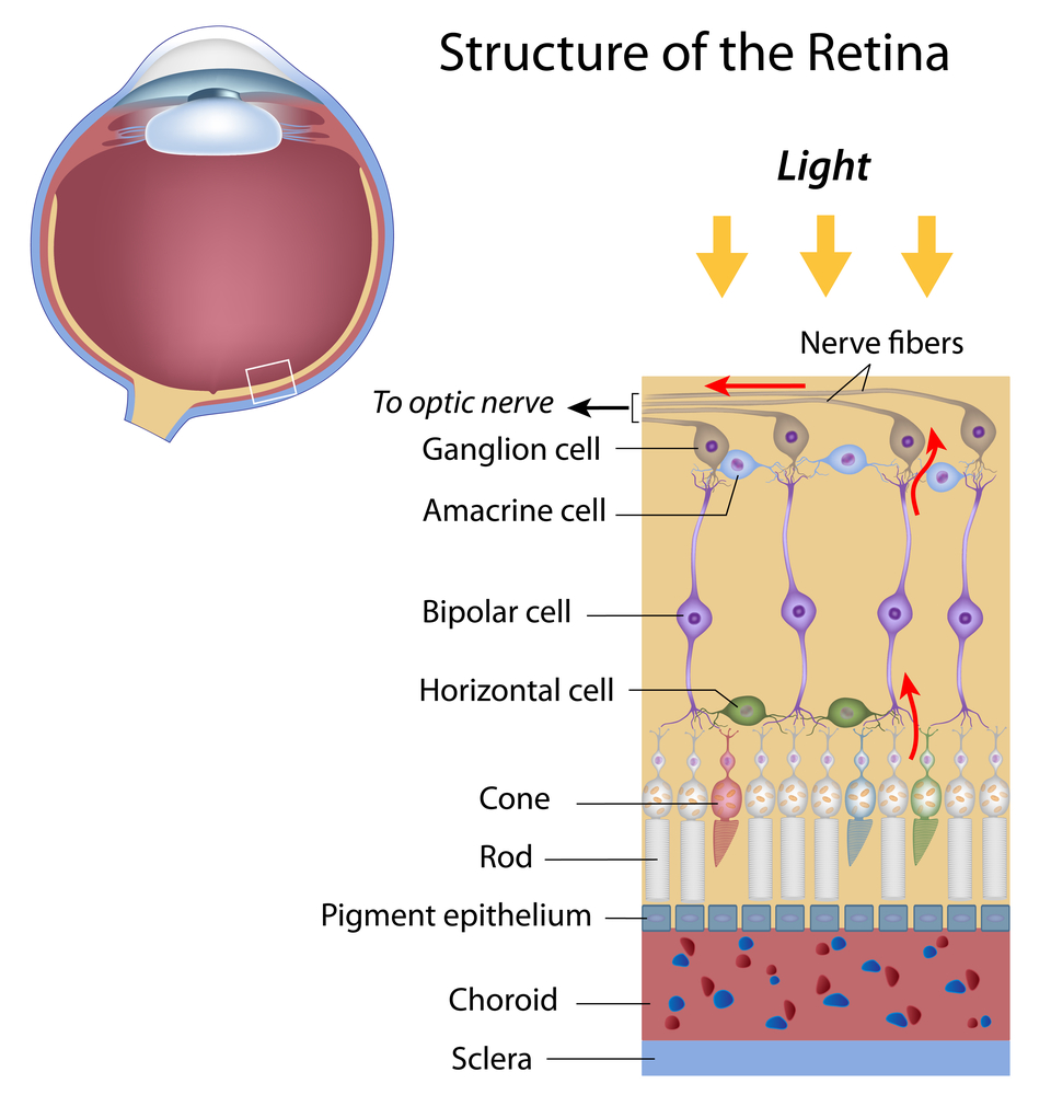
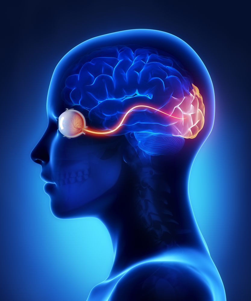
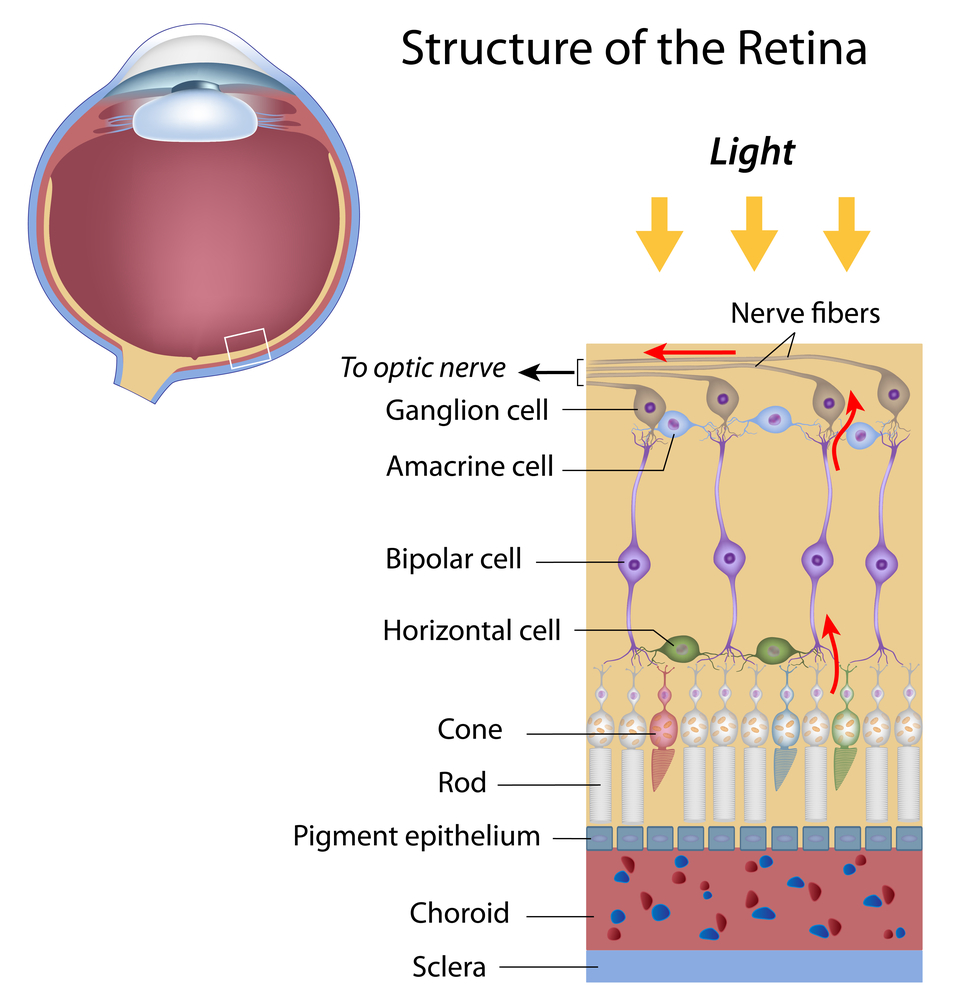
The optic nerve, a cable–like grouping of nerve fibers, connects and transmits visual information from the eye to the brain. The optic nerve is mainly composed of retinal ganglion cell (RGC) axons. In the human eye, the optic nerve receives light signals from about 125 million photoreceptor cells (known as rods and cones) via two intermediate neuron types, bipolar and amacrine cells. In the brain, the optic nerve transmits vision signals to the lateral geniculate nucleus (LGN), where visual information is relayed to the visual cortex of the brain that converts the image impulses into objects that we see.
Optic Nerve
In the retinal tissues of the eye, more than 23 types of RGCs vary significantly in terms of their morphology, connections, and responses to visual stimulation. Those visual transmitting RGCs are the neuronal cells. They all share the defining properties of:

  1. possessing a cell body (soma) at the inner surface of the retina
  2. having a long axon that extends into the brain via the optic chiasm and the optic tract
  3. synapsing with the LGN. The RGCs form multiple functional pathways within the optic nerve to mediate the visual signal

Human beings can see three primary colors: red, green, and blue. This is due to our having three different kinds of color sensitive cone cells: red cones, green cones, and blue cones.

The RGCs connecting to the red and green cones are midget RGCs. They are mainly located at the center of the retina (known as fovea). A single midget RGC communicates with as few as five photoreceptors. They transmit red-green color signals to the parvocellular layer in the LGN (see Figure). The midget-parvocellular pathway responds to color changes, but has little or no response to contrast change. This pathway has center-surround receptive fields, and slow conduction velocities. Because of this pathway, we can see objects precisely in detail and in full color.
retina and optic nerve
The bistratified RGCs are likely involved in blue color vision. Bistratified cells receive visual information input originally from an intermediate numbers of cones and rods. The bistratified RGCs connect to the koniocellular layers in the LGN (see Figure). The koniocellular neurons form robust layers throughout the visual hemifield and have moderate spatial resolution, moderate conduction velocities, and can respond to moderate-contrast stimuli. They have very large receptive fields that only possess on-center regions (no off-surround regions).

Objects can be seen in the dark with motion and coarse outlines accentuated due to the parasol RGCs. At the periphery of the retina, a single parasol RGC connects to many thousands of photoreceptors (many rods and few cones). The parasol RGCs project their axons to the magnocellular layers of the LGN (see Figure) and are primarily concerned with visual perception. They have fast conduction velocities, can respond to low-contrast stimuli, but are not very sensitive to changes in color.

Finally, humans can see objects in three-dimension courtesy of the crossing over of optic nerve fibers at the optic chiasm. This anatomic structure allows for the human visual cortex to receive the same hemispheric visual field from both eyes (see Figure), thus making it possible for the visual cortex to generate binocular and stereoscopic vision.

Recently, a new type of RGC, called photosensitive RGCs, was discovered. The photosensitive RGCs contribute minimally to our vision, but play a key role in vision regulation. Photosensitive RGCs axons do not have connections to the LGN, but form the retino-hypothalamic tract, and synapse to three other locations in the brain for specific vision regulation functions:

  1. Pretectal nucleus: involved in reflexive eye movements, thereby helping to target what we want to see
  2. Midbrain nuclei: involved in controlling the size of the pupil, thus helping to adjust the brightness of objects; and coordinating movement of the eye for focusing
  3. Suprachiasmatic nucleus: involved in regulating the sleep-wake cycle

A fully functional optic nerve is essential for vision. Obviously, any damage of the optic nerve will sever the precise transmission of visual information between the retina and brain, directly leading to vision distortion and/or vision loss. Damage to the optic nerve can result from:

  1. Direct/indirect physical damage (e.g. ocular trauma)
  2. Acute/sub-acute physiological lesion (e.g. infection or inflammation, or malignancy (cancer))
  3. Chronic neuronal degeneration (e.g. glaucoma, a most common cause of optic nerve damage)

Moreover, the optic nerve is also a very important vivo model for studying central nervous protection and regeneration. At the cell biology level, the RGC axons are covered with myelin produced by oligodendrocytes (rather than Schwann cells of the peripheral nervous system) after exiting the eye on their way to the LGN and thus part of the central nervous system. Scientists have recently acquired more and more evidence that certain types of damage to the optic nerve may be reversible in the future. Therefore, the optic nerve provides a potential window to explore more complicated neuronal degenerative diseases, such as Alzheimer’s disease and Huntington disease.

3/12/15

Jun Lin, MD, PhD
Assistant Professor,
Department of Ophthalmology
New York Eye and Ear Infirmary of Mount Sinai
Icahn School of Medicine at Mount Sinai

—–

James Tsai, MD, MBA
Chair, National Eye Health Education Program Glaucoma
Subcommittee President, New York Eye and Ear Infirmary of Mount Sinai Chair
Department of Ophthalmology
Icahn School of Medicine at Mount Sinai

Understanding and Treating Corneal Scratches and Abrasions

Corneal Scratches and Abrasions

Call it a scratch, an abrasion or erosion; no matter how you describe it or what the cause, damage to the cornea most always causes pain.

So what exactly is the cornea and why can even a small scratch hurt so much? The cornea is the clear dome at the very the front of the eye. Its primary job is to surface the tears and with them, focus light into the eye. It then passes through the crystalline lens and on to the retina where it is transformed into electrical impulses that are ultimately transformed by the brain into sight.

Because vision is so essential for survival and the cornea so critical to seeing, it is among the most richly innervated and exquisitely sensitive of all tissues. Even the smallest piece of dust that finds its way into the eye and touches the cornea can cause significant discomfort, irritation and copious tearing in an attempt to wash it away. A healthy cornea is transparent and consists of several layers that give the cornea its smooth dome like shape. The outermost layer, the epithelium, is designed to break away to protect the delicate deeper layers if scratched or abraded.
cornea layers - corneal scratches and abrasions

Looking For the Cause

The most common causes of corneal scratches are accidents. Tiny infant fingers and fingernails are a common cause of abrasions in young parents, tree branches are a frequent source of abrasions in hikers and lovers of the outdoors, and makeup brushes are a typical cause in women. Scratches can also be caused by foreign objects that get into the eye and then work their way on to the inside of the upper lid – causing a scratch that occurs with each blink. That’s why its important to carefully investigate the cause of every corneal scratch.

A scratch pr abrasion usually produces near instantaneous pain and tearing as the eye tries to wash away the irritant. Light sensitivity soon follows and can be so intense that the eye can involuntarily shut. This is actually nature’s way of “patching” the eye to facilitate healing.

To confirm you have a scratched cornea, a doctor or other health care professional will often apply a wetted fluorescein strip to the inside lid or white of the eye. Fluorescein is a dye that glows bright green when exposed to black light. The dye is absorbed by damaged areas, clearly showing the area if the scratch or abrasion.

Getting On the Mend

The good news is that most scratches will rapidly heal on their own, especially smaller and more superficial ones. The confocal microscope, a high tech device that provides extreme magnification views of living tissue, has been used to observe corneal healing in real time. The video captures are breathtaking as individual corneal cells can be seen literally stretching over each other to mend and seal the corneal surface.

If an abrasion is larger or deeper it may require patching to help healing. The traditional eye patch applied with tape to keep the eye shut has largely been replaced by the bandage contact lens which is far more comfortable and allows some vision and easier observation during follow up examination. It also allows medication to be applied if needed. Because there is a risk of infection whenever the outer boundaries of the body are breached, topical antibiotics are often used as a precaution in treating scratches of the cornea and ocular surface.

Most commonly the cornea heals quickly and completely, but not always. In rare cases damaged areas of the cornea may not heal fully, leaving the outer layers of the cornea susceptible to coming off again for no apparent reason. This is thought to be more common after scratches caused by organic material such as a tree branch. Called recurrent corneal erosions, they often occur during sleep waking the person with a sudden sharp pain and excessive tearing. There are a variety of treatments for recurrent corneal erosion.

Conclusion

Most people will sooner or later experience a scratched cornea. Most scratches will be minor and will resolve with minimal treatment. However, some can be serious and have significant consequences. The best way to avoid problems is to be aware that they can occur and take measures to protect the eyes in situations where the risk of eye trauma is higher. This includes: wearing safety glasses while working with power tools, or sports where eye contact is possible. This includes cycling and sport shooting.

Be aware of active infants with little fingers that seem to have a magnetic attraction of their parents eyes. If you use eye makeup, leave enough time to properly apply it without rushing and potentially scratching your cornea in the process.

Finally, if you experience a scratched cornea and the pain doesn’t rapidly abate, see an eyecare specialist. Urgent care centers are fine for most things, but when it comes to the eyes finding a knowledgeable eye care professional is wise.

2/10/15

AArthur B. Epstein, OD, FAAO
co-founder of Phoenix Eye Care
and the Dry Eye Center of Arizona
Fellow of the American Academy of Optometry
American Board of Certification in Medical Optometry
Chief Medical Editor of Optometric Physician™

Pupils Respond To More Than Light

Everyone knows that your pupils will change size according to the amount of light you are experiencing. With more light, the pupil constricts and becomes smaller. Less light and your pupil dilates, letting more light into the back of the eye. It is the muscles of the iris working with your autonomic nervous system (ANS) to adjust the iris so the right amount of light enters the eye – like the aperture of a camera.

The iris is made up of two types of muscle:

  • Sphincter muscles that are like concentric rings that constrict the pupil to as small as two millimeters across
  • Dilator muscles that are laid out like the spokes of a bicycle wheel and can expand the pupil up to eight millimeters across

dilated pupils respond
But the ANS is not only concerned with light reflex, it also reveals emotional and mental responses. The sympathetic branch of the ANS responds to a person being under stress, triggering the “fight or flight” response, which will cause the pupil to dilate. On the other hand, the parasympathetic branch known for “rest and digest” will cause pupil constriction. At any given time, your pupil is balancing between both the light and emotional reactions.

Here are some of examples of mental responses:

Princeton University psychologist Daniel Kahneman demonstrated that pupil size increases in proportion to the difficulty of the task being performed. Calculating 8 x 21 will cause your pupil to dilate slightly, however calculating 8 x 47 will cause them to dilate even more. Whatever the problem, they will remain dilated until you come up with the answer or give up.

Even memory recall creates a pupil response. When subjects were instructed to remember and recite a series of seven digits, their pupils would grow steadily as they learned each number, but reduce as steadily when they recited back each of the numbers.

Wolfgang Einhauser-Treyer, a neurophysicist at Philipps University Marburg in Germany, found that “pupil dilation can betray an individual’s decision before it is openly revealed.” He asked people to push a button at any point during a span of 10 seconds. Dilation began about one second before they pressed the button and continued to peak one to two seconds after the push.

This study of pupil size is known as pupillometry and is used to investigate a wide range of psychological phenomena including sleepiness, introversion, sexual interest, racial bias, schizophrenia, moral judgment, autism and depression. Kahneman said he has “never done any work in which the measurement is so precise.” And while “nobody really knows for sure what these changes do,” according Stuart Steinhauer, director of Biometric Research Lab at the University of Pittsburgh, pupillometry is a valuable tool for psychological research.

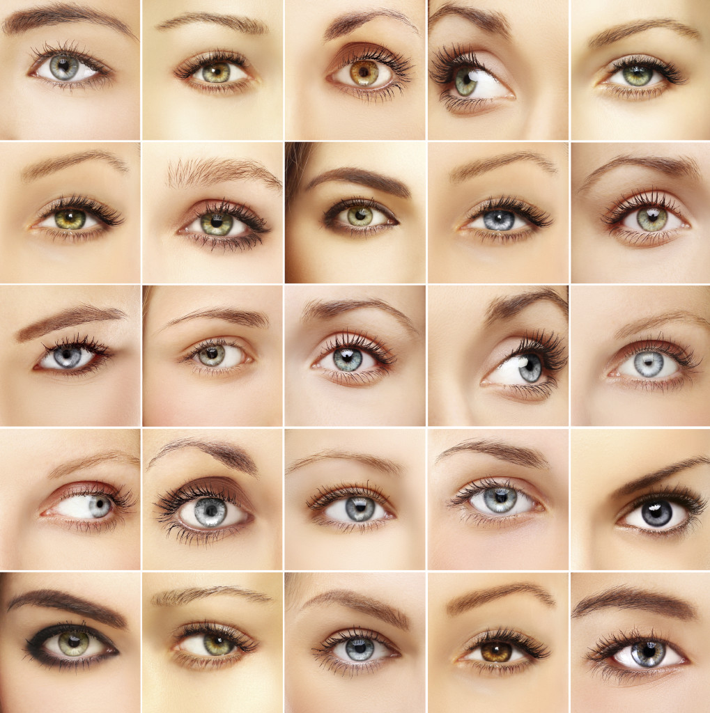
So the next time you look into someone’s eyes, know that you have the potential to see more than just their eye color. You might have a clue as to what is going on in their mind.

2/3/15


Susan DeRemerSusan DeRemer, CFRE
Vice President of Development
Discovery Eye Foundation

Uveitis Explained

12/18/14

Uveitis is defined as inflammation of the uveal tissue. The uvea includes the iris, ciliary body, and the choroid of the eye. The iris is located in the anterior compartment of the eye and acts like the aperture of the camera, precisely filtering the amount of light entering the eye. The ciliary body, which is attached posteriorly to the iris, is involved in both the production of the aqueous fluid in the eye as well as the accommodation of the lens apparatus. The choroid is a dense layer of blood vessels that sits underneath the retina on the back wall of the eye, helping to nourish and remove metabolic waste products from the retina. Inflammation of any of these structures will consequently cause disruption of the visual pathway and over the long term can cause permanent visual loss. In fact, uveitis is the third most common cause of preventable blindness in the developed world.
uveitis explained
Symptoms of uveitis include blurry vision, ocular pain, photophobia, redness, and floaters. These can be acute in nature, lasting a few days to weeks, and in some cases can be chronic, lasting weeks or months. Anyone with any of these symptoms should see their eye care provider as soon as possible, as faster treatment of uveitis has shown to result in better long term visual outcomes.

Uveitis can affect virtually any part of the eye, from front to back. Anterior uveitis or iridocyclitis is confined to the iris, ciliary body, anterior chamber, and cornea. Inflammation affecting the vitreous is termed intermediate uveitis, or pars planitis, and any inflammation affecting only the retina or choroid is termed posterior uveitis. The term panuveitis may be used when multiple layers of the eye are affected.

There are many possible causes of uveitis, including infection, inflammatory diseases, autoimmune diseases, and trauma. However, the majority of cases of uveitis, approximately half, are considered idiopathic, where no etiology is ever found. Trauma is the next most common cause of intraocular inflammation, accounting for approximately 20% of all cases. The remaining cases are secondary to a systemic disorder or localized ocular condition. Systemic etiologies can include inflammatory disorders such as sarcoidosis, infections such as tuberculosis and syphilis, as well as autoimmune diseases such as rheumatoid arthritis and lupus.

Treatment of uveitis is aimed at both blunting the intraocular inflammation as well as addressing any underlying systemic etiology. The most common treatment is the use of corticosteroids. These can be taken orally, or used topically as eye drops. In some cases, corticosteroids can be injected in or near the eye as well. If the uveitis is caused by an infection, such as tuberculosis or syphilis, the patient is also given antibiotics. Systemic corticosteroids can have major side effects when taken chronically, such as weight gain, hair loss, osteoporosis, hypertension, secondary diabetes, psychosis, and reduced growth in children. Because of these potential problems, the chronic use of systemic corticosteroids is not recommended. In cases of chronic uveitis that require long term treatment, immunosuppressive agents with less known side effects such as methotrexate, cyclosporine, and mycophenolate mofetil (Cellcept) are more commonly used. However, these biologic agents have their own set of potential side effects and therefore, it is recommended that a rheumatologist should also be involved in the care of the patient when using these agents. Topical and intraocular steroids localized to the eye can cause elevated intraocular pressure as well as cataracts. In most cases, elevated intraocular pressure can be controlled with topical glaucoma drops, but in some cases surgical intervention is required to prevent severe glaucomatous damage.

The most common type of uveitis is acute anterior uveitis or iridocyclitis. Many cases of anterior uveitis are idiopathic though almost half of all cases are associated with the HLA- B27 haplotype. Systemic diseases associated with HLA-B27 include psoriatic arthritis, ankylosing spondylitis, reactive arthritis, and inflammatory bowel syndrome. Signs of anterior uveitis include redness of the eye, sometimes termed ciliary flush. The conjunctiva can become extremely red, and when associated with ocular pain and photophobia, is a strong indicator of anterior uveitis. Inflammatory cells found in the anterior chamber are the hallmark of anterior uveitis, sometimes deposited on the corneal endothelium (keratic precipitates) or iris (Bussaca nodules). Patients with anterior uveitis are typically treated with topical corticosteroid and cycloplegic eye drops. A laboratory workup for systemic etiologies is usually not necessary unless the patient experiences a recurrent episode.

Inflammation affecting primarily the vitreous cavity is known as intermediate uveitis or pars planitis. Inflammatory cells in the vitreous, known as vitritis, are typically bilateral, and when severe, can be found clumped in the vitreous cavity (snowballs) or deposited on the inferior pars plana (snowbanking). Intermediate uveitis is typically idiopathic though sarcoidosis, multiple sclerosis, and Lyme disease are also possible causes. Certain malignancies such as lymphoma can also ‘masquerade’ as intermediate uveitis, and when seen in older patients, should be suspected and ruled out.

Posterior uveitis involves the retina, choroid, and/or the retinal vasculature, and usually is more difficult to treat than anterior uveitis.

Uveitis Explained
This patient with Cat-scratch disease, caused by infection with Bartonella henselae, is an example of posterior uveitis. Note the characteristic star-like pattern of exudate in the macula along with optic nerve swelling.

In many cases, patients with posterior uveitis will exhibit characteristic exam findings that help narrow the differential diagnosis. For instance, an area of active retinitis next to an old pigmented chorioretinal scar is highly suggestive of toxoplasmosis. The most common symptom in patients with posterior uveitis is blurred vision. One of the more typical findings in posterior uveitis is macular edema, which is usually treated with periocular or intraocular corticosteroids.

In summary, uveitis is a visually threatening inflammatory condition that should be diagnosed and treated immediately. It is important to determine as best as possible the etiology of the uveitis and treat appropriately. In general, most patients with uveitis have good visual recovery with the proper management. However, in some cases, severe damage can occur, either due to the inflammation itself (usually chronic) or as a side effect of therapy (corticosteroids).

RichardRoeMD-ThumbnailRichard H. Roe, MD, MHS
Retina-Vitreous Associates Medical Group

Posterior Vitreous Detachment

10/30/14

Have you ever noticed floaters in your vision? Perhaps they looked like a bunch of small dots or maybe a cobweb swaying back and forth in your visual field. Were the floaters associated with flashing lights that made you think there was a lightning storm coming your way? These are typical symptoms of a posterior vitreous detachment (PVD), and if you have had these symptoms you are far from alone.
Floaters Posterior vitreous detachment
PVD is a natural process that occurs in the majority of people usually over the age of 50. The vitreous is a jelly-like substance that occupies the back portion of the eye. The vitreous is comprised primarily of water, which accounts for 99% of its volume, and the remaining 1% includes proteinaceous substances such as collagen fibers as well as hyaluronic and ascorbic acids. The collagen fibers act as a scaffold to allow the vitreous to maintain a formed shape as well as provide a means for the vitreous to attach to the retina, which is the light-sensitive tissue that lines the inner back wall of the eye and is critical for vision. As we age, changes in these fibers cause the vitreous to lose its shape and eventually pull away from the retina. When the vitreous separates from the retina, this is called a PVD.

As we age, the collagen components of the vitreous can clump together and are free to float in the eye. When the vitreous separates from the retina during the development of a PVD, the floaters may become more noticeable or numerous. It is common for patients to describe floaters of different shapes and sizes, and patients may notice just one or in some cases many. In many people, a PVD develops slowly and there may be no symptoms or just a few annoying floaters. In others, a PVD may occur abruptly and cause more dramatic symptoms that can be very anxiety provoking.

Since the normal process of PVD development involves the vitreous tugging on the retina until it can fully separate, this tugging can result in flashing lights that can commonly appear in the peripheral, or side, vision. These flashing lights are sometimes described as lightning streaks, and patients may notice them more readily in settings with low ambient light. The flashes of light typically resolve once the vitreous has fully separated from the retina and the tugging has ceased.

The good news is that PVD is usually harmless in the vast majority of cases, and the annoying floaters will become less bothersome over time. In approximately 5-10% of cases, the vitreous can tug too hard on the retina as it tries to separate and it may pull a hole or tear in the retina. Tears in the retina can predispose to retinal detachment, which is a serious condition that can lead to permanent vision loss.
It is important to recognize that the typical symptoms of a regular PVD are often similar to a PVD with an associated tear. For this reason, it is recommended that all patients with the new onset of floaters or flashes have a dilated eye exam. If a retinal tear or detachment is discovered, early treatment can help prevent loss of vision.

Treatment for PVD usually involves simple observation. With time, the flashes will go away, and the floaters will become less noticeable. More recently, few providers have claimed that floaters can be treated with a laser in order to make them less noticeable. I would caution that this is not mainstream therapy at the current time, and I do not advise my patients to pursue this option. Another treatment possibility is vitrectomy surgery, where the vitreous gel is removed as part of a surgical procedure. Due to safety advances in vitrectomy surgery, this is now a potential option for the rare patient who has floaters that are so numerous and bothersome that they are negatively impacting their activities of daily living. For the vast majority of patients this is not necessary.

When I see a patient with a PVD, I often recommend one follow-up visit in 4-6 weeks to make sure there are no retinal holes or tears that have developed in the interim. If the other eye has not had a PVD yet, I will counsel them that a PVD will most likely develop in that eye within the next few years, and when it does they need to be examined. I will also discuss the retinal detachment warning signs. Patients with retinal detachment will not only have symptoms similar to PVD, including flashes and floaters, but in addition they may also notice what looks like a black shade or curtain that starts in the peripheral vision and extends towards the central vision. My patients are taught that this symptom requires an immediate examination.

In conclusion, PVD is a natural process that the majority of people will experience in their lives. The symptoms can range from having no symptoms at all to many floaters with associated lightning flashes. In the majority of patients, there is no damage to the eye or threat to the vision. A dilated exam is recommended to look for possible holes or tears in the retina, and if these are uncovered, prompt treatment can prevent vision loss.

Dr. Esmaili posterior vitreous detachmentDaniel D. Esmaili, MD
Retina Vitreous Associates Medical Group

What Are A Macular Pucker and Macular Hole?

10/9/14

What is the macula?
The eye is very much like a camera, taking light from the outside world and converting it into picture information that our brains perceive as vision. The retina is the light sensitive layer in the back of the eye that is very much like the film in that camera. The central retina, also known as the macula, is essential for crisp, high definition vision. Conditions that damage or distort the macula can therefore result in blurred or distorted vision. Two common conditions that affect the macula are macular puckers and macular holes.

What is a macular pucker or macular hole?
A macular pucker is a thin layer of scar tissue that forms on top of the retina. The amount of scar tissue can range from mild to severe. Mild macular puckers may be barely noticeable during an eye exam and resemble a fine layer of cellophane resting on the macula. More severe macular puckers can cause wrinkling or distortion of the macula. In contrast to a macular pucker, a macular hole is a small gap that extends through the entire thickness of the macula.

What are the symptoms of a macular pucker or a macular hole?
At first, a macular pucker may lead to mild blurring of the central vision. Because the problem involves the back of the eye, glasses will not completely restore vision. More severe macular puckers may result in wavy or distorted vision. For instance, objects that normally appear straight, such as venetian blinds or a printed line of text, might appear to have a dip or bend in the center. Small macular holes can cause similar symptoms of blurring or distortion. Larger macular holes often result in a central blind spot. This can also result in straight lines appearing broken or having a piece missing in the middle. Patients with a macular pucker or hole do not normally experience difficulty with peripheral vision.

What can cause a macular pucker or macular hole?
Recall that a macular pucker is a scar tissue. Anything that causes scar tissue, such as trauma or inflammation in the eye, can result in scar tissue and hence a macular pucker. Certain diseases that affect the retinal blood vessels such as diabetes can also cause a macular pucker to form. However, one of the most common causes of macular pucker is simple aging of structures within the eye. As the eye ages, the clear jelly that fills it, called the vitreous gel, shrinks. When enough shrinkage occurs, the vitreous gel detaches from its normal position adjacent to the retina. This process of vitreous detachment can cause microscopic damage or inflammation leading to macular pucker formation. In some cases, the vitreous gel does not detach cleanly from the retina. Instead it can put traction on the macula, pulling its delicate structures apart in the center, resulting in a macular hole.

How are macular puckers and macular holes diagnosed?
A simple examination from an ophthalmologist or retina specialist is often enough to diagnose a macular pucker or hole. However, additional testing is often useful in diagnosing subtle cases or monitoring eyes for changes. An optical coherence tomography (OCT) scan is a specialized photograph that allows your physician to look for microscopic changes in the contour of the macula. The following figures show an OCT of a normal macula, a macular hole, and a macular pucker. Note that the normal macula has a central dip known as the fovea, shown in Figure 1. In Figure 2, the dip is replaced by a gap which is a macular hole. Finally, Figure 3 shows a macular pucker where the dip is no longer visible. This is because the macular pucker, seen as a thin white line is distorting the normal shape of the macula.

Normal - Macular Pucker and Macular Hole
Figure 1: Normal Macula
Hole - Macular Pucker and Macular Hole
Figure 2: Macular Hole
Pucker - Macular Pucker and Macular Hole
Figure 3: Macular Pucker

What treatments are available for macular puckers and macular holes?
Macular puckers can be quite mild. For mild cases in patients with minimal symptoms, periodic monitoring may be all that is required. When blurred vision due to a macular pucker begins to affect activities such as driving or reading, treatment in the form of surgery can be considered. Surgery for a macular pucker is known as a vitrectomy. Vitrectomy surgery is usually done under local anesthesia and as an outpatient procedure. During the surgery, fine instruments are used to remove the scar tissue from the surface of the macula. After surgery, patients usually experience an improvement in the blurring and distortion as the eye recovers gradually over a period of months. Some residual waviness can be normal. Vitrectomy is generally very safe although there is a chance of increased cataract growth and a small risk of infection or retinal detachment.

For patients with small macular holes, close monitoring can also be an option since some macular holes can close on their own. For larger holes, there are two options. In select cases where the vitreous gel is actively pulling on the macula, an injection of medication into the eye may cause the gel to release cleanly, allowing the hole to close. In other cases, vitrectomy is recommended. During the surgery, any pulling on the macula is relieved and a gas bubble is placed in the eye to help the hole close. After surgery, patients are asked to look down for a several days to allow the bubble to float up against the hole. Once the body absorbs the bubble, vision is usually significantly improved.
In summary, both macular puckers and holes are common causes of blurry or distorted central vision. If treatment or surgery by a retina specialist is needed, the results are generally quite good and lead to significant restoration of vision.

Liao - Macular Pucker and Macular HoleDavid Liao, MD, PhD
Retina-Vitreous Medical Group