 Harsh weather conditions can reduce the natural moisture in your eyes and the irritation usually results in a burning or itching sensation that often leads to rubbing or scratching your eyes which can worsen the symptoms. Sometimes it feels like there is a foreign object in your eye and for some, dry eyes can even cause excessive tearing, as your eyes try to overcompensate for their lack of protective tears. Prolonged, untreated dry eyes can lead to blurred vision as well. Between the harsh winter winds outside and the dry heat radiating inside, our eyes are very quickly irritated and dried in the winter months. The result is itchy, dry eyes that may cause pain, blurred vision, a burning sensation, or even watery vision as our eyes try to compensate for the dryness.
What Are The Symptoms?
- Uncomfortable, stingy, burning or scratchy feeling.
- Stringy mucus in or around your eyes
- Increased eye irritation from smoke or wind
- Eye fatigue
- Sensitivity to light
- Eye redness
- A sensation of having something in your eyes
- Difficulty wearing contact lenses
- Periods of excessive tearing
- Blurred vision, often worsening at the end of the day or after focusing for a prolonged period
10 TIPS TO KEEP YOUR EYES COMFORTABLE DURING THE WINTER MONTHS
Whatever the symptoms, dry eyes can cause significant discomfort during the long winters and relief can seriously improve your quality of life.
- To keep eyes moist, apply artificial tears/eye drops a few times a day. If you have chronic dry eyes, speak to your eye doctor about the best product for your condition.
- Drink a lot of fluids – keeping your body hydrated will also help maintain the moisture in your eyes.
- If you spend a lot of time indoors in heated environments, use a humidifier to add some moisture back into the air.
- Try to situate yourself away from sources of heat, especially if they are blowing. While a nice cozy fire can add to the perfect winter evening, make sure to keep your distance so dry eyes don’t ruin it.
- Staring at a computer or digital device for extended amounts of time can further dry out your eyes. If you spend a lot of time staring at the screen, make sure you blink often and practice the 20/20/20 rule – every 20 minutes, look 20 feet away for 20 seconds. Use artificial tears often to lubricate eyes during long periods of using your eyes.
- Avoid air blowing in your eyes. Don’t direct hair dryers, car heaters, air conditioners or fans toward your eyes. In your car, direct heat to floor vents and away from your eyes once your windshield is defrosted.
- Stop smoking and avoid smoky environments.
- Don’t rub your eyes! This will only increase irritation and can also lead to infections if your hands are not clean.
- Give your eyes a break and break out your glasses. If your contact lenses are causing further irritation, take a break and wear your glasses for a few hours or days. Also talk to your optometrist about switching to contacts that are better for dry eyes.
- Protect your eyes. If you know you are going to be venturing into harsh weather conditions, such as extreme cold or wind, make sure you wear protection. Try large, 100% UV protective eyeglasses and a hat with a visor to keep the wind and particles from getting near your eyes. If you are a winter sports enthusiast, make sure you wear well-fitted ski goggles.
If you find that after following these tips you continue to suffer, contact your eye doctor.
 I can only imagine my wife’s beautiful face. Oh sure, I’ve touched it and kissed it many times. I’ve felt the lines with the tips of my fingers, tracing our lives together, and I’ve heard her smile. I understand that’s not really seeing it. It’s not seeing her eyes as they sparkle with something funny I said; or, when she looks at me with love reserved only for those who are truly in love.
 Tom Sullivan
 Costume Contact Lenses such as cat eyes or zombie may make your Halloween costume a bit more frightful although wearing those lenses without a prescription can be more terrifying, as it could result in vision loss or even blindness.
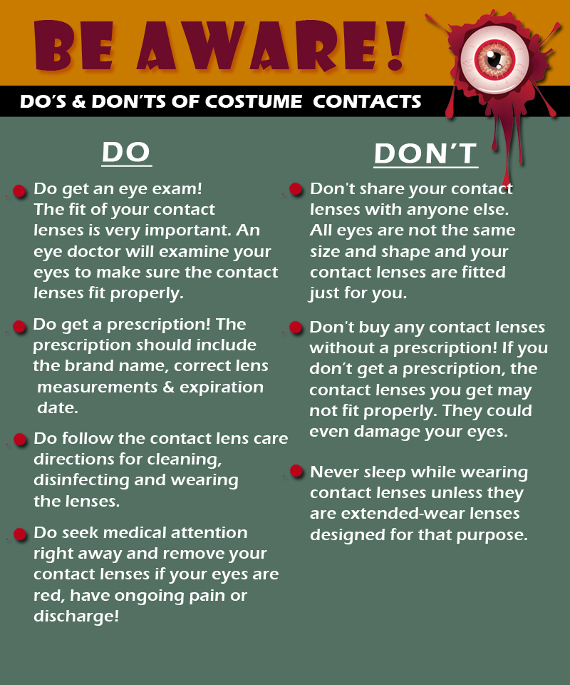
 I could hear Charlie rubbing his wife’s shoulders and telling her that everything would be alright. But, Rose kept saying “I know we’ll have to sell the house and move into something smaller, and I am going to be blind Charlie. Blind.”
 Summer time is officially here and everyone enjoys a dip in a nice, cool pool during the summer months. While swimming is a great form of exercise and a relaxing way to cool down, the water can be hard on your eyes.
 Wear Goggles – Wear a pair of swim goggles every time you swim. Goggles keep pool chemicals out of your eyes.
 Wash Your Eyes – Immediately after swimming, splash your closed eyes with fresh tap water. This washes chlorine and other chemicals off your eyelids and eyelashes.
 Use Eye Drops – Use over-the-counter lubricating eye drops before and after swimming to keep the tear film balanced and eyes comfortable.
 Stay Hydrated – Don’t forget to drink plenty of water. Staying well hydrated is an important part of keeping your eyes moist and comfortable.
 The Difference Between an
 tic nerve, a part of the central nervous system that carries visual information from the eye to the brain.