Famous People with Vision Loss – Part II

Here is part two of our look at famous people with vision loss. The focus this time is the creative minds of artists, actors and musicians.

Artists

Georgia O’Keeffe (1887-1986) – Best known for her paintings of enlarged flowers, New York skyscrapers, and New Mexico landscapes, she was recognized as the “Mother of American modernism”. In 1972, O’Keeffe’s eyesight was compromised by age-related macular degeneration (AMD), leading to the loss of central vision and leaving her with only peripheral vision. She stopped oil painting without assistance in 1972, but continued working in pencil and charcoal until 1984.

Claude Monet (1840-1926) – A French impressionist painter who had cataracts that blurred his vision and caused a severe loss of color perception. After complaining about his cataracts for ten years, he had cataract surgery in 1923. Those paintings before the surgery have more of a reddish tone, while those painted before his cataracts and after the surgery show more blue tones.

Edgar Degas (1834-1917) – A French artist known for his paintings, sculptures, prints, and drawings. He had a chronic and progressive retinal disease that made him sensitive to light and caused him to lose his central vision. Frustrated by these limitations he switched to oil pastels because less precision was needed. He finally resorted to sculpting to be able to utilize his sense of touch and feeling in order to continue to create art.

Actors

Johnny Depp (1963-) – Known for his many movie roles, Depp doesn’t let his vision loss stop his career. Besides correcting his blurred vision, the tinted lenses of the glasses he wears also help with Meares-Irien ‘visual stress’ syndrome, which causes dizziness, discomfort and eye pain. He is also nearly blind in his left eye.

Issac Lidsky (1979-) – A child actor known for his part in the Saved by the Bell: The New Class TV series, he was diagnosed with retinitis pigmentosa (RP) at 13 and was completely blind by 25 years of age. He changed the course of his career, becoming a lawyer. He was a law clerk for Justices Sandra Day O’Connor and Ruth Bader Ginsburg in 2008-09, making him the the first blind US Supreme Court clerk.

Jon Weller (1975-) – A TV actor who has performed on many shows, he is best known for his recurring role of toxicologist Henry Andrews on CSI: Crime Scene Investigation. He has been diagnosed with RP.

Dame Judith Dench (1934-) – A actress has had a long career in theater and film, receiving many awards including ten BAFTAs, six Olivier Awards, two Screen Actors Guild Awards, two Golden Globes, an Academy Award, and a Tony Award. She is well-known for the part of M in the James Bond series of films. In 2012 She announced that she has age-related macular degeneration (AMD), but has no intentions of slowing down. Scripts are now provided to her in a larger font.

Musicians

Ray Charles (1930-2004) – He was born with congenital glaucoma which resulted in blindness by the age of 7. This American singer, songwriter, musician and composer, received multiple Grammys, was inducted into the Rock and Roll Hall of Fame and a Kennedy Centers Honoree.

Stevie Wonder (1950-) – He is a singer-songwriter, multi-instrumentalist and record producer who signed with Motown Records at the age of 12. He has won 25 Grammys and received the Grammy Lifetime Achievement Award. He is blind due to retinopathy of prematurity due to excessive oxygen in his incubator when he was born.

Andrea Bocelli (1958-) – An Italian classical tenor, he was born with congenital glaucoma, but lost his vision at the age of 12 following a soccer accident in which he was hit in the head. He started playing piano when he was six, also learning the saxophone and flute. Bocelli has recorded fourteen solo studio albums, of both pop and classical music, three greatest hits albums, and nine complete operas, selling over 75 million records worldwide, making him one of the best-selling music artists of all time.

Ella Fitzgerald (1917-1996) – An American jazz singer often referred to as the First Lady of Song. She also appeared on TV and in movies. She won 14 Grammy Awards, National Medal of Arts and the Presidential Medal of Freedom. She was diagnosed later in life with diabetic retinopathy due to her advanced diabetes, which also cost her both legs.

Do you know of any creative famous people You could add to this list? Please share them below in the comments.

4/30/15

Susan DeRemerSusan DeRemer, CFRE
Vice President of Development
Discovery Eye Foundation

Famous People with Vision Loss – Part I

With all the recent news about Rosanne Barr’s vision loss due to age-related macular degeneration (AMD) and glaucoma, I thought it would be interesting to see other famous people with vision loss who didn’t let it stop their impact on the world around them. Today’s post will focus on writers, politicians, business and military leaders, scientists. Thursday we will explore artists, actors and musicians.

Authors

Harper Lee (1926-) – Best known for her 1960 Pulitzer Prize winning book To Kill a Mockingbird, she has been diagnosed with AMD. Her second novel, Go Set a Watchman, which was actually written before To Kill a Mockingbird, will be published this July.

John Milton (1608-1674) – English poet who wrote the poem Paradise Lost, among others, and became blind at the age of 43.

Alice Walker (1944-) – American author and activist who wrote The Color Purple which won the Pulitzer Prize and the National Book Award in 1983. She was blinded in one eye as a child when shot with a BB gun.

Joseph Pulitzer (1847-1911) – American newspaper publisher who established the prestigious journalism award, the Pulitzer Prize. He became blind at the age of 42 due to a retinal detachment.

James Thurber (1894-1961) – American humorist who switched his attention from sports to writing when his brother shot him in the eye with an arrow while recreating the legend of William Tell shooting the apple off his son’s head.

James Joyce (1882-1941) – Irish novelist and poet who had numerous eye surgeries for various conditions starting with iritis.

Stephen King (1947-) – American author of contemporary horror, supernatural fiction, suspense, science fiction, and fantasy. He has been diagnosed with AMD.

Leaders

Horatio Nelson (1758-1805) – British naval admiral lost an eye as a young seaman. He was said to have used this to his advantage by raising his telescope to his blind and then claim not to see the flags of surrender being raised by enemy ships.

Thomas Gore (1870-1949) – Blinded as a child, he became the first senator from Oklahoma and the first blind member of the US Senate.

Steve Wynn (1942-) – A well-known business leader having helped build up Las Vegas, and the owner of The Wynn and The Encore resorts, he was diagnosed with retinitis pigmentosa in 1971 and declared legally blind in 2010.

David Alexander Paterson (1954-) – He was the first African American Governor of New York and the second legally blind governor of any state, after Bob Riley of Arkansas. Paterson became blind at the age of three months when an ear infection spread to his optic nerve.

Willie Brown (1934-) – He spent over 30 years in the California State Assembly and served as the first African American Mayor of San Francisco for eight years. He has retinitis pigmentosa.

Hellen Keller (1880-1968) – She was an American activist, lecturer and author. She was the first deaf/blind person to graduate from college. At the age of 19 months she came down with infection that left her deaf and blind. She inspired the Lions Clubs International to become “knight of the blind,” leading them to focus their community service efforts toward vision-related causes.

Science & Medicine

Dr. Jacob Bolotin (1888-1961) – Was the first congenitally blind person to receive a medical license. This Chicago physician’s specialties were diseases of the heart and lungs.

John Glenn (1921-) – He was the first man to orbit the earth in 1963 on the Friendship 7 mission. He suffers from glaucoma.

Joseph Plateau (1801-1883) – Belgian physicist who invented an early stroboscopic device, the phenakistiscope, in 1836 that allowed still images to create an animated effect. It eventually led to the development of cinema. He performed an experiment in which he gazed directly into the sun for 25 seconds, leading to his eventual blindness.

Join us next Tuesday for part two.

4/28/15

Susan DeRemerSusan DeRemer, CFRE
Vice President of Development
Discovery Eye Foundation

Your Comprehensive Eye Exam

Your Comprehensive Eye Exam

Being able to see clearly is important to all of us. But it is also something we have a tendency to take for granted until we notice changes in our vision. The point of the yearly comprehensive exam is to monitor your eyes before any problems arise, and address any concerns that could affect your vision later. Here is what to expect at your next comprehensive eye exam.

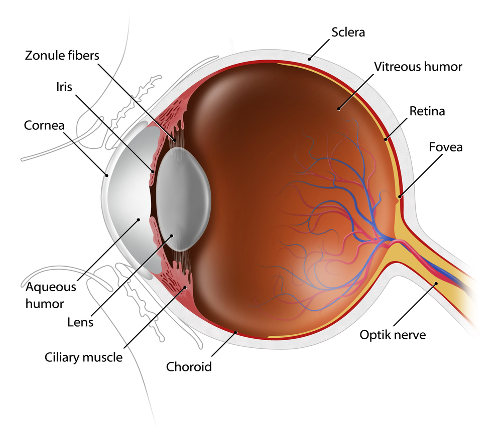
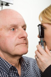
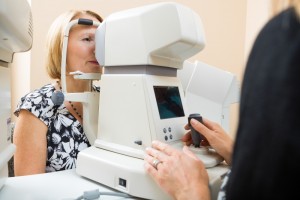
An eye exam involves an external examination of your eyes followed by a series of tests designed to evaluate your vision and check for eye diseases. Each test evaluates a different aspect of your vision and includes specific tests for visual acuity, pupil function, muscle function, visual fields, eye pressure and viewing the back of the eye through a dilated pupil.
cross section of eye - eye exam
External Exam
The external examination consists of inspecting the eyelids, surrounding tissues and the eyeball including the sclera (white part of the eye), iris and cornea.

Visual Acuity & Fields
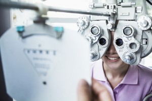
Visual acuity is your eye’s ability to detect fine details and see an in-focus image at a certain distance. A Snellen chart and a phoropter are used. The standard definition of normal visual acuity is 20/20. The term 20/20 comes from even sized objects that can be seen by a “person of normal vision” atvisual acuity eye exam the specified distance. For example, if a person can see at a distance of 20 feet an object that normally can be seen at 20 feet, then they have 20/20 vision. If they can see at 20 feet what a normal person can see at 40 feet, then they have 20/40 vision. For the visual acuity test each eye is tested separately to gauge your side or peripheral vision.

Pupil Functionpupil function eye exam
An examination of the pupil begins with inspecting your pupils for equal size, regular shape, reaction to light, and direct and consensual reaction (meaning the pupil of one eye constricts when the other eye is exposed to light).

Eye Muscle Function
Eye movement is assessed two ways. First by having you move your eye quickly to a target at the far right, left, top and bottom. Then by slow tracking which uses the ‘follow my finger’ test, which tests all the muscles that move your eye.

tonometer eye examEye Pressure Measurement
Intraocular pressure, or IOP, is measured using a tonometer to determine the fluid pressure inside your eye. This test provides information regarding your potential for glaucoma.

Viewing the Back of the Eyedilated eye - eye exam
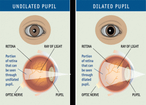
Increasing the size of your pupil with eye drops (known as dilating your eyes) allows the doctor to have a larger view of the back of your eye, including the retina, as demonstrated by this diagram. This is very important for diagnosis and tracking of macular degeneration and diabetic retinopathy.

Eye Structureslit lamp eye exam
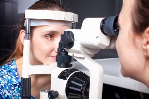
A special, high-powered microscope, called a slit-lamp, is used to view the structures of your eye clearly and in detail, enabling early diagnosis of a variety of eye conditions such as cataracts, presbyopia and corneal injury.

Taking care of your eyes, especially as you become older, is very important. Changes in vision may be gradual, or fast, but in both cases, early diagnosis is key to successful treatment and retaining your vision. It is suggested that individuals 40 and over have a comprehensive eye exam every one to two years. Exceptions would be if you have diabetes, in which case you should see your eye doctor yearly; or there is a family history of eye diseases such as glaucoma, macular degeneration, or corneal diseases, which may require more frequent visits to your eye doctor. If you have any degree of sudden vision loss, eye pain, or significant irritation, contact your eye doctor immediately.

4/23/15

Susan DeRemerSusan DeRemer, CFRE
Vice President of Development
Discovery Eye Foundation

Driving and Age-Related Macular Degeneration

Driving and Age-Related Macular Degeneration

The issue of driving and age-related macular degeneration is a particularly sensitive one for seniors losing their vision. Driving means independence and most people want to hold on to their cars as long as possible. When is it time to stop?
Driving and Age-related Macular Degeneration
A research survey by the Massachusetts Institute of Technology (MIT) Age-Lab and The Hartford Financial Services Group involved 3,824 drivers over age 50, asking them how and why they limited their driving.

The study found that two-thirds of the drivers self-regulated their activities in the car, restricting their driving for certain condition. Time of day was a common factor, with some people choosing to stay home at night or dusk. Bad weather conditions and heavy traffic were other conditions. Over time, drivers developed conscious strategies to compensate for failing vision, slower reflexes and stiffer joints.

Statistically, older drivers are actually very safe drivers, although over age 75, the accident rate per mile increases. The study found that health and medical conditions contributed far more to driving restrictions than age alone.

About ten percent of the nation’s drivers are over 65. However, by 2030, when one in five Americans are over age 65, this percentage will skyrocket. Consider that 23-40% of people over age 65 have macular degeneration – that ís a lot of drivers with a potential visual impairment.

Making the Decision

If your macular degeneration is causing a problem when you drive, you are most likely aware of it. Or, perhaps a friend or family member has pointed it out to you. Does this mean you should immediately stop driving? Not necessarily.

What you should do immediately is ask yourself some critical questions. How are you functioning when you drive during the day? What about dusk, dawn and cloudy days? Bright sunlight? At night?

Here are six important questions:

  1. Do you have difficulties reading clearly and rapidly all the instruments on a carís dashboard?
  2. Do you have difficulties reading road signs, or if you are currently driving, do you notice and understand the signs in time to react to them with comfort?
  3. Do other cars on the road appear to “pop” into and out of your field of vision unexpectedly?
  4. While on the road, do you drive well below the speed limit and slower than most cars around you?
  5. Do you have difficulties positioning yourself on the road, with respect to other cars, lane markers, curves, sidewalks, parking spaces, etc.?
  6. Do you find yourself feeling confused and/or disoriented on the road?

If you answered yes to any of the above questions, you may want to suspend your driving until you consult a specialist. If your answers indicate that you may have a problem under certain conditions (i.e., dim light or night) you may want to suspend your driving under those conditions until you consult a specialist further.

This questionnaire is from an excellent book, “Driving With Confidence, A Practical Guide to Driving With Low Vision” by Eli Peli and Doron Peli. Dr. Eli Peli is a Senior Scientist at the Schepens Eye Research Institute and Professor of Ophthalmology at Harvard Medical School. Their book contains a practical program to help you maximize your chances of retaining your driving privileges. It also provides a detailed description of driving vision regulations in every state as does the AAA website.

Other Useful Resources

AARP Driver Safety Program – Largest classroom driver refresher course specially designed for motorists age 50 and older. It is intended to help older drivers improve their skills while teaching them to avoid accidents and traffic violations.

AAA Safety Foundation for Traffic – Tips on driving and resources for other transportation options.

Summary

There are many ways to stay safe and maintain your independence. Just be attentive to your own abilities and find out all you can about your options.

4/21/15


Judi Delgado - age-related macular degenerationJudith Delgado
Executive Director
Macular Degeneration Partnership
A program of Discovery Eye Foundation

Do I Need Vision Insurance?

With the rising costs of health insurance, many people are looking for ways to reduce their costs. Since not all insurance packages include vision insurance, many people wonder, do I need vision insurance?
vision insurance

Standard Vision Insurance

Vision insurance is a type of health insurance that entitles you to specific eye care benefits such as routine eye exams and other procedures, as well as a specified dollar mount or discount for the purchase of eyeglasses and contact lenses. It only supplements regular health insurance and is designed to help reduce your costs for routine preventative eye care and eyewear.

You can get vision insurance as part of a group, such as your employer, an association, etc., through a government program such as Medicare or Medicaid, or as an individual. It is often a benefit linked to your regular HMO (health maintenance organization) or PPO (preferred provider organization) health insurance.

There are two primary vision insurance plans available:

  • Vision Benefits Package – provides free eye care services and eyewear within a fixed dollar amount for which you pay an annual premium or membership fee and a small co-pay. It may also include a deductible.
  • Discount Vision Plan – provides eye care and eyewear at a discounted rate after you pay an annual premium or membership fee.

Both insurance plans generally include:

  • Annual eye exams
  • Eyeglass frames (usually once every 24 months)
  • Eyeglass lenses (usually once every 24 months)
  • Contact lenses (usually once every 24 months)
  • Discounted rates for LASIK and PRK

Here is where you can check for a list of some vision insurance providers.

Medicare and Medicaid

Different kinds of vision care are included in the US government programs, Medicare and Medicaid. These programs are for qualifying American age 65 and older, individuals with specific disabilities and people with low income.

The Types of Medicare For Vision:

    • Medicare Part A (Hospital Insurance) –Medical eye problems that require a hospital emergency room attention, but routine eye exams are NOT covered.
    • Medicare Part B (Medical Insurance) – Visits to an eye doctor that are related to an eye disease, but routine eye exams are NOT covered.
    • Medicare Part D (Prescription Drug Coverage) – Will help pay for prescription medications for eye diseases.

If you have Medicare Parts A & B you are generally eligible for the following vision coverage, however, there is a deductible before Medicare will start to pay, at which point you will still be paying a percentage of the remaining costs.

  • Cataract surgery – covers many of the cost including a standard intraocular lens (IOL). If you chose a premium IOL to correct your eyesight and reduce your need for glasses, you must pay for this added cost out-of-pocket.
  • Eyewear after cataract surgery – one pair of standard eyeglasses OR contact lenses.
  • Glaucoma screening – an annual screening for people at high risk for glaucoma, including people with diabetes or a family history, and African-Americans whom are 50 or older.
  • Ocular prostheses – costs related to the replacement and maintenance of an artificial eye.

There is also Medicare Supplement Insurance (Medigap) which is sold by private insurance companies to supplement only Medicare Parts A & B. It is intended to cover your share of the costs of Medicare-covered services including coinsurance, co-payments and deductibles. For more details about Medicare plans and coverage check their website or call 800-633-4227.

Medicaid is the US health program that gives medical benefits to low-income people who may have no or inadequate medical insurance. A person eligible for Medicaid may be asked to make a co-payment at the time medical service is provided. Vision benefits for children under the age of 21 include eye exams, eyeglass frames and lenses. Each state determines how often these services are provided and some states offer similar vision services to adults. To learn more about Medicaid eligibility requirements and vision benefits call your state’s Medicaid agency or visit their website.
vision dial - vision insurance

Defined Contribution Health Plans

A way to lower your vison care costs is to take part in a defined contribution health plan (DCHP). You are given a menu of health care benefits to choose from where a portion of the fees you receive for health coverage come from money that is deducted from our paycheck before federal, state and social security taxes are calculated. Four types of DCHP are:

Cafeteria Plans – your employer takes a portion of your salary and deposits it into a non-taxable account for health care spending. The amount taken depends on the number and costs of the benefits you select.

Flexible Spending Accounts (FSA) – your employer takes a predetermined portion of your pre-tax salary and deposits it into health care account for you to pay medical expenses. But generally preventative care such as routine eye exams and are not reimbursable. Nor are eyeglasses and contact lenses reimbursable. You would need to verify with your employer. If you do not use all the money at the end of a 12 month period, the money goes back to your employer.

Health Reimbursement Arrangement (HRA) – this is similar to an FSA except you can use it for preventative care like eye exams and you do not lose the money if it isn’t spent within a certain time period as it can be carried from year to year.

Health Savings Account (HSA) – it can be employer-sponsored of you can set up one independently; however you must purchase a high-deductible health insurance plan to open an HSA and you cannot exceed the annual deductible of your health insurance plan. You cannot be enrolled in Medicare of be a depended on someone else’s tax return. You can use it for preventive care such as eye exams. You can learn more about HSAs by visiting the US Treasury’s website.

There are a variety of options when it comes to vision insurance. You just need to determine your needs and ask providers the correct questions.

4/14/15


Susan DeRemerSusan DeRemer, CFRE
Vice President of Development
Discovery Eye Foundation

6 Ways Women Can Stop Vision Loss

Women account for 53% of the world’s population. However, 2/3 of the blind and visually impaired people in the world are women. While 80% of these women are in developing countries, women in developed countries like the US are still more likely to face vision loss than men.
women can stop vision loss

Why are women more prone to eye disease than men?

Women are the caregivers in families, taking care of the health of family members over themselves. In addition, with many having jobs outside the home, they don’t feel they have the time to go to the doctor until something major happens, especially related to vision.

Women live longer than men are at greater risk for age-related eye diseases such as age-related macular degeneration (AMD), diabetic retinopathy, glaucoma and cataracts.

Women are more likely to develop several autoimmune diseases that can affect their eyes including, multiple sclerosis, lupus, rheumatoid arthritis and Sj?gren’s syndrome.

75% of new breast cancer diagnosed each year is estrogen-sensitive. A common part of estrogen-sensitive breast cancer treatment includes the prescription of tamoxifen. More studies are being done, but cataracts due to tamoxifen have been identified in about 10% of the patients taking the drug.

What can women do to lessen their chances for eye disease?

Know your family history as genetics play and important role in your eye health, so know what eye diseases run in your family. Let your eye doctor know so he can look for early warning signs that can help prevent of lessen the conditions in you.

Get routine comprehensive, dilated eye exams starting at the age of 40, to create a baseline for your doctor to work from. After that you can go every 2-4 years until the age of 60. At 60+ have a compressive, dilated exam every two years if you are symptom-free and low risk.

Eat healthy and exercise. It is important to maintain a healthy weight to reduce the risk of some eye diseases. Eating fresh fruits and vegetables is also important as they can contain carotenoids an some antioxidants that appear to help with vision retention. It should also be noted that in many studies, supplements did not show the same beneficial effects as whole foods.

Stop smoking! You not only increase your risk for cancer and heart disease, but smoking is the only thing besides advancing age that has been proven to be directly related to AMD.

Avoid ultraviolet light by wearing sunglasses (with wide-brimmed hats) and indoor glasses with UV protection. While everyone knows the sun is a source of UV light, so are electronic screens such as your TV, computer, tablet or smartphone. Prescription glasses and readers can have a clear UV coating put on them that will not distort your color vision. If you don’t need vision correction, there is eyewear with no correction that is coated to protect your eyes to avoid dry eye and retinal damage.

Use cosmetics and contacts safely. Always wash your hands first. Throw away old makeup and lens solutions. Do not share cosmetics or apply while driving. Make sure to clean your lenses thoroughly before putting them in your eyes.

Because women are relied upon to take care of the family, vision loss that can impact that responsibility can be devastating to the entire family. And later in life, when they may have outlived a spouse, the isolation and depression can destroy their quality of life as they try to cope on their own.

Reach out to women you know and remind them to take an active part in their own healthcare. Especially with regards to their vision, when women are at a higher risk of vision loss than men.

4/7/15


Susan DeRemerSusan DeRemer, CFRE
Vice President of Development
Discovery Eye Foundation

Three Generations of Saving Vision

In Discovery Eye Foundation’s spring e-newlsetter there was an article entitled Surgery for the Surgeon, where a leading ophthalmologist talks about convincing himself to have cataract surgery. That eye doctor was Dr. Nesburn, who was willing to share with us his family’s long tradition of saving vision.

Brainwashed by Medicine

“I was brainwashed from the age of 5,” says Dr. Anthony Nesburn, medical director of The Discovery Eye Foundation (DEF). “My dad would take me on rounds at the hospital and to his office. He introduced me to medicine at a really early age.”

Nesburn saving vision
Dr. Anthony Nesburn in UC Irvine lab
Dr. Henry Nesburn was an ophthalmologist in Los Angeles for more than four decades, and he passed his love of the specialty to his son. “I really looked up to my dad,” the younger Nesburn says. “He loved ophthalmology for the same reasons I do: You get to do medical diagnosis; you get to do wonderful surgery, where you keep people from going blind or restore vision; and you can work with people from newborns to the very elderly — they all need eye care.”

Nesburn received a telegram while he was an undergrad at UCLA, telling him he’d been accepted to Harvard Medical School. His mother started crying: “You’re going to Boston! We’re not going to see you anymore!” While he “was loathe to leave Southern California,” Harvard was too good to pass up.

Drafted by the Army out of his ophthalmology residency at Harvard in 1960, Nesburn joined the Navy instead. He followed in his father’s footsteps again, becoming a Navy flight surgeon. (Henry had volunteered during World War II.)

He went on to a Boston Children’s Hospital fellowship in infectious disease, working with Nobel laureate Dr. John Enders, whose work led to the polio vaccine and changed the face of virology. Nesburn then did his residency at Massachusetts Eye and Ear. “I was part of a special program that allowed us to do research, and I was running a research laboratory while I was a resident,” he says. “It gave me the start I needed.”

“At Mass Eye and Ear, I worked for an up-and-coming ophthalmologist and researcher to prove there was a substance that could treat herpes eye infections. We wrote a paper that included the very first antiviral ever described, and it was against herpes virus. It is the basis for today’s herpes antivirals,” Nesburn says. “I was hooked.”

He went back to Los Angeles and received NIH funding to continue his research on ocular herpes. In 1968, he joined his father’s practice half-time, spending the rest of his time doing research.

Two years later, he received a generous offer, when Rita and Morris Pynoos started DEF to fund his research. The Pynooses were grateful to Nesburn for diagnosing their son, Jon, with keratoconus (KC). “I was a second-year resident at Mass Eye and Ear, and Jon Pynoos was an undergraduate at Harvard. His parents went to see my dad, because Jon couldn’t see well, and no one could figure out what was wrong with his vision. My dad said, ‘Send him over to Tony; he’ll figure out what’s going on!’ I said to myself, ‘Holy mackerel! What happens if he has something really complicated? I’m just a newbie!’” Nesburn remembers. “Jon came in; I looked at him, and the keratoconus was so clear and easy to spot. I couldn’t imagine how his doctors didn’t see it. We got him contact lenses, and he was able to see again. When I came back to LA, the Pynooses wanted to do something to help.”

At first, DEF research focused on KC and the herpes research Nesburn was working on at the time. It soon broadened to include macular degeneration and retinal disease.

“My dad had to retire from the practice of ophthalmology at the age of 70, because of bad age-related macular degeneration (AMD). His mother and older sister had had it, as did several cousins. There was no treatment back then that helped,” Nesburn says.

“AMD is the most common cause of permanent vision loss in the elderly in the developed world. I could see where the need was,” he says. “We moved forward at DEF with two driving mantras: We wanted to do something significant in macular degeneration research and to find the cause of keratoconus.”

As a virologist in research and a corneal surgeon, Nesburn realized he needed a corneal biochemist to help with the KC research. He met Dr. Cristina Kenney at an Association for Research in Vision and Ophthalmology meeting. She joined DEF, and within 15 years, they found the chemical cause of keratoconus; they also got married.

Now nearly 80 years old, Nesburn spends most of his time “wearing three hats”: fundraising for DEF, lab research and clinical practice. His daughter, Kristin, is the third generation to join the family ophthalmology practice.

“While I’m still able, I want to try to make a difference in medicine, particularly in macular degeneration,” Nesburn says. “Macular degeneration affects so many people. This is where I want to put my energy. Luckily, as strong as it is in our family, I don’t have it … yet.

“As a researcher, my interest in putting together a program for macular-degeneration diagnosis and treatment has been because it’s a great public health problem. Yes, if I should ever get it, it might be able to help me or my family, but the first thing, as a scientist, is to try to get something to help humankind. I know it sounds sappy, but it’s true.”

3/26/15


Anthony B. Nesburn, MD, FACSAnthony B. Nesburn, MD, FACS
Medical Director, Discovery Eye Foundation
Professor & Vice Chairman for Research, Ophthalmology
Gavin Herbert Eye Institute, University of CA, Irvine

Medical Research Funding Needs Individual Donors

The Need for Medical Research Funding

About 1.75 million U.S. residents currently have advanced age-related macular degeneration with associated vision loss, with that number expected to grow to almost 3 million by the year 2020.

About 8.4 million individuals worldwide are blind from primary open-angle glaucoma, with that number expected to grow to almost 11 million by the year 2020.

About 22 million Americans have cataracts affecting their vision, with that number expected to grow to more than 30 million by the year 2020.

The economic impact of this increase of people with vision loss will be tremendous.  But right now scientist are working on ways to treat and eventually cure many eye diseases.  The only problem is the funding necessary to support this sight-saving research. Here is a look at the decline of medical research funding in the US and what you can do to help.
medical research funding

3/17/15


Susan DeRemerSusan DeRemer, CFRE
Vice President of Development
Discovery Eye Foundation

10 Tips For Healthy Eyes

In honor of Save Your Sight Week, here are 10 tips for healthy eyes. There are some simple things you can begin to do today to help your retain your vision. Also there are no warning signs for many of the leading eye diseases (age-related macular degeneration, glaucoma, cataracts), yet most people don’t see an eye doctor until they have a problem seeing. Unfortunately, by that time, irreparable damage has been done.
10 Tips For Healthy Eyes

3/3/15


Susan DeRemerSusan DeRemer, CFRE
Vice President of Development
Discovery Eye Foundation

February Is Low Vision Awareness Month

Help for People With Vision Loss

Here’s eye-opening news: Currently, 4.2 million Americans ages 40 and older are visually impaired. Of these, 3 million have low vision. By 2030, when the last baby boomers turn 65, the number of Americans who have visual impairments is projected to reach 7.2 million, with 5 million having low vision. For the millions of people who currently live or will live with low vision, the good news is there is help.
Low vision awareness
But first, what is low vision? Low vision is when even with regular glasses, contact lenses, medicine, or surgery, people have difficulty seeing, which makes everyday tasks difficult to do. Activities that used to be simple like reading the mail, shopping, cooking, and writing can become challenging.

Most people with low vision are age 65 or older. The leading causes of vision loss in older adults are age-related macular degeneration, diabetic retinopathy, cataract, and glaucoma. Among younger people, vision loss is most often caused by inherited eye conditions, infectious and autoimmune eye diseases, or trauma. For people with low vision, maximizing their remaining sight is key to helping them continue to live safe, productive, and rewarding lives. The first step is to seek help.

“I encourage anyone with low vision to seek guidance about vision rehabilitation from a low vision specialist,” advises Paul A. Sieving, MD, PhD, director of the National Eye Institute (NEI), one of the National Institutes of Health (NIH) and the federal government’s principal agency for vision research.

What is a low vision specialist? A low vision specialist is an ophthalmologist or optometrist who works with people who have low vision. A low vision specialist can develop a vision rehabilitation plan that identifies strategies and assistive devices appropriate for the person’s particular needs. “A vision rehabilitation plan helps people reach their true visual potential when nothing more can be done from a medical or surgical standpoint,” explains Mark Wilkinson, O.D., a low vision specialist at the University of Iowa Hospitals and Clinics and chair of the low vision subcommittee for the National Eye Health Education Program (NEHEP).

Vision rehabilitation can include the following:

  • Training to use magnifying and adaptive devices
  • Teaching new daily living skills to remain safe and live independently
  • Developing strategies to navigate around the home and in public
  • Providing resources and support

There are also many resources available to help people with low vision. NEI offers a 20-page, large-print booklet, titled What You Should Know About Low Vision, and companion DVD, featuring inspiring stories of people living with low vision. This booklet and DVD, among other resources, are available at
www.nei.nih.gov/lowvision.

With the aging of the population, eye diseases and vision loss have become major public health concerns in the United States. NEI is committed to finding new ways to improve the lives of people living with visual impairment. Aside from making information and resources readily available, NEI has dedicated more than $24 million to research projects on low vision, including learning how the brain adapts to vision loss; strategies to improve vision rehabilitation; and the development of new technologies that help people with low vision to read, shop, and find their way in unfamiliar places. Research like this will help people with low vision to make the most of their remaining vision and maintain their independence and quality of life.

2/17/15

NEHEPsq75The National Eye Health Education Program (NEHEP) of the National Eye Institute (NEI), a part of NIH, offers a 20-page, large-print booklet, titled What You Should Know About Low Vision, and a series of videos featuring patient stories about living with low vision. These and other resources are available at http://www.nei.nih.gov/lowvision.