Spring is truly here. With Passover and Easter later this week, it is time to rediscover some of the amazing produce that is at its peak during the months of April, May and June. These 7 spring fruits and vegetables are not only delicious; they are good for you . . . and your vision. Here are some recipes for you to try and enjoy the bounty of spring. Some even include more than one of the seven fruits and vegetables listed below.
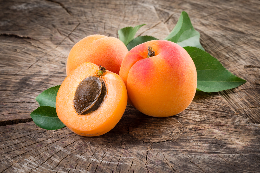
Apricots – Apricots should be firm, but not hard, with a nice fruit scent when sniffed. They are best purchased locally so they aren’t picked too early and have a tree-ripened sweetness.
Apricot & Orange Breakfast Smoothie from Discovery Eye Foundation’s Eye Cook
Spicy Apricot Wings from Food & Wine
Fresh Apricot Chutney from Cooking Light
Chicken Tagine with Apricots & Almonds from Gourmet
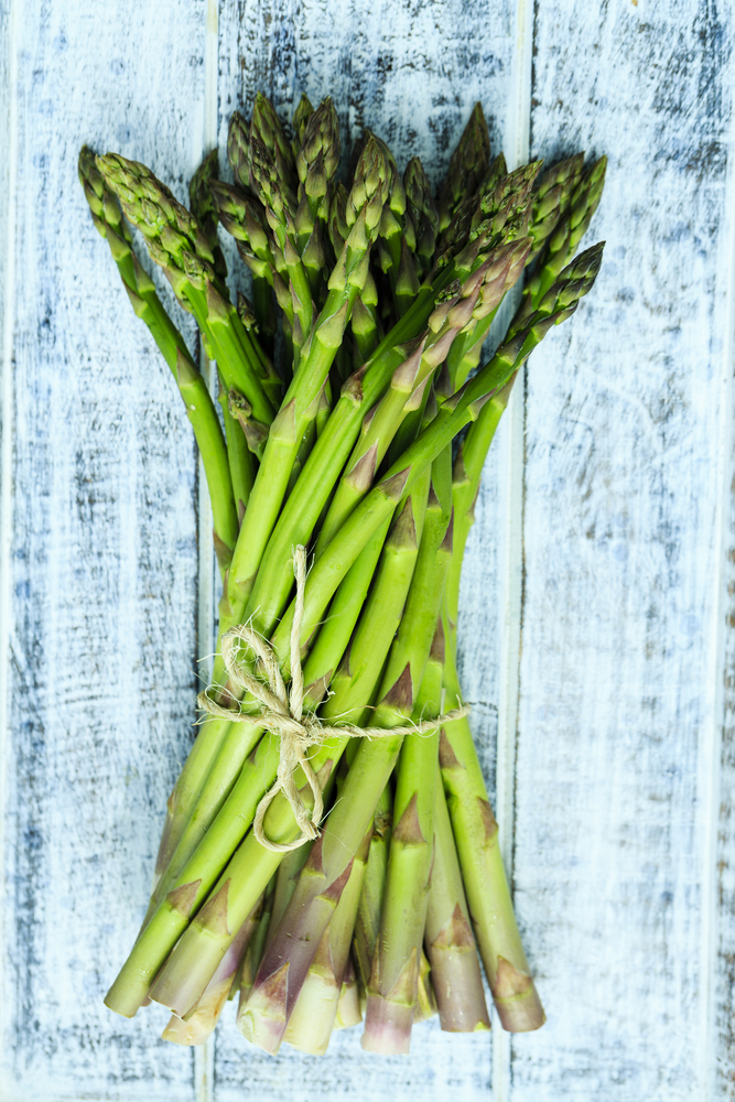
Asparagus – Look for firm stalks, from the tips down to the base of stalks. Once asparagus are harvested they deteriorate quickly, so place them in cool storage to retain freshness and their nutrition value.
Asparagus and Strawberry Salad from Discovery Eye Foundation’s Eye Cook
Asparagus with Watercress and Brown Butter Potatoes from Food & Wine
Grilled Asparagus with a Caper Vinaigrette from Cooking Light
Asparagus, Tomato & Red Pepper French Bread Pizza from the Mayo Clinic
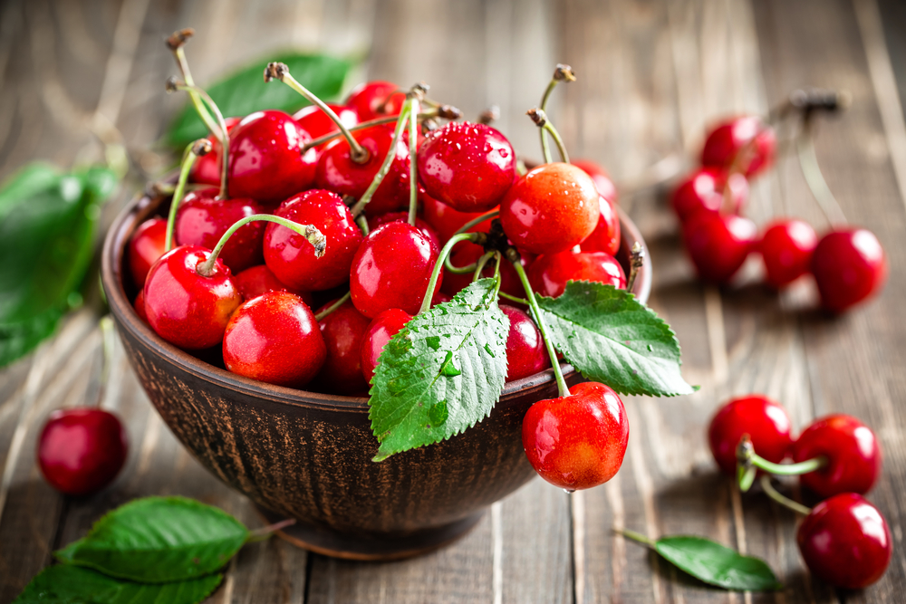
Sweet Cherries – The best cherries are an inch or more in diameter, plump, firm, and rich in color.
Cherry Pie from Cooking Light
Cherry Tortoni from Gourmet
Cherry Tart from Bon Appétit
Easy Almond & Dried Cherry Cookies from Discovery Eye Foundation’s Eye Cook
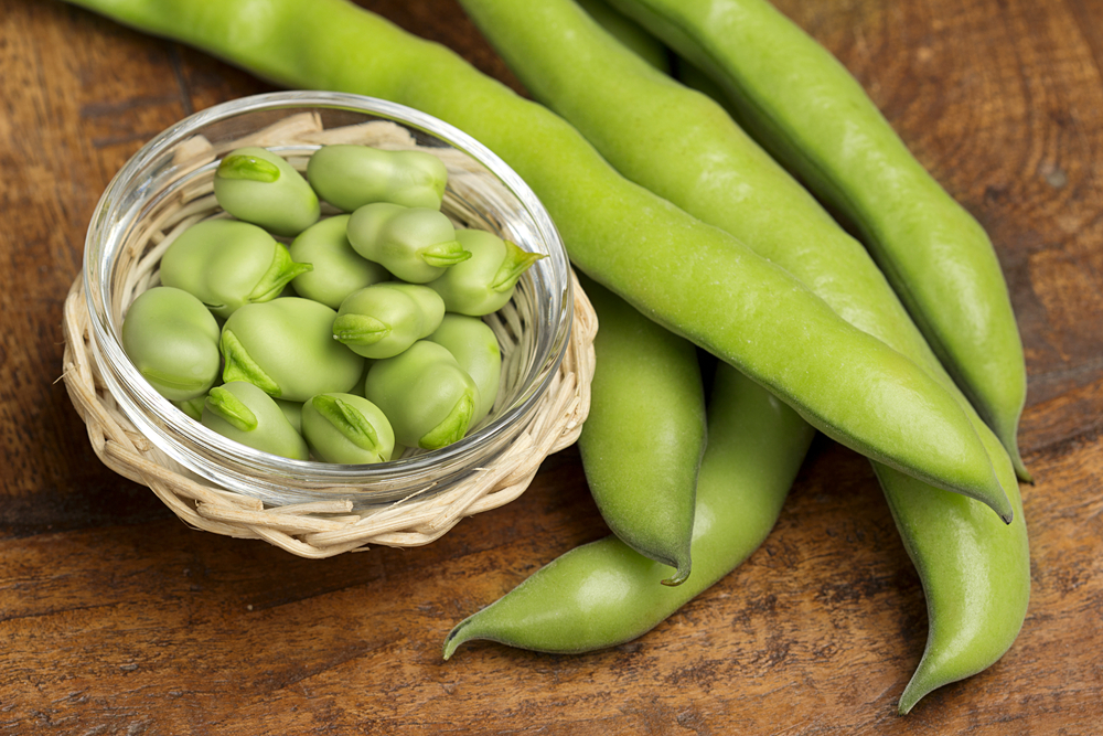
Fava Beans – Young fava beans can be shelled and eaten either raw or cooked, but more mature favas need to be shelled and skinned, since their skins are too tough to eat.
Quinoa Salad with Grilled Scallions, Favas & Dates from Food & Wine
Sliced Filet Mignon with Fava Beans, Radishes & Mustard Dressing from Bon Appétit
Arugula & Fava Bean Crostini from Gourmet
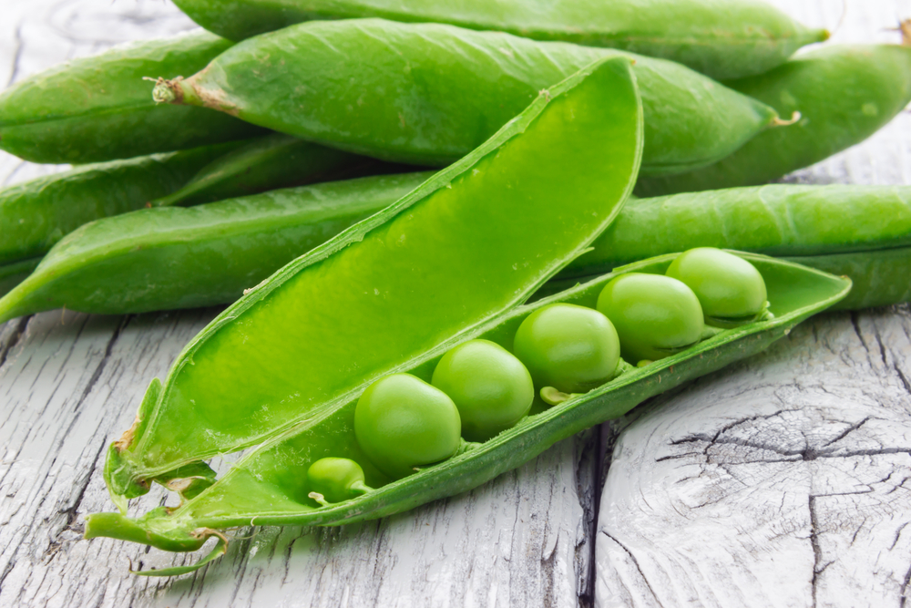
Green Peas – Fresh green peas include sugar snap peas, snow peas, and green peas. Look for bright green pods that are firm.
Fava, Sweet Pea & Sugar Snap Salad from Cooking Light
Salmon with Sweet Chili Glaze, Sugar Snap Peas & Pea Tendrils from Bon Appétit
Strawberry, Almond & Pea Salad from Bon Appétit
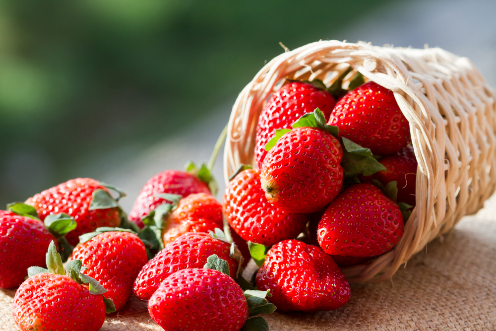
Strawberries – For the best flavor, you are best looking for strawberries grown close to home since they more are likely to be fresh and not be damaged in transit. They should be plump, firm, well-shaped, and uniformly colored.
Carrot & St rawberry Tea Bread from Discovery Eye Foundation’s Eye Cook
Strawberries Romanoff from Cooking Light
Strawberry and Cream Cheese Crepes from the Mayo Clinic
Pink Grapefruit, Strawberry & Champagne Granita from Bon Appétit
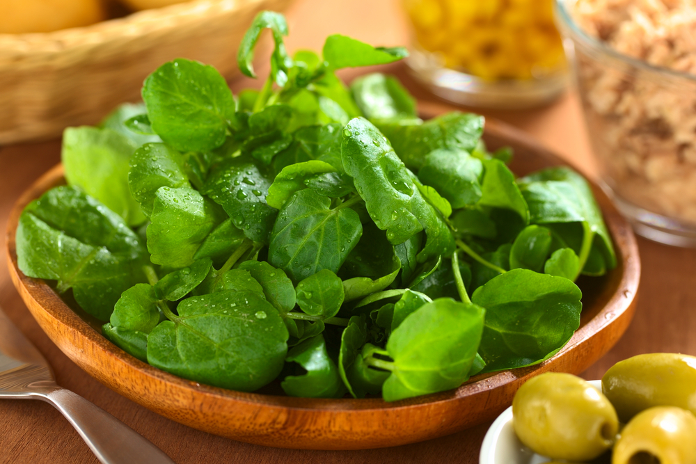
Watercress – Look for uniformly dark green leaves and sniff for a fresh, spicy scent. Watercress has a short shelf life and should be kept in a plastic bag in the refrigerator for no more than three days.
Watercress, Orange & Avocado Salad from Gourmet
Watercress Salad with Verjus Vinaigrette from Food & Wine
Watercress Salad with Pan-Seared Mahimahi from Cooking Light
These are some great ways to enjoy what spring has to offer. Do you have any spring recipes you want to share?
3/31/15
 Susan DeRemer, CFRE
Vice President of Development
Discovery Eye Foundation