Driving and Age-Related Macular Degeneration

Driving and Age-Related Macular Degeneration

The issue of driving and age-related macular degeneration is a particularly sensitive one for seniors losing their vision. Driving means independence and most people want to hold on to their cars as long as possible. When is it time to stop?
Driving and Age-related Macular Degeneration
A research survey by the Massachusetts Institute of Technology (MIT) Age-Lab and The Hartford Financial Services Group involved 3,824 drivers over age 50, asking them how and why they limited their driving.

The study found that two-thirds of the drivers self-regulated their activities in the car, restricting their driving for certain condition. Time of day was a common factor, with some people choosing to stay home at night or dusk. Bad weather conditions and heavy traffic were other conditions. Over time, drivers developed conscious strategies to compensate for failing vision, slower reflexes and stiffer joints.

Statistically, older drivers are actually very safe drivers, although over age 75, the accident rate per mile increases. The study found that health and medical conditions contributed far more to driving restrictions than age alone.

About ten percent of the nation’s drivers are over 65. However, by 2030, when one in five Americans are over age 65, this percentage will skyrocket. Consider that 23-40% of people over age 65 have macular degeneration – that ís a lot of drivers with a potential visual impairment.

Making the Decision

If your macular degeneration is causing a problem when you drive, you are most likely aware of it. Or, perhaps a friend or family member has pointed it out to you. Does this mean you should immediately stop driving? Not necessarily.

What you should do immediately is ask yourself some critical questions. How are you functioning when you drive during the day? What about dusk, dawn and cloudy days? Bright sunlight? At night?

Here are six important questions:

  1. Do you have difficulties reading clearly and rapidly all the instruments on a carís dashboard?
  2. Do you have difficulties reading road signs, or if you are currently driving, do you notice and understand the signs in time to react to them with comfort?
  3. Do other cars on the road appear to “pop” into and out of your field of vision unexpectedly?
  4. While on the road, do you drive well below the speed limit and slower than most cars around you?
  5. Do you have difficulties positioning yourself on the road, with respect to other cars, lane markers, curves, sidewalks, parking spaces, etc.?
  6. Do you find yourself feeling confused and/or disoriented on the road?

If you answered yes to any of the above questions, you may want to suspend your driving until you consult a specialist. If your answers indicate that you may have a problem under certain conditions (i.e., dim light or night) you may want to suspend your driving under those conditions until you consult a specialist further.

This questionnaire is from an excellent book, “Driving With Confidence, A Practical Guide to Driving With Low Vision” by Eli Peli and Doron Peli. Dr. Eli Peli is a Senior Scientist at the Schepens Eye Research Institute and Professor of Ophthalmology at Harvard Medical School. Their book contains a practical program to help you maximize your chances of retaining your driving privileges. It also provides a detailed description of driving vision regulations in every state as does the AAA website.

Other Useful Resources

AARP Driver Safety Program – Largest classroom driver refresher course specially designed for motorists age 50 and older. It is intended to help older drivers improve their skills while teaching them to avoid accidents and traffic violations.

AAA Safety Foundation for Traffic – Tips on driving and resources for other transportation options.

Summary

There are many ways to stay safe and maintain your independence. Just be attentive to your own abilities and find out all you can about your options.

4/21/15


Judi Delgado - age-related macular degenerationJudith Delgado
Executive Director
Macular Degeneration Partnership
A program of Discovery Eye Foundation

How to Help a Blind or Visually Impaired Person with Mobility

You as a Guide
As the spouse, partner or friend of a person that has low or no vision, it is hard to know when to step and help. You don’t want to offend them by jumping in and making them feel incapable, nor do you want to stand by and have them stumble along, possibly hurting themselves.
blind or visually impaired person with mobility
Here are few pointers to help a blind or visually impaired person with mobility so you can be supportive and considerate.

  • First and most importantly, never assume the person wants or needs your help. Always ask first and never force the person to accept your help.
  • If your offer of help is accepted, ask if the person would like you on their right or left, then the tap them with your appropriate elbow so the person needing help can grasp slightly above the elbow.
  • Relax and walk at a comfortable, consistent pace, about a half step ahead.
  • When navigating obstacles, guiding signals, along with verbal cues, are helpful. Examples:
    Curb – brief pause and state “we are approaching a curb and it is slanted upwards.”
    Narrow door or passage – you enter first moving your guiding arm behind the small of your back and let them know you are moving through a doorway or narrow space and which direction the door opens so they can move to the appropriate side.
  • Stairs – stop at the edge of the first step and let the other person know if the steps are up or down, where the railing is (make sure it is on the side of their free hand), and if there is anything special about the steps (they are uneven, very steep, have an overly wide tread, etc.). If there are just a few steps let them know, “you will take 4 steps down.” The will follow one step behind you, with one hand on the handrail and the other holding your arm. Pause after completing the stairs.
  • Escalators and revolving doors – Use similar guiding cues as you do for the stairs. Let them know when to step onto the escalator and when to get ready to get off. If the person you are guiding is uncomfortable with escalators or revolving doors, use the elevator and regular doors which all buildings are required to have.
  • Chair – when approaching a chair, place the hand of the person being guided on the back or side of the chair, letting them know which direction the chair is facing.
  • Never leave a person who is blind or visually impaired in “free space.” Make sure they are in contact with a wall, railing or some other stable object until you return.

If They Use a Guide Dog
Guide dogs are invaluable to people that are blind or partially sighted. They allow their owners a sense of independence. But how should you respond to a guide dog who is working?blind or visually impaired person with mobility

  • Never distract the dog from its duty, so don’t pet the dog without asking.
  • Before asking a questions of a person handling a dog, allow them to complete the task at hand.
  • Remain calm when you approach, never teasing or speaking to the dog.
  • Do not offer the dog food or other treats. They are fed on a schedule and follow a specific diet to keep them healthy. Deviations from their routine can disrupt their regular and ad relieving schedules, seriously inconveniencing their handlers.
  • Do not offer toys to a guide dog. Though they are treated as pets when they are not in their harnesses, they are only allowed specific toys. In their harness they don’t play with toys.
  • Do not call out to the guide dog or obstruct its path, as it can break the dog’s concentration which could prove to be dangerous to its handler.
  • In some cases the person with low or no vision may prefer to take your arm above the elbow and allow their dog to heel instead of lead. Follow the same instructions as in the first part of this blog. When approaching stairs, ask how the person how they wish to proceed, as they will be holding your arm with one hand and the guide dog with the other, making it impossible to grasp a handrail.

Giving Directions To Someone Who is Blind or Visually Impaired
If a person is on their own with a guide dog or white cane, giving complete accurate directions is necessary. While you may be used to pointing or saying “it is over there,” or “go around the next corner,” if you can’t see you have no idea where “there” is or the “next corner.”

  • Always refer to a specific direction from the perspective of the person you are advising. Your right is their left.
  • Indicate the approximate distance in addition to the direction.
  • Give the approximate number of streets to cross to reach the destination. Even if you are off a block or two, it gives the person an idea of when to stop and ask for further instructions if needed.
  • If possible, provide information about landmarks on the way. Remember that sounds, scents and ground textures can be landmarks. You can hear an escalator, smell the scent of fresh brewed coffee and feel the difference between grass and a sidewalk.

4/16/15


Susan DeRemerSusan DeRemer, CFRE
Vice President of Development
Discovery Eye Foundation

Do I Need Vision Insurance?

With the rising costs of health insurance, many people are looking for ways to reduce their costs. Since not all insurance packages include vision insurance, many people wonder, do I need vision insurance?
vision insurance

Standard Vision Insurance

Vision insurance is a type of health insurance that entitles you to specific eye care benefits such as routine eye exams and other procedures, as well as a specified dollar mount or discount for the purchase of eyeglasses and contact lenses. It only supplements regular health insurance and is designed to help reduce your costs for routine preventative eye care and eyewear.

You can get vision insurance as part of a group, such as your employer, an association, etc., through a government program such as Medicare or Medicaid, or as an individual. It is often a benefit linked to your regular HMO (health maintenance organization) or PPO (preferred provider organization) health insurance.

There are two primary vision insurance plans available:

  • Vision Benefits Package – provides free eye care services and eyewear within a fixed dollar amount for which you pay an annual premium or membership fee and a small co-pay. It may also include a deductible.
  • Discount Vision Plan – provides eye care and eyewear at a discounted rate after you pay an annual premium or membership fee.

Both insurance plans generally include:

  • Annual eye exams
  • Eyeglass frames (usually once every 24 months)
  • Eyeglass lenses (usually once every 24 months)
  • Contact lenses (usually once every 24 months)
  • Discounted rates for LASIK and PRK

Here is where you can check for a list of some vision insurance providers.

Medicare and Medicaid

Different kinds of vision care are included in the US government programs, Medicare and Medicaid. These programs are for qualifying American age 65 and older, individuals with specific disabilities and people with low income.

The Types of Medicare For Vision:

    • Medicare Part A (Hospital Insurance) –Medical eye problems that require a hospital emergency room attention, but routine eye exams are NOT covered.
    • Medicare Part B (Medical Insurance) – Visits to an eye doctor that are related to an eye disease, but routine eye exams are NOT covered.
    • Medicare Part D (Prescription Drug Coverage) – Will help pay for prescription medications for eye diseases.

If you have Medicare Parts A & B you are generally eligible for the following vision coverage, however, there is a deductible before Medicare will start to pay, at which point you will still be paying a percentage of the remaining costs.

  • Cataract surgery – covers many of the cost including a standard intraocular lens (IOL). If you chose a premium IOL to correct your eyesight and reduce your need for glasses, you must pay for this added cost out-of-pocket.
  • Eyewear after cataract surgery – one pair of standard eyeglasses OR contact lenses.
  • Glaucoma screening – an annual screening for people at high risk for glaucoma, including people with diabetes or a family history, and African-Americans whom are 50 or older.
  • Ocular prostheses – costs related to the replacement and maintenance of an artificial eye.

There is also Medicare Supplement Insurance (Medigap) which is sold by private insurance companies to supplement only Medicare Parts A & B. It is intended to cover your share of the costs of Medicare-covered services including coinsurance, co-payments and deductibles. For more details about Medicare plans and coverage check their website or call 800-633-4227.

Medicaid is the US health program that gives medical benefits to low-income people who may have no or inadequate medical insurance. A person eligible for Medicaid may be asked to make a co-payment at the time medical service is provided. Vision benefits for children under the age of 21 include eye exams, eyeglass frames and lenses. Each state determines how often these services are provided and some states offer similar vision services to adults. To learn more about Medicaid eligibility requirements and vision benefits call your state’s Medicaid agency or visit their website.
vision dial - vision insurance

Defined Contribution Health Plans

A way to lower your vison care costs is to take part in a defined contribution health plan (DCHP). You are given a menu of health care benefits to choose from where a portion of the fees you receive for health coverage come from money that is deducted from our paycheck before federal, state and social security taxes are calculated. Four types of DCHP are:

Cafeteria Plans – your employer takes a portion of your salary and deposits it into a non-taxable account for health care spending. The amount taken depends on the number and costs of the benefits you select.

Flexible Spending Accounts (FSA) – your employer takes a predetermined portion of your pre-tax salary and deposits it into health care account for you to pay medical expenses. But generally preventative care such as routine eye exams and are not reimbursable. Nor are eyeglasses and contact lenses reimbursable. You would need to verify with your employer. If you do not use all the money at the end of a 12 month period, the money goes back to your employer.

Health Reimbursement Arrangement (HRA) – this is similar to an FSA except you can use it for preventative care like eye exams and you do not lose the money if it isn’t spent within a certain time period as it can be carried from year to year.

Health Savings Account (HSA) – it can be employer-sponsored of you can set up one independently; however you must purchase a high-deductible health insurance plan to open an HSA and you cannot exceed the annual deductible of your health insurance plan. You cannot be enrolled in Medicare of be a depended on someone else’s tax return. You can use it for preventive care such as eye exams. You can learn more about HSAs by visiting the US Treasury’s website.

There are a variety of options when it comes to vision insurance. You just need to determine your needs and ask providers the correct questions.

4/14/15


Susan DeRemerSusan DeRemer, CFRE
Vice President of Development
Discovery Eye Foundation

6 Ways Women Can Stop Vision Loss

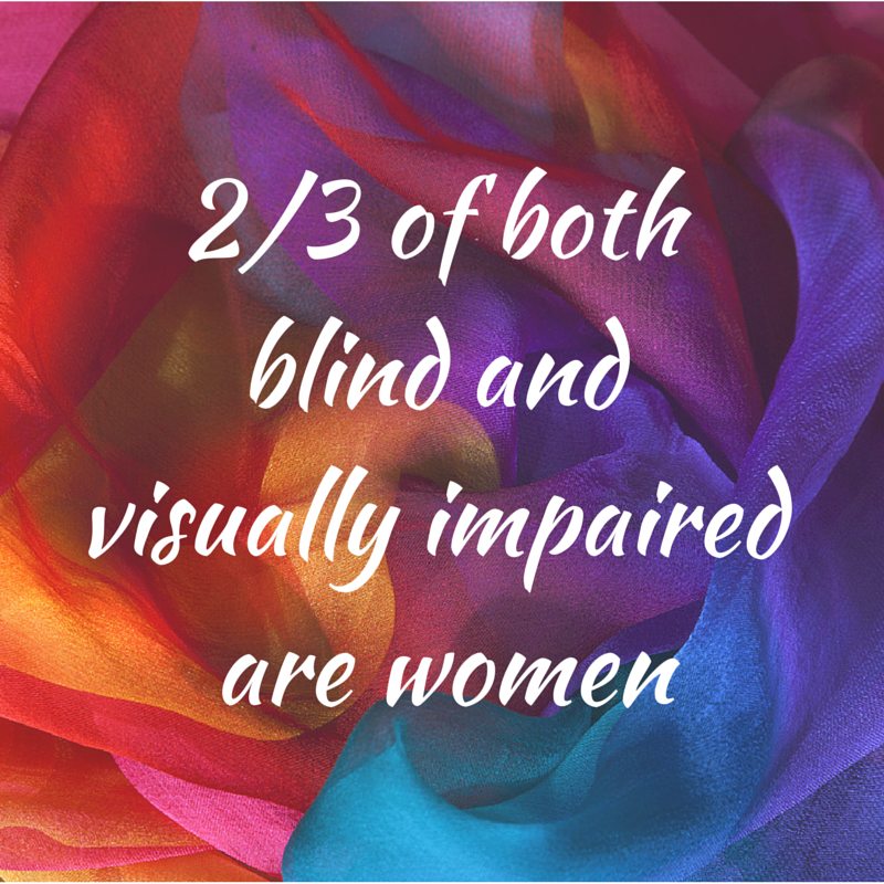
Women account for 53% of the world’s population. However, 2/3 of the blind and visually impaired people in the world are women. While 80% of these women are in developing countries, women in developed countries like the US are still more likely to face vision loss than men.
women can stop vision loss

Why are women more prone to eye disease than men?

Women are the caregivers in families, taking care of the health of family members over themselves. In addition, with many having jobs outside the home, they don’t feel they have the time to go to the doctor until something major happens, especially related to vision.

Women live longer than men are at greater risk for age-related eye diseases such as age-related macular degeneration (AMD), diabetic retinopathy, glaucoma and cataracts.

Women are more likely to develop several autoimmune diseases that can affect their eyes including, multiple sclerosis, lupus, rheumatoid arthritis and Sj?gren’s syndrome.

75% of new breast cancer diagnosed each year is estrogen-sensitive. A common part of estrogen-sensitive breast cancer treatment includes the prescription of tamoxifen. More studies are being done, but cataracts due to tamoxifen have been identified in about 10% of the patients taking the drug.

What can women do to lessen their chances for eye disease?

Know your family history as genetics play and important role in your eye health, so know what eye diseases run in your family. Let your eye doctor know so he can look for early warning signs that can help prevent of lessen the conditions in you.

Get routine comprehensive, dilated eye exams starting at the age of 40, to create a baseline for your doctor to work from. After that you can go every 2-4 years until the age of 60. At 60+ have a compressive, dilated exam every two years if you are symptom-free and low risk.

Eat healthy and exercise. It is important to maintain a healthy weight to reduce the risk of some eye diseases. Eating fresh fruits and vegetables is also important as they can contain carotenoids an some antioxidants that appear to help with vision retention. It should also be noted that in many studies, supplements did not show the same beneficial effects as whole foods.

Stop smoking! You not only increase your risk for cancer and heart disease, but smoking is the only thing besides advancing age that has been proven to be directly related to AMD.

Avoid ultraviolet light by wearing sunglasses (with wide-brimmed hats) and indoor glasses with UV protection. While everyone knows the sun is a source of UV light, so are electronic screens such as your TV, computer, tablet or smartphone. Prescription glasses and readers can have a clear UV coating put on them that will not distort your color vision. If you don’t need vision correction, there is eyewear with no correction that is coated to protect your eyes to avoid dry eye and retinal damage.

Use cosmetics and contacts safely. Always wash your hands first. Throw away old makeup and lens solutions. Do not share cosmetics or apply while driving. Make sure to clean your lenses thoroughly before putting them in your eyes.

Because women are relied upon to take care of the family, vision loss that can impact that responsibility can be devastating to the entire family. And later in life, when they may have outlived a spouse, the isolation and depression can destroy their quality of life as they try to cope on their own.

Reach out to women you know and remind them to take an active part in their own healthcare. Especially with regards to their vision, when women are at a higher risk of vision loss than men.

4/7/15


Susan DeRemerSusan DeRemer, CFRE
Vice President of Development
Discovery Eye Foundation

Vision and Special Needs Children

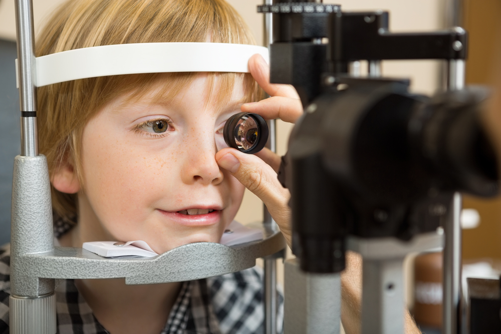
In the United States, special needs is a term used in clinical diagnostic and functional development to describe individuals who require assistance for disabilities that may be medical, mental, or psychological. Different types of special needs vary in severity. People with autism, Down syndrome, dyslexia, blindness, ADHD, or cerebral palsy, for example, may be considered to have special needs. Statistics tell us that among children ages 3 to 17, nearly 15 percent have one or more developmental disabilities. For many of these children, the kinds of disabilities they experience may require special approaches to providing care, education and/or other accommodations.
eye exam - vision and special needs children
The way a special needs child behaves or reacts can sometimes be unexpected because he/she processes sensory information differently than other children. Having the right cues in our environment can mean the difference between participation and non-participation in medical care for many of these children. Our work environment should always be arranged in a way to achieve physical and emotional comfort for the child. Patience, attentive listening, caring, and building a trusting relationship with a family and child who may be undervalued in other settings are all critical to providing good medical care.

Vision and Special Needs Children

Children with special needs are referred to pediatric ophthalmologists on a regular basis. Many of these children do not respond appropriately to standard vision screening procedures. Unidentified vision problems in this special population can further impact growth and development. The role of pediatric ophthalmologists is to ensure that the child does not have any eye conditions that could potentially interfere with his/her learning opportunities, personal development and/or overall wellbeing.

Down syndrome:
Down syndrome continues to be the most common chromosomal disorder. Each year, about 6,000 babies are born with Down syndrome, which is about 1 in every 700 babies born. It is estimated that more than 80% of these patients have some clinically significant ocular pathology. Such conditions include refractive error requiring glasses (70%), strabismus (45%), and nystagmus (35%). Other common eye problems include blepharitis, blocked tear duct, eyelid ptosis, cataracts, and keratoconus (irregularly shaped cornea). The improved quality of medical care and educational resources have allowed for a more productive life and a longer life expectancy for people with Down syndrome. Their quality of life can be further enhanced by the proper assessment and correction of eye problems.

Autism:
In 2014, the Centers for Disease Control and Prevention (CDC) released new data on the prevalence of autism in the United States. This surveillance study identified 1 in 68 children (1 in 42 boys and 1 in 189 girls) as having autism spectrum disorder (ASD). Autistic behaviors may include visual components such as lack of eye contact, starring at light or spinning objects, fleeting peripheral glances, side viewing and difficulty attending visually. An eye examination is essential in order to detect an eye condition that could potentially explain these visual behaviors. According to a study published in the June 2013 issue of Strabismus, more than 40 percent of children with autism have strabismus, or crossed eyes. This work is limited in that the researchers did not use a representative sample of children with autism. Still, the prevalence of eye problems in autism appears to be several times higher than that of the general population.
boy eye exam - vision and special needs children
Learning Disabilities:
Dyslexia is the most common neurobehavioral disorder affecting children. Visual abnormalities have not been found to affect the brain’s ability to process visual stimuli and children with learning disabilities have no increased incidence of ophthalmologic disease. However, ophthalmologic consultation should be provided to children who fail vision screening tests. This allows for diagnosis and therapy of treatable ocular conditions such as refractive errors and eye muscle imbalances.

Children with ADHD, cerebral palsy, or any other neurodevelopmental disorders should also have periodic vision screenings. Children who do not pass the vision screening should be referred to an ophthalmologist with experience in the care of children.

Physicians are not the only adults involved in the care of special needs children. At increasing rates, children with special needs are being provided with the same life experiences as their non-disabled peers. Taking part in a sport or joining a group like the Scouts, are popular activities for children. Children with disabilities are encouraged to join in such activities to help improve their health and give them opportunities to make friends. As a result, more and more adults in the community are finding themselves working with these children on a regular basis.

When considering ways to work with special needs children, we have to keep in mind that every child is different. A positive attitude and patience are probably the two most important qualities for anyone who works with these children. Parents of special needs children focus on helping their child to be “the best he/she can be”. The devotion and care these parents provide for their child have been an inspiration, and I always look forward to my visits with them.

4/2/15


Dr. Chantal BoisvertChantal Boisvert, OD, MD
Assistant Clinical Professor
Gavin Herbert Eye Institute, UC Irvine
Pediatric Ophthalmology & Strabismus
Neuro-Ophthalmology

7 Spring Fruits and Vegetables

Spring is truly here. With Passover and Easter later this week, it is time to rediscover some of the amazing produce that is at its peak during the months of April, May and June. These 7 spring fruits and vegetables are not only delicious; they are good for you . . . and your vision. Here are some recipes for you to try and enjoy the bounty of spring. Some even include more than one of the seven fruits and vegetables listed below.
apricots - 7 Spring Fruits & Vegetables
Apricots – Apricots should be firm, but not hard, with a nice fruit scent when sniffed. They are best purchased locally so they aren’t picked too early and have a tree-ripened sweetness.

Apricot & Orange Breakfast Smoothie from Discovery Eye Foundation’s Eye Cook

Spicy Apricot Wings from Food & Wine

Fresh Apricot Chutney from Cooking Light

Chicken Tagine with Apricots & Almonds from Gourmet
asparagus - 7 Spring Fruits & Vegetables
Asparagus – Look for firm stalks, from the tips down to the base of stalks. Once asparagus are harvested they deteriorate quickly, so place them in cool storage to retain freshness and their nutrition value.

Asparagus and Strawberry Salad from Discovery Eye Foundation’s Eye Cook

Asparagus with Watercress and Brown Butter Potatoes from Food & Wine

Grilled Asparagus with a Caper Vinaigrette from Cooking Light

Asparagus, Tomato & Red Pepper French Bread Pizza from the Mayo Clinic
cherries - 7 Spring Fruits & Vegetables
Sweet Cherries – The best cherries are an inch or more in diameter, plump, firm, and rich in color.

Cherry Pie from Cooking Light

Cherry Tortoni from Gourmet

Cherry Tart from Bon Appétit

Easy Almond & Dried Cherry Cookies from Discovery Eye Foundation’s Eye Cook
fava beans - 7 Spring Fruits & Vegetables
Fava Beans – Young fava beans can be shelled and eaten either raw or cooked, but more mature favas need to be shelled and skinned, since their skins are too tough to eat.

Quinoa Salad with Grilled Scallions, Favas & Dates from Food & Wine

Sliced Filet Mignon with Fava Beans, Radishes & Mustard Dressing from Bon Appétit

Arugula & Fava Bean Crostini from Gourmet
green peas - 7 Spring Fruits & Vegetables
Green Peas – Fresh green peas include sugar snap peas, snow peas, and green peas. Look for bright green pods that are firm.

Fava, Sweet Pea & Sugar Snap Salad from Cooking Light

Salmon with Sweet Chili Glaze, Sugar Snap Peas & Pea Tendrils from Bon Appétit

Strawberry, Almond & Pea Salad from Bon Appétit
strawberries - 7 Spring Fruits & Vegetables
Strawberries – For the best flavor, you are best looking for strawberries grown close to home since they more are likely to be fresh and not be damaged in transit. They should be plump, firm, well-shaped, and uniformly colored.

Carrot & St rawberry Tea Bread from Discovery Eye Foundation’s Eye Cook

Strawberries Romanoff from Cooking Light

Strawberry and Cream Cheese Crepes from the Mayo Clinic

Pink Grapefruit, Strawberry & Champagne Granita from Bon Appétit
watercress - 7 Spring Fruits & Vegetables
Watercress – Look for uniformly dark green leaves and sniff for a fresh, spicy scent. Watercress has a short shelf life and should be kept in a plastic bag in the refrigerator for no more than three days.

Watercress, Orange & Avocado Salad from Gourmet

Watercress Salad with Verjus Vinaigrette from Food & Wine

Watercress Salad with Pan-Seared Mahimahi from Cooking Light

These are some great ways to enjoy  what spring has to offer.  Do you have any spring recipes you want to share?

3/31/15


Susan DeRemerSusan DeRemer, CFRE
Vice President of Development
Discovery Eye Foundation

Three Generations of Saving Vision

In Discovery Eye Foundation’s spring e-newlsetter there was an article entitled Surgery for the Surgeon, where a leading ophthalmologist talks about convincing himself to have cataract surgery. That eye doctor was Dr. Nesburn, who was willing to share with us his family’s long tradition of saving vision.

Brainwashed by Medicine

“I was brainwashed from the age of 5,” says Dr. Anthony Nesburn, medical director of The Discovery Eye Foundation (DEF). “My dad would take me on rounds at the hospital and to his office. He introduced me to medicine at a really early age.”

Nesburn saving vision
Dr. Anthony Nesburn in UC Irvine lab
Dr. Henry Nesburn was an ophthalmologist in Los Angeles for more than four decades, and he passed his love of the specialty to his son. “I really looked up to my dad,” the younger Nesburn says. “He loved ophthalmology for the same reasons I do: You get to do medical diagnosis; you get to do wonderful surgery, where you keep people from going blind or restore vision; and you can work with people from newborns to the very elderly — they all need eye care.”

Nesburn received a telegram while he was an undergrad at UCLA, telling him he’d been accepted to Harvard Medical School. His mother started crying: “You’re going to Boston! We’re not going to see you anymore!” While he “was loathe to leave Southern California,” Harvard was too good to pass up.

Drafted by the Army out of his ophthalmology residency at Harvard in 1960, Nesburn joined the Navy instead. He followed in his father’s footsteps again, becoming a Navy flight surgeon. (Henry had volunteered during World War II.)

He went on to a Boston Children’s Hospital fellowship in infectious disease, working with Nobel laureate Dr. John Enders, whose work led to the polio vaccine and changed the face of virology. Nesburn then did his residency at Massachusetts Eye and Ear. “I was part of a special program that allowed us to do research, and I was running a research laboratory while I was a resident,” he says. “It gave me the start I needed.”

“At Mass Eye and Ear, I worked for an up-and-coming ophthalmologist and researcher to prove there was a substance that could treat herpes eye infections. We wrote a paper that included the very first antiviral ever described, and it was against herpes virus. It is the basis for today’s herpes antivirals,” Nesburn says. “I was hooked.”

He went back to Los Angeles and received NIH funding to continue his research on ocular herpes. In 1968, he joined his father’s practice half-time, spending the rest of his time doing research.

Two years later, he received a generous offer, when Rita and Morris Pynoos started DEF to fund his research. The Pynooses were grateful to Nesburn for diagnosing their son, Jon, with keratoconus (KC). “I was a second-year resident at Mass Eye and Ear, and Jon Pynoos was an undergraduate at Harvard. His parents went to see my dad, because Jon couldn’t see well, and no one could figure out what was wrong with his vision. My dad said, ‘Send him over to Tony; he’ll figure out what’s going on!’ I said to myself, ‘Holy mackerel! What happens if he has something really complicated? I’m just a newbie!’” Nesburn remembers. “Jon came in; I looked at him, and the keratoconus was so clear and easy to spot. I couldn’t imagine how his doctors didn’t see it. We got him contact lenses, and he was able to see again. When I came back to LA, the Pynooses wanted to do something to help.”

At first, DEF research focused on KC and the herpes research Nesburn was working on at the time. It soon broadened to include macular degeneration and retinal disease.

“My dad had to retire from the practice of ophthalmology at the age of 70, because of bad age-related macular degeneration (AMD). His mother and older sister had had it, as did several cousins. There was no treatment back then that helped,” Nesburn says.

“AMD is the most common cause of permanent vision loss in the elderly in the developed world. I could see where the need was,” he says. “We moved forward at DEF with two driving mantras: We wanted to do something significant in macular degeneration research and to find the cause of keratoconus.”

As a virologist in research and a corneal surgeon, Nesburn realized he needed a corneal biochemist to help with the KC research. He met Dr. Cristina Kenney at an Association for Research in Vision and Ophthalmology meeting. She joined DEF, and within 15 years, they found the chemical cause of keratoconus; they also got married.

Now nearly 80 years old, Nesburn spends most of his time “wearing three hats”: fundraising for DEF, lab research and clinical practice. His daughter, Kristin, is the third generation to join the family ophthalmology practice.

“While I’m still able, I want to try to make a difference in medicine, particularly in macular degeneration,” Nesburn says. “Macular degeneration affects so many people. This is where I want to put my energy. Luckily, as strong as it is in our family, I don’t have it … yet.

“As a researcher, my interest in putting together a program for macular-degeneration diagnosis and treatment has been because it’s a great public health problem. Yes, if I should ever get it, it might be able to help me or my family, but the first thing, as a scientist, is to try to get something to help humankind. I know it sounds sappy, but it’s true.”

3/26/15


Anthony B. Nesburn, MD, FACSAnthony B. Nesburn, MD, FACS
Medical Director, Discovery Eye Foundation
Professor & Vice Chairman for Research, Ophthalmology
Gavin Herbert Eye Institute, University of CA, Irvine

Protective Eyewear for Home, Garden & Sports

Spring is here and it is always a good time to review important ways to protect your eyes now that you will be spending more time outdoors, enjoying sports, gardening or just basking in the warm spring sunshine.

Protective Eyewear for Home, Garden & Sports

protective eyewear - sunglasses
Sunglasses and wide brimmed hats are the first things to consider as you go outdoors. The damage from UVA/UVB rays from sunlight is ever present, even on cloudy days. It is also cumulative and can lead to cataracts and age-related macular degeneration. Don’t forget to use sunscreen generously, helping to prevent a painful sunburn and skin cancer. If you perspire a great deal, think of a waterproof sunscreen that will not run into your eyes causing blurry vision and irritation.
protective eyewear - goggles
In your backyard or garden it is wise to use safety glasses or goggles when operating a chain saw, axe or hedge clipper. They will help to prevent small flying objects, dirt and debris from getting into your eyes. Tree sap and plant secretions can also be hazardous to your eyes. Wearing gloves should make you think twice about rubbing your eyes, or at least you can remove them if you can’t resist.

Home maintenance and spring cleaning offer some of the same threats as gardening. Beware of using any regular or power tools, paints and chemicals without protective eyewear because of flying debris, drips, splashes and sprays. Besides the general eye irritations and painful corneal scratches, you could permanently impact your vision. Also take care if your children are helping or playing nearby, they could also be at risk.
protective eyewear - sports
Spring is a great time to get outdoors and enjoy your favorite sports, but if you engage in any activities that involve throwing and catching balls, “flying” arms and elbows (such as karate), swinging bats, sticks or clubs, or anything that involves shooting (such as paintball or airsoft), you need protective goggles that wrap around and protect you from all angles. Not every threat will be coming from directly in front of you.

For these sports and recreational activities prescription eyeglasses, sunglasses and even occupational safety glasses are not enough to protect your eyes. You will need a highly impact-resistant polycarbonate to avoid a lens that can shatter and cause additional danger to your eyes. Consult your eye care professional to choose the right kind of eye protection for your warm weather activities.

3/24/15


Susan DeRemerSusan DeRemer, CFRE
Vice President of Development
Discovery Eye Foundation

Do You Have a Pterygium?

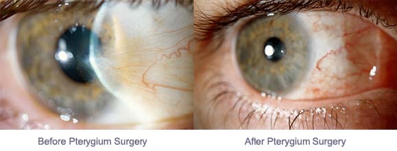
A pterygium (pronounced tur-IJ-ee-um) is a wing-shaped, fleshy growth of tissue that grows from the conjunctiva (the clear thin covering of the sclera or white part of the eye) onto the cornea (the clear front of the eye). The word pterygium comes from the Greek, pterygos, which means little wing. Pterygia are quite common and can be seen in surfers (hence the common reference as surfer’s eye), sunbathers, landscape architects and other outdoor people who do not adequately protect their eyes from the damaging ultraviolet radiation from the sun. Furthermore, people living closer to the equator have been shown to have a higher incidence of pterygia. Prolonged exposure to eye irritants such as wind and dust are also risk factors for the development of pterygia.
pterygium before & after
Pterygia usually grow slowly or follow an indolent course and can be asymptomatic or can flare up from irritation. Early on, pterygia may not be even noticed and may not be serious. Or, there can be signs of dry eyes, such as mild burning, tearing and itching due to irregular tear wetting of the surface of the eye. As the pterygium grows in size, the lesion will become more noticeable and may become cosmetically unpleasant, irritating to the patient and may give the sensation of a foreign body in the eye. Pterygia may cause visual symptoms if they induce astigmatism as they approach the center of the cornea or visual axis. Once the pterygium grows towards the visual axis, where vision could be compromised, various surgical procedures can be employed to remove it.

Having an ophthalmologist (an eye MD) examine your eyes is the first step to making the correct diagnosis and receiving the appropriate treatment, if it is necessary. Your ophthalmologist will examine the front of your eyes with a slit lamp, a specialized microscope for eye examination. Early in the disease process, most ophthalmologists will take a conservative approach and treat with lubricating eye drops and recommend protective eye wear (to prevent further UV exposure) and use of a wide-brimmed hat. If the pterygium enlarges and grows onto the cornea towards the visual axis, the ophthalmologist will consider surgical intervention to halt any more damage and scarring to the eye. Sometimes, a more aggressive pterygium may induce astigmatism on the cornea (causing the cornea to become more oval or football-like in shape as compared to more round in shape like a basketball). Astigmatism may cause blurred vision and, if left untreated, may not be reversed even with surgical intervention. Long-lasting pterygia may cause irreversible scarring of the underlying cornea, which may leave the cornea with a thin white film, or scar, even after the removal of the pterygium. If this scar is out of the visual axis, then vision may not be affected.

There are several different surgical techniques that are commonly used to remove aggressive pterygia. Microsurgical techniques can be done safely and comfortably on an outpatient basis in an eye surgery center using local anesthesia with mild sedation. The most common surgical technique involves surgical removal of the pterygium, placement of a preserved amniotic membrane graft (from the placenta) or a small graft of the patient’s own conjunctiva taken from the superior (upper) part of the conjunctiva to cover the area where the pterygium was removed, and using fibrin glue and/or small sutures to hold the graft in position. This procedure usually takes 30 minutes to one hour to perform, depending its complexity, with little discomfort for the patient. Post-operatively, the patient usually wears a patch for one day and can return to work after a few days (avoiding eye rubbing, swimming and hot tubs). Topical antibiotic and steroid drops and/or ointments are used for several weeks or months on a tapering dosage schedule per your ophthalmologist. Steroid medications reduce inflammation and chance of recurrence and should be used as directed. Careful follow-up for a year after surgery is recommended.

The main risk of pterygium surgery is recurrence of the pterygium after removal with a regrowth of abnormal tissue onto the cornea. When an amniotic membrane graft or conjunctival auto-graft is not used, the recurrence rate is quite high and has been reported from 25% to over 50%. However, when these grafts are used, the recurrence rate can be as low as 5 to 10%. For recurrent pterygium removal, however, the recurrence rate can be much higher and other adjunctive therapy may be utilized. Intraoperative mitomycin C (MMC) is often used in addition for recurrent pterygium excision to inhibit the regrowth of the pterygium. Some ophthalmologists use MMC eye drops after excision. Discuss these options with your ophthalmologist before your surgery. Be sure to see your ophthalmologist if you have a pterygium so that it can be properly assessed and appropriate treatment can be performed.

3/19/15

Ronald Gaster, MD, FACSRonald N. Gaster, MD, FACS
Gaster Eye Center
Beverly Hills and Huntington Beach, CA