Living With the Argus II

5/27/14

Last year Discovery Eye Foundation spoke to Dean Lloyd, who lost his sight to retinitis pigmentosa (RP), about his experiences with the Argus II “bionic eye.”  After FDA approval, when the article came out, more people across the country have been fitted with the Argus II, using its 60-electrode system to help them regain some part of their vision.  Researchers continue to try and improve upon the “bionic eye” such as this research for a 24-electrode version from Australia.  Here is that article:

Dean Lloyd wearing the ArRgus II
Dean Lloyd wearing the Argus II

As one of only 30 people in the world with a “bionic retina,” Dean Lloyd has gained a bit of notoriety of late. The Argus II Retinal Prosthesis System received US market approval from the FDA on Feb. 14, and Lloyd has been part of the clinical trial since he was implanted with the device in 2007.


Lloyd’s vision difficulties began in the early 1960s, while he was in medical school at the University of South Dakota. He realized he wasn’t seeing the same thing as his classmates when looking through a high-powered microscope. He was misdiagnosed with Usher Syndrome, a rare genetic disorder that can result in deafness, blindness and dementia. While he was later correctly diagnosed with the less-dire x-linked retinitis pigmentosa (RP), he was asked to leave medical school due to his vision impairment.

“I had a moment of self-pity, then I needed to do some introspection to figure out how to find my future,” he says. “I decided I can’t give up on life. My brain works well … If your brain works, it can solve a lot of difficult problems. I realized I would survive.”

Seeing Stars

Armed with a BS in chemistry and an MS in bio-chemistry, Lloyd became a research chemist, then a software engineer. At the time, RP was having only one real effect on his life: reduced night vision. “I always thought people were seeing more stars than I saw. They’d see the big dipper and little dipper, and I didn’t see any dippers,” he recalls. Other than being extra-careful when driving at night, Lloyd’s daily life remained relatively unaffected until 1974, when he developed cataracts at age 34.

Lloyd’s wife decided she did not want to deal with any impact the disability would have on his ability to support their family. “She left and got the best divorce lawyers,” he says. “The court thought, because I was visually impaired, I wouldn’t be able to take care of my children. There were a lot of issues around disability and parent-ability in the mid- to late 70s.” Because of his vision loss, Lloyd lost custody of his two children. Believing disabled people were not treated fairly and seeing “the power of the court over people in their everyday lives,” Lloyd decided to go to law school. He passed the bar in 1982, “as a disabled person in a closed room,” and has been practicing law for more than 30 years.

He underwent two cataract surgeries, and a pair of convex lenses he wore on his nose was used in place of the actual lenses that had been removed from his eye. He had no difficulty with daytime mobility for nearly 14 years. But in 1989, he lost image formation. “I got an edema within the macula area, which didn’t go away,” he says. “I lost the ability to form images — that’s the point when you lose the opportunity for safe mobility.”

Lloyd had already been practicing law for seven years, and RP didn’t stop him. “I had quite a bit of experience in the courtroom,” he says. “I am fortunate to have a good memory, and I memorized everything I used there. I usually work with a junior or associate attorney; I do all the talking, and they handle all the documents and things. It didn’t change my career.”

A cane helped with his mobility, and for a few years, he says, “A doctor at Stanford pulled me in for some non-FDA experiments, because he knew I was a four-eyed guinea pig and was willing to try anything that might help RP.” None worked.

Lloyd was involved with several vision-related organizations and followed the progression of research and experiments to see if any might benefit him. In early 2007, he attended a Foundation Fighting Blindness meeting run by his daughter, who has “the recessive expression for RP.” The speaker that day was Dr. Jacque Duncan of UCSF, who interviewed all the attendees. Soon after, she contacted Lloyd to ask him to participate in the Argus II clinical trial.

He agreed immediately: “I figured if I lost the eye, I wouldn’t lose much, because it didn’t work anyway.”
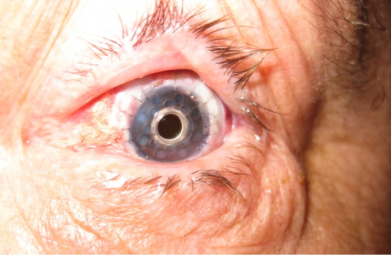
The Bionic Retina

The Argus II Retinal Prosthesis System consists of three parts: an implant, glasses and a belt-worn video-processing unit (VPU).

The implant is surgically implanted in and on the eye. It includes an antenna, an electronics case and an electrode array. The glasses contain a tiny video camera that captures a scene. The video is sent to the VPU, where it is processed and turned into electrical stimulations — instructions — that are sent back to the glasses through a cable. The glasses then transmit the instructions wirelessly to the implant antenna, which sends signals to the electrode array, which emits electric pulses. These pulses bypass damaged photoreceptors (RP damages photoreceptors and impedes image formation) and stimulate the retina’s remaining healthy cells, which transmit the information along the optic nerve to the brain. If all goes well, it creates the perception of patterns of light, which wearers can learn to interpret as visual patterns.

Chicken Dance

Duncan, working with another UCSF doctor, had Dr. Eugene de Juan put the Argus II implant in Lloyd’s right eye on July 17, 2007. A few weeks after an initial infection subsided, he did his first test walk with the device outside, looking for boundaries and points of light.

“Human eyes naturally move around, using a rapid ‘saccade’ movement,” Lloyd explains. “With the device, you have to physically move your head back and forth to find boundaries — like a chicken, which does not have moving eyes. They move their heads back and forth to see things. During my walk, the field-service worker would say, ‘Dean, Dean, more chicken movement.’”

After about a year and a half spending a half-day a week at UCSF for the trial, he had a second operation. “The device didn’t satisfy me,” he says. “I went through all that, and all I could see was a little point of light. Only nine of 60 electrodes were working. I said, ‘This isn’t going to cut it for me, because I want to see images.’ They reset the implant closer to the retinal tissue, and that dramatically improved the device.”

Lloyd conducted his own tests of the device at home. “I was one of first to use it to sort socks. I wear black socks to court and white socks to the gym. I went home after the implant and wanted to see how I could make it practically useful. So I went to my socks and found the device worked. I always got the white ones right!”

While he is generally pleased with the Argus II, the most he will say is, “It shows potential.”

“The improvement was not very good at first. I wasn’t very impressed. There was a rumor going around that the implant restored vision. My vision wasn’t restored at all; I was not ascertaining images,” he says. “I do see boundaries. I can create an image. I know what a car should look like. I know what a tree should look like. I know what houses should look like. I know what objects should look like, and I have those images as memories in my brain. With this device, you can start to create an image by going back and forth, checking the boundaries and borders. We don’t have natural saccade movement helping us. The camera is right above my nose in a fixed position. It doesn’t move. You can create an image, but it takes a lot of time and a lot of work. It’s a labor-intensive task, and you have to have a good memory.”

The Argus II was approved by the FDA in February, and Lloyd is actively involved with Second Sight, the company that makes the device: “I am helping with new improvements,” he says. “I want this device to work. I didn’t go through all this for nothing.” He hopes an improved version will be approved this coming September, and he doesn’t rule out getting a new implant in the future if the improvements are worth it. To be honest, he says, “I find it annoying to move like a chicken.”

Susan DeRemerSusan DeRemer, CFRE
Vice President of Development
Discovery Eye Foundation

New Hope for Corneal Scarring

5/22/14

There are several etiologies for limbal stem cell deficiency of the front of the eye. These include chemical and thermal burns, Steven-Johnson syndrome (which is an autoimmune severe allergic reaction that causes a burn from within), congenital aniridia, and a few other insults such as contact lens over-wear. All of these cause severe ocular surface scarring and problems with the cornea. Many eyes with these diseases have problems with corneal healing. They do not have the stem cells to support ocular surface health. The scarring can be so severe in many cases that severe corneal blindness can result.

Limbal stem cells from the human cornea, with a protein known as p63 stained yellow. Cell nuclei (which hold the DNA) are stained red.  From eurostemcell.org
Limbal stem cells from the human cornea, with a protein known as p63 stained yellow. Cell nuclei (which hold the DNA) are stained red. From eurostemcell.org

In these cases, a simple corneal transplant will quickly fail and not result in any visual improvement. The reason for this is that the stem cells of the ocular surface have been damaged or burned out.

Visual rehabilitation for these eyes usually requires a limbal-corneal stem cell transplantation. The stem cells can be taken from the other healthy eye of the same patient, a living related donor, and or cadaveric tissue. In most cases systemic immunosuppression medications need to be taken for 1 to 3 years following surgery in order to minimize risk of rejection. Management of these patients is done in conjunction with an immunologist or a transplant specialist who can co-manage and monitor for systemic toxicity while the patient is on the these immunosuppressive medications. As most of these eyes also have concomitant glaucoma and scarring of the eyelids to the globe, co-management with a glaucoma specialist and an oculoplastic specialist is also required.

For patients who cannot be on systemic immunosuppression for other health reasons such as diabetes or cancer, they may require an artificial corneal transplantation. The artificial corneal transplantation is reserved as a last step for visual rehabilitation in these eyes. The only artificial cornea that has shown potential, is the Boston keratoprosthesis. Even this artificial cornea carries a high risk for infection and glaucoma. Very close monitoring of eyes that have an artificial cornea is required to monitor for infection and glaucoma progression. However these eyes do not require systemic immunosuppression.

Eye with Boston keratoprosthesis


The management of eyes with severe ocular surface disease is a difficult one for the cornea specialist. A subspecialist in severe ocular surface disease and limbal stem cell transplantation is required to manage these very sick eyes. At the Gavin Herbert Eye Institute, we have developed a team approach for the management of severe ocular surface disease patients and have successfully treated and are managing many patients who have otherwise no place to go.

Farid 3.6.14Marjan Farid, MD
Director of Cornea, Cataract, and Refractive Surgery
Vice-Chair of Ophthalmic Faculty
Director of the Cornea Fellowship Program
Associate Professor of Ophthalmology
Gavin Herbert Eye Institute, University of California, Irvine

6 Summertime Tips for Children’s Vision

5/20/14

Summer vacation is around the corner and for children this means more time spent outside playing, swimming, or going to the beach. All of this outside activity increases their exposure to ultraviolet rays which is of particular concern because the lens of a child allows 70% more UV rays to reach the retina than in an adult. This may put them at increased risk of developing debilitating eye diseases such as cataracts or macular degeneration as adults.
Children with sunglasses
If you are wearing sunglasses to combat the bright sunlight, then your child should be wearing them, from babies on up. Wrap-around sunglasses provide more sun and eye protection. Wearing protective goggles during sports activities is also important as the National Eye Institute reports there are more than 100,000 sports-related eye injuries every year with 42,000 requiring emergency care.

While it may be hard to get them to leave them on, or if they keep falling off, invest in a strap that can range from $4.50-$10.00. They can be made of neoprene with fun designs like Croakies or they can use an adjustable cord like Chums. In any case it also helps cut down on lost sunglasses.

Pediatricians offer the following five suggestions for children to enjoy a fun and safe summer:

1. Wear sunglasses – especially younger children

During our lives, almost half of the time we spend outdoors is before the age of 12. Sunglasses for children don’t have to be expensive, but make sure they are rated to block both UVA and UVB radiation. Glasses should also have a polycarbonate lens to withstand shattering.

2. Wear protective eye gear for ball or shooting sports

Every year there are 18,000 sports-related eye injuries in US hospital emergency rooms. The American Academy of Ophthalmology recommends that children wear polycarbonate goggles for baseball, basketball and racket sports, including tennis. It becomes even more important with shooting games like air-soft where the projectiles are so small, but can do major damage to the eye. Regular glasses are not recommended if they cannot be secured to the head or are not made from polycarbonate. Also make sure the goggles have proper sun protection for outside sports.

3. Don’t rub if sand gets in the eyes

If a child gets sand into his eyes, take the child immediately to a sink with running water. Do not allow them to rub their eyes as this can scratch the outer layer of the eye known as the cornea. Use a clean cup to pour water over the eyes to remove sand. Encourage blinking and do not discourage crying, because tears remove eye irritants. If flushing and blinking does not work, seek immediate medical attention.

4. Use a non-irritating sunscreen

While you can use adult sunscreens for children, make sure it is PABA free, since that chemical can cause irritation in some people. If your child gets a rash from his sunscreen, review the ingredient’s list and choose a different one. UVA protection from titanium dioxide or zinc oxide tends to be less irritating than avobenzone, another common ingredient.

5. Wear a wide-brimmed hat

Don’t just rely on sunscreen.  Have your child wear a hat with a wide brim.  It not only provides additional protection against sunburn on susceptible areas like the nose, neck and ears, but it also helps to protect their eyes from harmful UV rays.  Not all sunlight enters the eye direct from the front.  Wrap-arounds may help protect light from coming in the sides, but they do not stop sunlight from coming in the top or reflective glare from coming up from the bottom.

6. Check chlorine levels in your pool

Too little chlorine in a swimming pool can allow algae and other bacteria to grow, which can lead to eye infections. On the other end of the spectrum, be sure to check the levels of chloramines and the pH of the pool to avoid stinging and redness. Swim goggles are helpful to keep pool water from entering the eye. If redness and irritation persist after swimming, it could be a sign of a more serious infection and medical attention is needed.

Susan DeRemerSusan DeRemer, CFRE
Vice President of Development
Discovery Eye Foundation

May is Healthy Vision Month

When it comes to our health, we may visit our doctors and nurses regularly to make sure our bodies are healthy. But what about our eyes? They’re not always top of mind, but they’re just as important! HVM Logo-02 May is Healthy Vision Month, an observance coordinated by the National Eye Institute to empower Americans to make their eye health a priority and educate them about steps they can take to protect their vision. What do those steps include?

Get a dilated eye exam. When it comes to common vision problems, many people don’t realize their vision could be improved with glasses or contact lenses. In addition, many common eye diseases have no symptoms. A dilated eye exam is the only way to detect these diseases in their early stages.

Live a healthy lifestyle. Almost everything we do affects our eyes. To keep our vision healthy, it is important to:

    • Eat healthy foods, especially dark leafy greens such as spinach and kale and fish high in omega-3 fatty acids, including salmon, halibut, and tuna.
    • Maintain a healthy weight. Being overweight or obese can increase your risk of developing diabetes and other systemic conditions, which can lead to vision loss.
    • Don’t smoke. It’s as bad for your eyes as it is for the rest of your body.
    • Manage chronic conditions. Many conditions, including diabetes, hypertension, and multiple sclerosis, can greatly impact vision and result in inflammation of the optic nerve, diabetic retinopathy, glaucoma, and even blindness.

Know your family history. Eye health can be hereditary, so it’s important to talk to your family members about their eye health history. Knowing your family history will help you determine whether you are at higher risk for developing an eye disease.

Use protective eye wear. Eye injuries can happen at work, while playing sports, and when doing chores around the house. Prevent those injuries from happening by wearing protective eyewear! Look for safety glasses, goggles, safety shields, or eye guards made of polycarbonate, which is 10 times stronger than other plastics.

Wear sunglasses. The sun’s rays can damage your eyes, so make sure to wear your shades! When purchasing sunglasses, look for ones that block out 99 to 100 percent of both UVA and UVB radiation.

Celebrate Healthy Vision Month by taking these steps today! Then encourage your family and friends to make their vision a priority by helping NEI spread the word. Here are some ideas:

We hope you’ll join in the celebration! And don’t forget to protect your eyes today to see well for a lifetime.

Susan DeRemerSusan DeRemer, CFRE
Vice President of Development
Discovery Eye Foundation

Beyond Eating Green

Beyond Eating Green

Spinach and kale aren’t the only things to watch in your diet. Eating low glycemic foods may slow the development and progression of AMD. The glycemic index measures how quickly carbohydrates get glucose (sugar) into the blood stream. People with diabetes will find this familiar. High glycemic foods like potatoes, white rice, processed foods like cakes and crackers and cereal raise the blood glucose level. Low glycemic foods include vegetables and beans.

from www.crossfithoboken.com
Several years ago researchers at Tufts University found that mice fed a low glycemic diet developed fewer and less severe age-related lesions in the retina than mice fed the higher GI diet. When lesions like this develop after age 60 in humans, they are the earliest warning sign of age-related macular degeneration. Earlier studies in humans resulted in similar observations.

*Here are some tips from Harvard University for low-glycemic eating.

1. Eat a lot of non-starchy vegetables, beans, and fruits such as apples, pears, peaches, and berries. Even tropical fruits like bananas, mangoes, and papayas tend to have a lower glycemic index than typical desserts.

2. Eat grains in the least-processed state possible: “unbroken,” such as whole-kernel bread, brown rice, and whole barley, millet, and wheat berries; or traditionally processed, such as stone-ground bread, steel-cut oats, and natural granola or muesli breakfast cereals.

3. Limit white potatoes and refined-grain products, such as white breads and white pasta, to small side dishes.

4. Limit concentrated sweets – including high-calorie foods with a low glycemic index, such as ice cream – to occasional treats. Reduce fruit juice to no more than one-half cup a day. Completely eliminate sugar-sweetened drinks.

5. Eat a healthful type of protein, such as beans, fish, or skinless chicken, at most meals.

6. Choose foods with healthful fats, such as olive oil, nuts (almonds, walnuts, pecans), and avocados, but stick to moderate amounts. Limit saturated fats from dairy and other animal products. Completely eliminate partially hydrogenated fats (trans fats), which are in fast food and many packaged foods.

7. Have three meals and one or two snacks each day, and don’t skip breakfast.

8. Eat slowly and stop when full.

*Adapted from Ending the Food Fight, by David Ludwig with Suzanne Rostler (Houghton Mifflin, 2008)

Judi Delgado headshotJudith Delgado
Executive Director
Macular Degeneration Partnership

When You See Things That Aren’t There

5/8/14

Charles Bonnet Syndrome


“Do you ever see anything you know is not there but looks real anyway?” I asked Sam Weinberg when he came to the Low Vision Living program.

“No.” he said, looking at his wife, Rachel, and fidgeting with his sweater.

“Oh”, I said casually, “I just asked because many people with macular degeneration see things they know are not there. I call it phantom vision, but the technical term is Charles Bonnet Syndrome.”

“Is this syndrome an early sign of Alzheimer’s?” Sam asked pointedly, still looking at Rachel. . .

“Absolutely not”, I said firmly. “Charles Bonnet Syndrome has nothing to do with mental agility or stability. When you have phantom vision, your mind is fine; it is your eyes that are playing tricks on you. It’s a side effect of low vision.”

“Well,” Sam admitted quickly, “I see little monkeys with red hats and blue coats playing in the front yard. I’ve seem them for eighteen months.”

“What!” Rachel’s eyes about popped out of her head. “Little monkeys in the front yard?”

“Well. . .um,” Sam continued, “sometimes I see them in the living room too.”

What is Charles Bonnet Syndrome (CBS)?
Charles Bonnet was an eighteenth century Swiss naturalist and philosopher. . . who described his grandfather’s curious experience of seeing men, women, birds and buildings that he knew were not there. Later in his life, Bonnet’s own vision deteriorated and he experienced phantom visions similar to his grandfather’s. . . .Charles Bonnet’s discovery didn’t capture medical attention at the time. But 150 years later, in the 1930’s, his files were dusted off, and he was credited with being the first person to describe the syndrome that came to be named for him.

Image seen by someone with CBS

How common is CBS?
This syndrome is very common. Studies place the number somewhere between 10 and 40 percent of people with low vision. Twenty percent of my low vision patients have Charles Bonnet Syndrome. . . To determine whether or not you are experiencing phantom vision: Do the images that appear to you have the following six characteristics?

  1. They occur when you are fully conscious and wide awake, often during broad daylight
  2. They do not deceive you; you are aware that they are not real.
  3. They occur in combination with normal perception. For example, you may see a sidewalk clearly but find it covered with dots, flowers, or faces.
  4. They are exclusively visual and do not appear in combination with any sounds or bizarre sensations.
  5. They appear and disappear without obvious cause.
  6. They are amusing or annoying but not grotesque.
An image described by a person with CBS

What do people with CBS see?
My patients. . . have reported seeing cartoon characters, flowers in the bathroom sink, hands rubbing each other, waterfalls and mountains, tigers, maple trees in vibrant autumn foliage, yellow polka dots, row houses, a dinner party and brightly colored balloons. . . One of the most remarkable qualities of these figures is that they almost always wear pleasant expressions. . . Menacing behavior, grotesque shapes and scenes of violent conflict are not, to my knowledge, a part of this syndrome.

Usually the same image or set of images reappears to each person. Sam’s monkeys usually materialized around sunset. . .They stayed for 10 or 20 minutes several times a week for two years and then began to appear less frequently. Some times the images change of multiple images appear. . .

Little girls dancing in the yard

Dolly Kowalski’s Little Girls with Pink Bows
‘I see little girls with pink bows playing in my yard. At first, there was only one little girl. But after a while, she had several playmates. Now they come almost every evening for fifteen minutes. . .They are so delightful, so cheerful, so active. Their little white dresses and pink bows blow in the wind. I see them so incredibly clearly, much more clearly than I see anything else now. . . .I know they aren’t real, but you wouldn’t believe how realistic they seem. . . . I wish you could see them the way I do.’”

Further note by Lylas Mogk, MD
Fortunately, most people, like Dolly, find the images of CBS largely untroubling and many actually find them amusing or enjoyable, as they are usually pleasant and they are crystal clear. There is no drug treatment for CBS, but it is associated with sensory deprivation, so the more active and engaged one is the less likely it is to occur. That’s one reason why vision rehabilitation to empower individuals to accomplish their daily activities in spite of vision loss.

Excerpts were used from Macular Degeneration: The Complete Guide to Saving and Maximizing Your Sight, by Lylas G. Mogk, MD and Marja Mogk, PhD, New York: Ballantine Books, 2003, Chapter 8, pp. 236-252.

Mogk_Lylas_11C[1]Lylas G. Mogk, MD
Director, Center for Vision Rehabilitation and Research
Henry Ford Health System

The Costs of Eye Care

5/6/14

**Update 6/16/14 – New estimate on the cost of vision problems just came out in the new Prevent Blindness America report showing they could reach $717 billion by 2050.

This past week I attended a dinner where a topic of discussion was the cost of chronic disease on healthcare – 75% of healthcare dollars go to the treatment of chronic diseases. This led me to explore the costs chronic eye diseases such as age-related macular degeneration, cataract, glaucoma and diabetic retinopathy.
PBA 2012 booklet
In 2007 Prevent Blindness America (PBA) published a report, The Economic Impact of Vision Problems that was updated in 2012, which explored the economic burden related to people 40 and older and the conditions of, age-related macular degeneration (AMD), cataract, diabetic retinopathy, primary open-angle glaucoma and refractive error, as well as the economic impact of vision problems and blindness.

They looked at direct medical costs including inpatient, outpatient and prescription costs; direct non-medical costs, which include nursing home care, guide dogs, and government programs for the blind and visually impaired; and loss of productivity, which includes lower labor force participation and lower wages for those with vision problems as opposed to the same age group with normal vision.

Direct Medical Costs $16.2 billion
Direct Non-medical Costs $11.2 billion
Productivity Losses $8.0 billion
TOTAL ECONOMIC IMPACT $35.4 billion

Looking more closely at the direct medical cost with regards to age-related eye diseases, the costs per patient broke down as follows:

AMD Cataract Diabetic Retinopathy Glaucoma
Medical Costs $797 $12,625 $6,012 $3,552

A year later, the NORC at the University of Chicago presented their findings, but also included the age groups of children 0-17 and adults 18-39 in a publication titled, Cost of Vision Problems: The Economic Burden of Vision Loss and Eye Disorders in the United States. It was interesting to see how the estimates varied in the two approaches when I only focused on the group of age 40 and older, as they did in the PBA study.
Noca booklet
The reclassification of the costs into two line items, from the previous three of the PBA report, presented a challenge and is one reason for discrepancies.  They went into more depth and were broken down as follows: direct costs included medical care, medical vision aids, low vision aids, special education, school screenings and Federal assistance programs; and indirect costs included loss of productivity for adults and caregivers, long-term care and transfer payments.

Direct Costs $52.58 billion
Indirect Costs $58.49 billion
TOTAL ECONOMIC IMPACT $111.07 billion

And when I looked more closely at the medical cost with regards to age-related eye diseases, the costs per patient broke down as follows:

AMD Cataract Diabetic Retinopathy Glaucoma
Medical Costs $4,457 $10,570 $4,005 $5,617

Regardless of the data sources and reporting classifications, one thing is clear – the direct medical costs of eye disease is high and growing each year.

Both reports contained a wide variety of information that is very interesting.  Links for both studies have been included so you can review the numbers and draw your own conclusions.

Susan DeRemerSusan DeRemer, CFRE
Vice President of Development
Discovery Eye Foundation

New Technology for Evaluating Contact Lenses

5/1/14

Successful management with contact lenses can sometimes be a frustrating process for those with keratoconus. The fitting and evaluation process involves numerous visits to the optometrist, out of pocket expenses as well as medical insurance co-pays. Luckily, the contact lens industry has responded to the need to have better contact lens materials for patients with keratoconus. It used to be that the only contact lenses available to manage keratoconus were gas-permeable (GP) lenses. Now, more and more patients are being fit with newer generation hybrid lenses (GP lens core with a skirt of soft lens material to aid in fit and comfort) and scleral lenses (large diameter GP lenses that do not rest on the cornea, only the outlying sclera). These newer designs are intended to vault over the central cornea and do not rest on the cornea at all. They have resulted in much more comfortable and wearable strategies for full-time use. As a result of the newer lens designs, the game has changed when it comes to the science of fitting and evaluating the lenses.

For years, optometrists have used corneal topography to guide their decision making on fitting keratoconic eyes. A corneal topographer is an instrument that maps the shape of the cornea, and gives information much like that of a topographical map for hiking. Corneal topography is still an absolutely mandatory part of evaluating the shape of the cone, the simulated corneal curvature, and monitoring for progression of the disease, and is not a standard part of a typical eye examination. However, doctors have a new tool at their disposal for fitting contact lenses on patients with keratoconus.

Optical Coherence Tomography (OCT) was once reserved for use in the back of the eye, or retina. OCT uses visible light passed through the clear structures of the eye to generate a cross-sectional image of the layers of the retina, much like an image generated by an MRI. Advances in OCT technology has improved the resolution to image the eye on the micrometer scale (one-thousandth of a millimeter). OCT technology is now commercially available not only for the retina, but the structures of the front part of the eye. The obvious application is to aid the doctor in the fitting and evaluating complex contact lenses that vault the cornea.

Figure 1.  OCT image of a scleral lens fit on a keratoconic patient.  The cornea is the opaque white band located at the bottom of this picture, the tear film reservoir is the middle clear band and the contact lens is the top band.  Using an electronic caliper tool, the precise amount of vault can be measured, leaving no doubt as to the precision of the fit.
Figure 1. OCT image of a scleral lens fit on a keratoconic patient. The cornea is the opaque white band located at the bottom of this picture, the tear film reservoir is the middle clear band and the contact lens is the top band. Using an electronic caliper tool, the precise amount of vault can be measured, leaving no doubt as to the precision of the fit.

OCT allows the optometrist to view a cross-sectional image of the contact lens on the eye in real time and to monitor the health of the cornea in the presence of the contact lens. This view is valuable for judging the vault of new designs of contact lenses over the cornea and judging where the lenses land on the eye. It is the most specific way to determine if the fit is acceptable and to troubleshoot if lenses are not fitting appropriately.

Figure 2.  OCT image of the periphery of a scleral lens on a patient with pellucid marginal degeneration.  The lens contacts the cornea over an area of 0.87mm long.  These types of measurements help guide decision making in modifying the lens fit and were impossible before the advent of this technology.
Figure 2. OCT image of the periphery of a scleral lens on a patient with pellucid marginal degeneration. The lens contacts the cornea over an area of 0.87mm long. These types of measurements help guide decision making in modifying the lens fit and were impossible before the advent of this technology.
Figure 3.  Hybrid lens on a highly irregular eye after corneal transplant.  The point of contact of the soft skirt with the cornea is visible to the right of the image.
Figure 3. Hybrid lens on a highly irregular eye after corneal transplant. The point of contact of the soft skirt with the cornea is visible to the right of the image.

Optometrists now have a much more powerful tool for evaluating and managing even the most challenging contact lens fits. It remains to be seen whether this technology has the ability to reduce the number of visits required for successful fit. But, the precision afforded by this technology does have the ability to improve patient outcomes.

Sonsino HeadshotJeffrey Sonsino, OD, FAAO
The Contact Lens Center at Optique Diplomate
Cornea, Contact Lens, and Refractive Therapies

Four Tips For Buying Sunglasses

4/29/14

May will be here this week, and in Southern California we are looking at bright, sun-filled days with temperatures in the upper 80s and low 90s. This means that thousands will be heading to the beaches or their own backyards to enjoy the warm weather.

Now is the perfect time to review one of the biggest contributing factors to vision loss – sun exposure. And it’s not just about sunglasses, but also brimmed hats.

from esty.com
from esty.com

First let’s talk about sunglasses. There are three things to think about when selecting your sunglasses:
1. Lens tint
2. UV protection
3. Glare
4. Frames

Lens Tint
There is a misconception that the darker your sunglass lens, the better protection for your eyes. No true. The color or darkness of your lens is personal preference and often based on the activity you are doing while wearing sunglasses or the sun conditions. At the beach in bright sunlight you are subject to more reflective light and may prefer dark amber, copper or brown lens, if you are on the ski slopes when the skies are overcast you may prefer yellow or orange lens to increase contrast and fight “flat light.” If you are looking to increase contrast on a partially cloudy day, and if you don’t mind distorted color perception, you might prefer amber or rose lenses.

Other considerations include mirrored sun lenses that can block 10-15% more of the sun’s visible rays, or photochromic lenses that darken automatically when you go outside and then quickly become lighter when you come inside.

UV Protection
While darker lenses don’t offer better eye protection, controlling the UV exposure does. Research has found links that extended exposure to UVA and UVB rays can result in eye damage such as cataracts, photokeratitis and macular degeneration. By wearing sunglasses that block these harmful rays your eyes should remain healthier as you age. Also know that some parts of the country receive more UV rays than others – here is a wonderful chart from The Vision Council to let you see how your location rates.

Glare
Another problem when out in the sun, and especially driving, is glare. Making sure your lenses are polarized is a great help. They work by only letting in specific amounts of light at certain angles and reducing the brightness of that light.

Because I am light sensitive I find I use polarized lenses when I am reading outside is helpful. The reflected light from the page of a book can cause me to squint or fatigue my eyes if I read for a long period of time. The only other option is using a paper-ink e-reader which also helps cut down on glare.

Another way to deal with glare is the use of an anti-reflective (AR) coating on your lenses. It reduces eye stain by preventing light from reflecting off lens surfaces. When applied to the back of your lenses it can help with problems when the sun is behind you or to your side.

Frames
Not all light hits your eyes from directly in front. It can come through the top, sides and bottom of your frames. The smaller the frames, the more unfiltered light makes its way to your eyes. This is where a brimmed hat can help keep the sun coming in from the top while also providing protection for your face.

Fitovers - Auroa in Claret
Fitovers – Auroa in Claret

To provide you with the maximum protection, “fit-over” sunglasses, that you can wear over your regular prescription glasses, are a great idea and more economical. Cocoons Eyewear and Fitovers Eyewear are two of several companies that make them. They filter the light from the top, sides and even below to give you the maximum protection and come in a wide variety of lens colors. It is also nice not to have to get new sunglasses when your eyeglass prescription changes.

Whatever frames you choose make sure they fit properly and will not keep sliding down your nose or fall of when being active. You may even want to purchase a band-style foamed neoprene retainer that attaches at both temples, sometimes known as a gator.

Also remember, it is not just the direct sunlight you need to worry about. Water reflects up to 100% of the harmful UV rays, dry sand and concrete up to 25% and even grass reflects up to 3%.

Susan DeRemerSusan DeRemer, CFRE
Vice President of Development
Discovery Eye Foundation

Dry Eye and Tear Dysfunction

4/24/14

With an aging population and increasing awareness of the condition, dry eye has become one of the hottest topics in eye care today. There have been numerous advances in our understanding of the condition as well as several breakthroughs in treatment.
eye drops in eye
Lets start with the most fundamental question. Why do we have tears? Tears serve many functions, but perhaps the most important is that they create a perfectly smooth optical surface at the front of the eye to allow sharp focusing. The sharper your vision the more quickly you can spot and run from a predator or find a supply of food. Hence, a stable tear film is critical for survival. As a result, the body has evolved many ways to help keep the tears healthy and intact.

When I describe the tears to a patient I describe it as a structure. Tears have a foundation that anchors them to the eye’s surface, an elastic protective support section not that different than the walls in a building and even a roof, just like a house has. Like a house, problems with any structural part can adversely impact the entire building.

Over the past few years the most important breakthroughs in understanding dry eye have focused on the outer portion of the tear structure – what you might think of as the roof. When a roof develops holes and it rains, water gets in which can destroy the entire structure of the house. With the tears, holes in the “roof” can let moisture evaporate causing the structure to collapse and become unstable.

The outer layer of the tears consist of lipid or oil produced by the meibomian glands – glands that run radially through the lids and express small amounts of complex oil with each blink. When these glands work properly, the oils they produce coat the tears, preventing evaporation and stabilizing and lubricating. When the glands become blocked – which can occur for a number of reasons including decreased blinking due to excessive computer use, the lack of oil can lead to a down spiral of damage to the eye’s surface with symptoms that can range from annoying to life altering.

Lets talk for a moment about symptoms. We know that dry eye is progressive and, if left untreated will worsen over time, so the sooner we know about it, the sooner treatment can begin. I am often surprised that even with increased awareness, most patients don’t recognize that they suffer from dry eye until the condition is fairly advanced. One reason is that many people think that the symptoms of dry eye are a normal and expected part of aging. For the most part they are not. If you experience, burning, irritation, unstable vision, grittiness or any change in your vision or eye comfort, you should bring this to the attention of your eye care practitioner.

One of the most common signs of dry eye is excessive tearing. Patients often complain that their eyes are too wet and I have to explain that the excessive tearing is the eye’s way of trying to correct for dryness and prevent further damage. Unfortunately excessive tearing further destabilizes the tears and makes matters worse.

Lipiview System
Lipiview System

Perhaps the greatest advance today is the recognition that meibomian gland dysfunction causes or contributes to nearly 90% of all dry eye. Many eye care providers are adding high tech tools like meibography which can show the actual state of the meibomian glands like an X-Ray and LipiView which measure the actual thickness of the lipid layer of the tears. As a result diagnostic ability has improved.

Lipiflow Device
Lipiflow Device

Recently introduced eyedrops like Alcon’s Systane Balance and Allergan’s Refresh Optive Advanced Formula drops can help restore absent or deficient oils in the tears. And new treatments including moist heat goggles like Tranquileyes by EyeEco can help improve comfort. LipiFlow, a technology pioneered by TearScience, offers a breakthrough treatment that can clear blocked and reset meibomian glands. It can produce amazing improvement.

There have also been significant advances for contact lens wearers suffering from dry eye. Scleral lenses are especially helpful for patients with advanced dry eye who also must wear contact lenses to see, such as those who have keratoconus. The large scleral lens serves as a barrier that protects the eye and provides sharp stable vision. In fact, scleral lens designs are used as a treatment for severe dry eye and ocular surface disease.

Finally, if you have been diagnosed with dry eye previously and are still suffering despite treatment, this may be a good time to return to your eye care provider to discuss your condition and explore the possibility of new treatment options.

AArthur B. Epstein, OD, FAAO
co-founder of Phoenix Eye Care
and the Dry Eye Center of Arizona
Fellow of the American Academy of Optometry
American Board of Certification in Medical Optometry
Chief Medical Editor of Optometric Physician™