Dry Eye Diagnosis

As covered earlier by Dr. Wade, the symptoms of dry eye disease (DED) can be variable. Simply put, dry eyes can be separated into two categories: aqueous tear deficient (ATD) or dysfunctional tear syndrome (DTS). More commonly there is a combination of the two that I like to refer to as ocular surface disease (OSD). Lucky for you, as clinicians, we have several tools that will allow us determine what type of DED you have.

Steps In A Dry Eye Diagnosis

First is a review of your symptomatology. This is crucial to determining if you 1) have DED, and 2) what type you have. This determination can drive our treatment plan that is individual to you. In addition, we utilize various questionnaires that can help us hone in on your OSD.

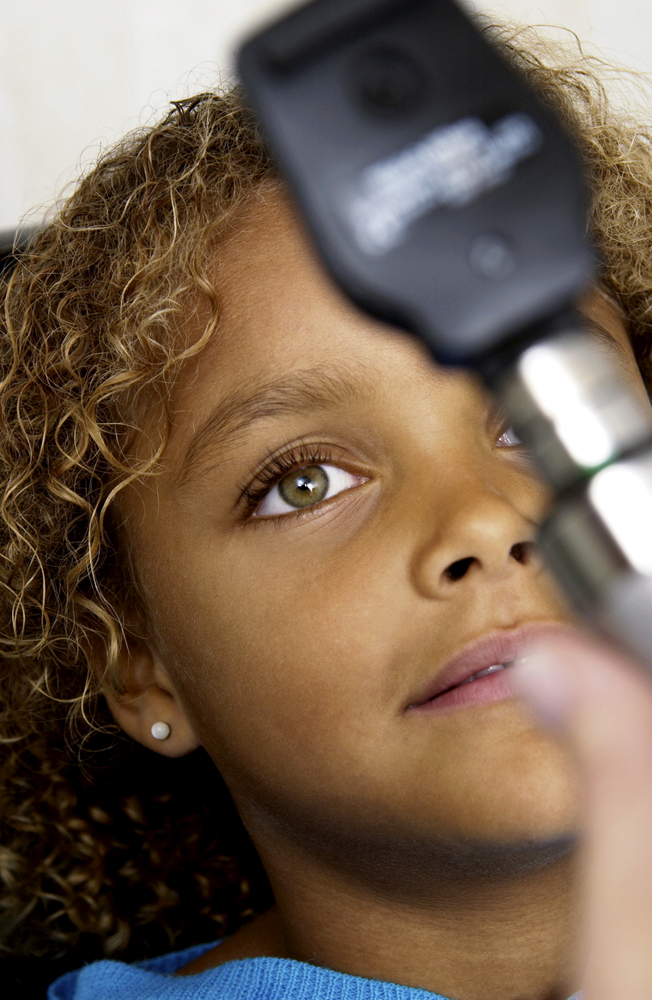
Second is the ocular examination. We use a microscope (slit-lamp) to carefully examine the surface of the eye. When we look at your tear film we are looking to see the amount and health of your tears, how well they are working, and what effect they are having on the ocular surface (conjunctiva and cornea). Not only do we look at your tears, but we pay special attention to your eyelids. In your eyelids, there are oil producing glands called Meibomian Glands. These glands are responsible for creating a key component to the tear film: lipid. Human tears are very complex, but simply put, tears have 3 main components – water, mucus, and oil. I like to describe tears like salad dressing. In order to have tasty salad dressing, there needs to be a balance of oil, vinegar, and spices. Human tears are very much similar. In order for your tears to work properly, There needs to be the proper balance of the aqueous component (water), lipid component (from the Meibomian glands), and mucus component (image 1).

dry eye diagnosis
Image 1 – A relatively healthy meibiomian gland examination

If there is an imbalance in your tears, this will reflect in their function, and ultimately cause signs and symptoms of ocular surface disease. To highlight the appearance and function of the tears on the ocular surface, clinicians often use special stains that can aid us in determining the amount and function of your tears. Two of the most common stains are fluorescein and lissamine green. Each of these stains has particular characteristics that help determine the severity and extent of your ocular surface disease. For example, if you have significant staining near the bottom part of your cornea, your eyes maybe slightly open when you sleep, and therefore you may benefit from using an ointment at nighttime. Alternatively, if your tears appear to break up very quickly on your ocular surface, there is likely an imbalance in the tear composition that may benefit from institution of warm compresses along with tear replacement in the form of artificial tears.

Third is the use of ancillary testing to help confirm our clinical diagnosis. We are fortunate to have access to several commercially available OSD diagnostics at the Gavin Herbert Eye Institute. A brief description of if you have these diagnostics follows.

    1) Schrimer Testing – This is a very simple and common method of determining whether a patient has hey aqueous tear deficiency. Essentially, the eye is numbed and a sterile piece of special paper is placed in the lower outer corner of the eye. After a specified amount of time, the amount of tears is recorded, and if under a threshold value (generally 10 millimeters at five minutes) there is a high suspicion of aqueous tear deficiency. Treatments for this subtype of OSD will be covered in the next blog.
    2) Tear osmolarity – The most available tear osmolarity system is from TearLab. With this test, we look at the integrity of the tears by determining the osmolarity – essentially the ultrastucture of the tears. If the tear osmolarity is high (hyperosmolar), we know that the tears are not functioning properly. With proper institution of treatment, the osmolarity can normalize indicating a healthier tear film. This test is very noninvasive, requiring only a tear sample of 50 nanoliters – less than the volume of a single tear!
    3) InflammaDry – Inflammation has long been accepted as a hallmark of dry eye disease/ocular surface disease. As such, many of our treatment modalities have focused on treating ocular surface inflammation (discussed in the next installment of this blog). Prior to having access to the InflammaDry test, we would have to assume that there was inflammation involved in an individual’s OSD. Now, however, we can test the ocular surface for inflammatory markers and have an answer within just a few minutes. This test not only allows us to custom tailor treatments to an individual, but also we are able to see if our treatments are working. Again, this test is minimally invasive requiring the small sample of tears for testing.
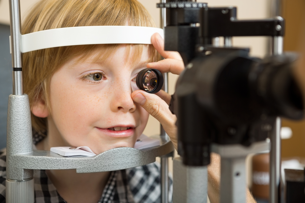
      4) LipiView II – This test allows us to Image of the structure and function of the meibomian glands in vivo. The images obtained allow for several things. First, we are able to determine the extent of meibomian gland dysfunction. Second we are able to determine the extent of meibomain gland drop out (image 2).
dry eye diagnosis
Image 2 – Significant dropout of meibomian glands
    And third we are able to educate our patients so they can see the importance of treatment of their MGD. Again, this information can help us custom tailor treatment options for the individual patient.

In conclusion, as you can see diagnosis of ocular surface disease can be quite intricate. We are fortunate to be in and age where there has been significant improvements in our tools to help us better diagnose our patients and use this information to individualize treatment options. Stay tuned for the next installment of this blog focusing on treatments for ocular surface disease.

8/20/15


Sam Garg, MDSumit “Sam“ Garg, MD
Medical Director and Vice Chair of Clinical Ophthalmology
Assistant Professor of Ophthalmology
Gavin Herbert Eye Institute – UC Irvine

Symptoms of Dry Eye Disease

Introduction

If you could be a fly on the exam room wall of your local ophthalmologist, you would hear patient after patient report symptoms of dry eye disease.

Some patients come in already knowing they have dry eyes. However, the variability of symptoms that can occur from dry eye disease is so wide many patients don’t even use the word “dry.” We will cover some of these symptoms in this article.

symptoms of dry eye
Redness often associated with dry eye

Underlying Factors

Dry eye disease has many underlying factors including an inadequate production of tears, rapid evaporation of tears, poor eyelid function and an imbalance in the tear composition of water, oil, and mucus. Dry eye disease can be associated with systemic conditions such as Sjogrens disease, Sarcoid disease and sleep apnea among many others. It is more common in females especially after hormonal changes such as menopause.

Exacerbating Influences

Many medications can exacerbate dry eye disease. Over the counter antihistamines are one example.

Environmental factors may also worsen dry eye symptoms. These include dry climates, windy weather conditions, smoky environments and the dry air found in airplanes.

Modern life includes hours and hours of focusing our eyes on everything from cell phones to computer screens to television. Prolonged focusing reduces the blink rate resulting in more tear evaporation and worsening of dry eye symptoms. Increased evaporation can also occur with exposure to heating, air conditioning, fans and rolling down the car windows while driving.

Symptoms Fluctuate

It is very common for dry eye symptoms (especially blurred vision) to wax and wane throughout the day. Symptoms can even change from blink-to-blink. Dry eye disease which is predominately due to insufficient tears tends to worsen throughout the day with symptoms worse at night. Dry eye disease that is more associated with blepharitis can be worse in the morning. Blepharitis is associated with burning and itching of the eyes.

Visual Symptoms

The front surface of the eye is the most powerful focusing surface of the eye. Thus, a dry ocular surface will produce visual symptoms. These symptoms can include:

Blurred vision: A decrease or fluctuation in visual acuity. This is manifested in the inability to see fine detail. Objects at both near and far may appear out of focus.

Sensitivity to light: Sensitivity to light is termed photophobia. It occurs because a dry ocular surface has more irregularities than a health surface. These irregularities scatter light entering the eye. This scattered light can cause significant discomfort. The inability to tolerate light may lead to squinting and headaches.

Difficulty with nighttime driving: During low light conditions, such as at night, the pupil enlarges and allows more light into the eye. When the ocular surface is dry, the incoming light becomes unfocused and scattered. Many of these abnormalities are filtered out by the small size of the pupil during the day. However, at night, the larger pupil size allows more light abnormalities to pass through to the retina. This results in nighttime glare and halos. Glare is a decreased tolerance of bright lights. Halos present as circles or auras around a bright source of light. Glare and halos from the headlights of oncoming traffic are especially troublesome.

Physical Symptoms

The front surface of the eye is richly supplied with nerve endings. As such, a dry ocular surface can result in significant symptoms of discomfort. In addition to feeling dry, these symptoms include:

Foreign body sensation: Patients may feel as if there is something present in the eye.

Redness of the eye: Enlarged blood vessels on the ocular surface cause the eye to look red.

Ocular and periocular pain: Pain from dry eye can be mild or severe. Pain from dry eye can be felt on the ocular surface. Pain can also be felt in structures around the eye such as the eyelids or scalp.

Periocular irritation: Stinging, burning, or itching sensations of the ocular surface and eyelids.

symptoms of dry eye
Watery eyes can be a symptom of dry eye

Watery eyes: Patients typically raise an eyebrow or two when I explain how the tearing they are experiencing is from dryness. “How can my eyes be dry if they are watering all of the time?” Although this may seem counter-intuitive, when the ocular surface is very dry it will overproduce the watery component of the tears as a protective mechanism.

Eye fatigue: A tired sensation of the eyes and heaviness of the eyelids.

Decreased tolerance of sustained visual focusing: As noted earlier, any activity that requires prolonged visual attention will decrease the blink rate and increase tear evaporation.

Discomfort while wearing contact lenses: Individuals may experience pain and irritation in the eyes while inserting or wearing contact lenses.

Inability to cry: Tears associated with emotional discomfort or watching a sad movie may be decreased in some types of dry eye disease.

Stringy discharge from the eye: A dry ocular surface can result in the overproduction of a sticky, mucus discharge.

Conclusion

There are many symptoms of dry eye disease. Some symptoms affect vision and others affect ocular comfort. If symptoms persist, an evaluation by your eye care provider can help clarify the cause and offer information on treatment options.

It is important to remember the symptoms of dry eye disease can overlap with the symptoms of other ocular conditions. One example is cataracts which, like dry eye disease, can also cause blurred vision and nighttime glare. The next post in this series will review how dry eye disease (and its sub-types) are diagnosed.

As a final note, while the name “dry eye disease” may sound innocuous, the symptoms of dry eye disease can be very severe in many patients. If you suffer from dry eye disease, you are not alone. Today there are many treatment options which can be very helpful. Significant research is underway to continue improving our ability to treat dry eye disease.


Matthew Wade, MD - toric intraocular lensesMatthew Wade, MD
Assistant Professor of Ophthalmology
Gavin Herbert Eye Institute


Minal Reddy was also a contributor to the is article.

Toric Intraocular Lenses after Cataract Surgery

Toric Intraocular Lenses for Post-Transplant Astigmatism

Corneal transplants can be very successful at replacing diseased or damaged corneas. However, vision after a corneal transplant is often limited by high amounts of astigmatism. Treating this astigmatism is often difficult. Typically the amount of astigmatism is higher than can be corrected with glasses. Rigid contact lenses are often required. LASIK, PRK and astigmatic incisions in the cornea (astigmatic keratotomy) have all been tried with varying success.

This month, doctors at the Gavin Herbert Eye Institute at the University of California, Irvine, published a paper describing the use of commercially available, FDA approved toric (astigmatism correcting) intraocular lenses (IOL) during cataract surgery in patients with previous corneal transplant surgery.

Good candidates for this procedure are those who have had all transplant sutures removed and had corneal astigmatism that was stable, and for the most part symmetric and regular. (Image 1A shows topography that is both regular and symmetric. Image 1B is regular but not symmetric and image 1C is irregular.)

toric intraocular lenses

The study showed improvement in uncorrected vision (post-treatment average 20/40) and vision corrected with glasses only (post-treatment average 20/25). The images below, 1D and 1F, illustrate how toric intraocular lenses are positioned along the axis of corneal astigmatism.

toric intraocular lenses

While any intraocular surgery after corneal transplant can decrease the life expectancy of the graft, no complications or graft failures were seen during the course of the study. Not all types of astigmatism can be treated with this procedure.

This study highlights an effective treatment for regular symmetric corneal astigmatism after corneal transplant in patients needing cataract surgery.

7/24/15

Matthew Wade, MD - toric intraocular lensesMatthew Wade, MD
Assistant Professor of Ophthalmology
Gavin Herbert Eye Institute
University of California, Irvine

When Is The Best Time For Cataract Surgery?

As you age, cataracts become a concern prompting the question – when is the best time for cataract surgery?

There are decades worth of old wives tales floating around regarding cataracts that often lead to unnecessary fear and apprehension for many patients. These myths involve concepts such as “ripeness”, having to wear eye patches afterwards, danger in “waiting too long, etc. Just as the techniques of cataract extraction have changed over the decades, so have the indications to proceed to surgery.
best time for cataract surgery - people
Firstly, cataracts are a normal part of the aging process. Patients should not be alarmed if they are told that they are developing cataracts, even as early as their fifties. As we age, the natural clear lens inside the eye becomes progressively harder, darker, and cloudier. This dark, cloudy lens is what is referred to as a cataract. Cataracts develop at different rates for different people, and even between the two eyes of the same person. It typically takes many years for the lens to become cloudy enough to impact the clarity of vision. There are many different types of cataracts depending of what area of the lens becomes cloudy, but the typical cataract related to normal aging results in a relatively uniform cloudiness with a denser central core, and is referred to as “Nuclear Sclerosis”. Other varieties of cataracts tend to grow more quickly, are relatively uncommon, and often result from certain conditions other than typical aging.
best time for cataract surgery
Regardless of what type of cataract the patient has, the treatment is the same: cataract extraction with an implant of an intraocular lens. There have been great advances in lens design over the years, and they now result in excellent, stable, predictable vision for the remainder of the patient’s lifetime and do not typically need to be changed once implanted.

Cataracts result in different symptoms that may be more of less relevant to a specific person’s needs, such as:

  • Glare with bright lights
  • Difficulty with fine print
  • Difficulty following the golf or tennis ball
  • Impairment in night driving
  • Difficulty with seeing street signs
  • Seeing the score or small print on the television
  • Fine visual tasks such as threading a needle, etc.

Although cataract surgery is an incredibly successful procedure with only about a 1-2% risk of complications, it still DOES have some risk. Therefore, cataract surgery should only be undertaken when there is something to gain. In other words, the BENEFITS MUST OUTWEIGH THE RISKS. This means that if your symptoms are mild and are not interfering with your activities of daily living, it is not time to accept the risks of surgery. Once your visual impairment progresses to the point that YOU feel your activities of daily living and enjoyment are impaired, this is the time to proceed to surgery. This threshold is very different between people. Some people feel impaired with vision of 20/25, and others still function within their scope of usual activities until they are 20/100! The best first-step in determining if it is time for your surgery is to get an up-to date refraction. This means a detailed check for new glasses. Often, cataract development will change a person’s glasses prescription, and updating this can improve the visual symptoms for months to years. When a new glasses prescription no longer improves the sight adequately, this is when surgery is indicated.

For the most part, putting off cataract surgery does not impact the final outcome. It will not harm you or your eye to leave the cataract alone until you are ready. There are of course certain exceptions to this rule, such as in Fuchs’ dystrophy, pseudoexfolation, untreated narrow-angle glaucoma, and some others. However, these are relatively rare conditions that your doctor will speak to you about if you have any of these diagnoses.

In summary, the time to proceed to cataract surgery is something that you as the patient determine. YOU assess your lifestyle needs and your vision performance within your scope of activities. When you feel you are impaired in these activities, the benefits will outweigh the risks, and it’s time to take them out. You should not feel any pressure to urgency in this process.

Once you have determined you are ready to have cataract surgery, your surgeon will discuss with you your options for intraocular lens implantation including astigmatism neutralizing lenses, standard distance or near-vision lenses, multiple focal distance lenses, accommodating lenses, and others. The current standard approach for cataract surgery is called “phacoemulsification” and uses ultrasound technology to remove the cataract. There are also laser devices that assist in making the incisions and breaking up the lens, which many surgeons now employ in addition to the phacoemulsification. In general cataract surgery only takes a few minutes, is performed with topical anesthesia, is pain-free, and has a very short recovery time. No pirate-patches are used these days! Most patients are very happy with the results, but this requires adequate discussion with the surgeon prior to the procedure to best assess the needs of the individual patient. A well- informed patient who participates in their care results in the best outcomes!

6/18/15

Sameh Mosaed, MD best time for cataract surgerySameh Mosaed, MD
Director of Glaucoma Services, Gavin Herbert Eye Institute, UC Irvine
Associate Professor, Cataract and Glaucoma Surgery, UC Irvine School of Medicine

Cataract Prevention

The more you know about cataracts, the easier it is to focus on cataract prevention.

What is a cataract?

At birth, with rare exceptions, most of us arrive in the world with a clear crystalline lens within each eye. The pathway of our visual images start with light passing through the cornea (the clear front window of the eye), through the pupil (the opening in the center of the iris, or colored portion of the eye) and through crystalline lens which functions to focus light onto the center of the retina (the film of the eye). cataract preventionThe retina, via the optic nerve, will then transmit visual images to the brain. When the crystalline lens becomes opacified (cloudy), this system becomes disrupted, and vision becomes impaired. Opacification of the crystalline lens is called “cataract”, and there are many variations in appearance and type and many causes and can present at any age. The word cataract originates from the Greek word “cataracta”, which means waterfall. The ancient Greeks used this term as they noticed a similarity in the appearance of the white opaque rushing water of a waterfall and the appearance of a white mature cataract.

To understand the different types of cataracts and causes, it is important to understand the anatomy of the lens. Using a metaphor, the lens anatomy can be compared to a Peanut M&M candy™. There is an outer candy coating (the lens capsule), a chocolate layer inside (the lens cortex), and a peanut in the center (the lens nucleus).

The most common cause of a cataract is an age related nuclear clouding which is due to long term accumulation of metabolic and oxidative waste products within the lens and possibly UV-B/Sunlight light exposure. Cortical clouding (within the cortex of the lens), due to similar causes, is also a common cause of an age related cataract.

Cataracts can occur earlier in life with poorly controlled diabetes resulting in cortical and nuclear cataract. Patients who are exposed to steroid medications in any form (orally, topically as eye drops, skin creams etc.) are at an increased risk to develop a posterior subcapsular (PSC) cataract which occurs on the posterior lens capsule. PSC cataracts can have a much more abrupt and earlier onset in life than nuclear or cortical cataract. Smoking has also been known to predispose patients to formation of a PSC cataract. Other less common varieties of cataract can occur with any trauma to the eye or even present at birth as a congenital cataract with a large variety of causes.

What can be done to prevent cataracts?

I often joke with patients that a cataract is such a common occurrence that just like birth, death, and taxes, it is an issue we must all face at some juncture in life (hopefully later than earlier). I am often asked if there are any dietary measures or vitamin supplementation to reduce the formation of a cataract, however this is not as well studied as the use of vitamins in the prevention of macular degeneration. Several scientific epidemiological studies following populations over many decades have shown some merit however that using multivitamins regularly (Vitamin B6 and B12, Vitamin C, beta carotene, antioxidants and possibly lutein and zeaxathin) can reduce the degree of lens opacification over time. As with all medications, you should consult with your physician before deciding to use any vitamin supplementation to clarify if you have any contraindication to using them.

There is conflicting evidence regarding the role of UV-B exposure in sunlight as a causative agent for cataracts. There is some support that using sunglasses on a regular basis to block UV-B light may help to reduce cortical cataract formation. Smoking cessation can also help to reduce the formation of cataract. If a patient is diabetic, strict blood sugar control is also an important measure to reduce the formation of a cataract. If possible, reducing or avoiding the use of steroid medication can reduce the formation of a PSC cataract.

What can be done if a cataract is worsenening and glasses cannot help improve vision significantly?

If you are experiencing gradual painless loss of vision, you should consult with your ophthalmologist as cataract can be a common cause. If you are found to have cataract formation, there is generally a shift in the glasses prescription in the early stage. Having your glasses prescription checked to see if your vision can be improved with glasses is the first step in determining how significant your cataract has become. If glasses are not able to sufficiently improve your vision and your daily activities are affected by the decrease in vision your experience, you may be a candidate to have cataract surgery.

Modern cataract surgery has improved a tremendous degree compared to decades earlier. It is the most common and successful surgery in the world, and is typically performed on an outpatient basis with topical anesthetic and often without any sutures or eye patch. Prior to surgery the pupil is dilated, and once in the operating room, a small self-sealing incision is made on the side of the cornea. The surgeon then makes a circular opening in the anterior lens capsule (the candy coating of the peanut M&M), and uses an ultrasound instrument to emulsify and vacuum out the nucleus (the central peanut), and remove the cortex (the chocolate layer). The inside of the lens capsule is polished and an intraocular lens is folded and introduced into the eye through the corneal incision and seated into the remaining lens capsule to conclude the surgery.

Prior to surgery, measurements are taken to determine the power of lens necessary to achieve the best vision after surgery based on the curvature of the cornea and anterior-posterior length of the eye. Intraocular lenses (IOLs) can potentially have several features depending on a patient’s needs. The most common IOL used is a monofocal lens, which does not typically require an additional out of pocket expense. This lens is chosen to have a point of focus either for distance vision (driving, TV) or near vision (reading), but not both. Typically patients who have the monofocal lens will choose to have distance focus and use reading glasses for near vision. There are multifocal/accommodating IOLs available for patients who are appropriate candidates, to allow the patient a larger range of vision at far, near and intermediate (computer) distance and may allow great independence from glasses. There are still other IOLs which can correct astigmatism (a special type of glasses prescription) at the time of cataract surgery. After discussion of the patient’s needs and preferences, the surgeon can best advise their patient regarding which type of IOL may best suit them.

6/11/15

Anand Bhatt, MD - cataract preventionAnand B. Bhatt, MD
Assistant Professor of Glaucoma and Cataract Surgery, Gavin Herbert Eye Institute
UC Irvine School of Medicine

Vision and Special Needs Children

In the United States, special needs is a term used in clinical diagnostic and functional development to describe individuals who require assistance for disabilities that may be medical, mental, or psychological. Different types of special needs vary in severity. People with autism, Down syndrome, dyslexia, blindness, ADHD, or cerebral palsy, for example, may be considered to have special needs. Statistics tell us that among children ages 3 to 17, nearly 15 percent have one or more developmental disabilities. For many of these children, the kinds of disabilities they experience may require special approaches to providing care, education and/or other accommodations.
eye exam - vision and special needs children
The way a special needs child behaves or reacts can sometimes be unexpected because he/she processes sensory information differently than other children. Having the right cues in our environment can mean the difference between participation and non-participation in medical care for many of these children. Our work environment should always be arranged in a way to achieve physical and emotional comfort for the child. Patience, attentive listening, caring, and building a trusting relationship with a family and child who may be undervalued in other settings are all critical to providing good medical care.

Vision and Special Needs Children

Children with special needs are referred to pediatric ophthalmologists on a regular basis. Many of these children do not respond appropriately to standard vision screening procedures. Unidentified vision problems in this special population can further impact growth and development. The role of pediatric ophthalmologists is to ensure that the child does not have any eye conditions that could potentially interfere with his/her learning opportunities, personal development and/or overall wellbeing.

Down syndrome:
Down syndrome continues to be the most common chromosomal disorder. Each year, about 6,000 babies are born with Down syndrome, which is about 1 in every 700 babies born. It is estimated that more than 80% of these patients have some clinically significant ocular pathology. Such conditions include refractive error requiring glasses (70%), strabismus (45%), and nystagmus (35%). Other common eye problems include blepharitis, blocked tear duct, eyelid ptosis, cataracts, and keratoconus (irregularly shaped cornea). The improved quality of medical care and educational resources have allowed for a more productive life and a longer life expectancy for people with Down syndrome. Their quality of life can be further enhanced by the proper assessment and correction of eye problems.

Autism:
In 2014, the Centers for Disease Control and Prevention (CDC) released new data on the prevalence of autism in the United States. This surveillance study identified 1 in 68 children (1 in 42 boys and 1 in 189 girls) as having autism spectrum disorder (ASD). Autistic behaviors may include visual components such as lack of eye contact, starring at light or spinning objects, fleeting peripheral glances, side viewing and difficulty attending visually. An eye examination is essential in order to detect an eye condition that could potentially explain these visual behaviors. According to a study published in the June 2013 issue of Strabismus, more than 40 percent of children with autism have strabismus, or crossed eyes. This work is limited in that the researchers did not use a representative sample of children with autism. Still, the prevalence of eye problems in autism appears to be several times higher than that of the general population.
boy eye exam - vision and special needs children
Learning Disabilities:
Dyslexia is the most common neurobehavioral disorder affecting children. Visual abnormalities have not been found to affect the brain’s ability to process visual stimuli and children with learning disabilities have no increased incidence of ophthalmologic disease. However, ophthalmologic consultation should be provided to children who fail vision screening tests. This allows for diagnosis and therapy of treatable ocular conditions such as refractive errors and eye muscle imbalances.

Children with ADHD, cerebral palsy, or any other neurodevelopmental disorders should also have periodic vision screenings. Children who do not pass the vision screening should be referred to an ophthalmologist with experience in the care of children.

Physicians are not the only adults involved in the care of special needs children. At increasing rates, children with special needs are being provided with the same life experiences as their non-disabled peers. Taking part in a sport or joining a group like the Scouts, are popular activities for children. Children with disabilities are encouraged to join in such activities to help improve their health and give them opportunities to make friends. As a result, more and more adults in the community are finding themselves working with these children on a regular basis.

When considering ways to work with special needs children, we have to keep in mind that every child is different. A positive attitude and patience are probably the two most important qualities for anyone who works with these children. Parents of special needs children focus on helping their child to be “the best he/she can be”. The devotion and care these parents provide for their child have been an inspiration, and I always look forward to my visits with them.

4/2/15


Dr. Chantal BoisvertChantal Boisvert, OD, MD
Assistant Clinical Professor
Gavin Herbert Eye Institute, UC Irvine
Pediatric Ophthalmology & Strabismus
Neuro-Ophthalmology

Three Generations of Saving Vision

In Discovery Eye Foundation’s spring e-newlsetter there was an article entitled Surgery for the Surgeon, where a leading ophthalmologist talks about convincing himself to have cataract surgery. That eye doctor was Dr. Nesburn, who was willing to share with us his family’s long tradition of saving vision.

Brainwashed by Medicine

“I was brainwashed from the age of 5,” says Dr. Anthony Nesburn, medical director of The Discovery Eye Foundation (DEF). “My dad would take me on rounds at the hospital and to his office. He introduced me to medicine at a really early age.”

Nesburn saving vision
Dr. Anthony Nesburn in UC Irvine lab
Dr. Henry Nesburn was an ophthalmologist in Los Angeles for more than four decades, and he passed his love of the specialty to his son. “I really looked up to my dad,” the younger Nesburn says. “He loved ophthalmology for the same reasons I do: You get to do medical diagnosis; you get to do wonderful surgery, where you keep people from going blind or restore vision; and you can work with people from newborns to the very elderly — they all need eye care.”

Nesburn received a telegram while he was an undergrad at UCLA, telling him he’d been accepted to Harvard Medical School. His mother started crying: “You’re going to Boston! We’re not going to see you anymore!” While he “was loathe to leave Southern California,” Harvard was too good to pass up.

Drafted by the Army out of his ophthalmology residency at Harvard in 1960, Nesburn joined the Navy instead. He followed in his father’s footsteps again, becoming a Navy flight surgeon. (Henry had volunteered during World War II.)

He went on to a Boston Children’s Hospital fellowship in infectious disease, working with Nobel laureate Dr. John Enders, whose work led to the polio vaccine and changed the face of virology. Nesburn then did his residency at Massachusetts Eye and Ear. “I was part of a special program that allowed us to do research, and I was running a research laboratory while I was a resident,” he says. “It gave me the start I needed.”

“At Mass Eye and Ear, I worked for an up-and-coming ophthalmologist and researcher to prove there was a substance that could treat herpes eye infections. We wrote a paper that included the very first antiviral ever described, and it was against herpes virus. It is the basis for today’s herpes antivirals,” Nesburn says. “I was hooked.”

He went back to Los Angeles and received NIH funding to continue his research on ocular herpes. In 1968, he joined his father’s practice half-time, spending the rest of his time doing research.

Two years later, he received a generous offer, when Rita and Morris Pynoos started DEF to fund his research. The Pynooses were grateful to Nesburn for diagnosing their son, Jon, with keratoconus (KC). “I was a second-year resident at Mass Eye and Ear, and Jon Pynoos was an undergraduate at Harvard. His parents went to see my dad, because Jon couldn’t see well, and no one could figure out what was wrong with his vision. My dad said, ‘Send him over to Tony; he’ll figure out what’s going on!’ I said to myself, ‘Holy mackerel! What happens if he has something really complicated? I’m just a newbie!’” Nesburn remembers. “Jon came in; I looked at him, and the keratoconus was so clear and easy to spot. I couldn’t imagine how his doctors didn’t see it. We got him contact lenses, and he was able to see again. When I came back to LA, the Pynooses wanted to do something to help.”

At first, DEF research focused on KC and the herpes research Nesburn was working on at the time. It soon broadened to include macular degeneration and retinal disease.

“My dad had to retire from the practice of ophthalmology at the age of 70, because of bad age-related macular degeneration (AMD). His mother and older sister had had it, as did several cousins. There was no treatment back then that helped,” Nesburn says.

“AMD is the most common cause of permanent vision loss in the elderly in the developed world. I could see where the need was,” he says. “We moved forward at DEF with two driving mantras: We wanted to do something significant in macular degeneration research and to find the cause of keratoconus.”

As a virologist in research and a corneal surgeon, Nesburn realized he needed a corneal biochemist to help with the KC research. He met Dr. Cristina Kenney at an Association for Research in Vision and Ophthalmology meeting. She joined DEF, and within 15 years, they found the chemical cause of keratoconus; they also got married.

Now nearly 80 years old, Nesburn spends most of his time “wearing three hats”: fundraising for DEF, lab research and clinical practice. His daughter, Kristin, is the third generation to join the family ophthalmology practice.

“While I’m still able, I want to try to make a difference in medicine, particularly in macular degeneration,” Nesburn says. “Macular degeneration affects so many people. This is where I want to put my energy. Luckily, as strong as it is in our family, I don’t have it … yet.

“As a researcher, my interest in putting together a program for macular-degeneration diagnosis and treatment has been because it’s a great public health problem. Yes, if I should ever get it, it might be able to help me or my family, but the first thing, as a scientist, is to try to get something to help humankind. I know it sounds sappy, but it’s true.”

3/26/15


Anthony B. Nesburn, MD, FACSAnthony B. Nesburn, MD, FACS
Medical Director, Discovery Eye Foundation
Professor & Vice Chairman for Research, Ophthalmology
Gavin Herbert Eye Institute, University of CA, Irvine

Cataract Surgery and Keratoconus

1/8/15

The eye works like a camera, specifically a digital camera. There is the front lens of the camera (cornea), the aperture (iris), the film (retina), and a cable to take the image to the brain (optic nerve). This “camera” also has an additional lens – the natural crystalline lens, which lies behind iris. This natural lens is flexible when we are young, allowing us to focus at distance then instantaneously up close. Around age 40-45, this natural lens starts to stiffen, necessitating the need for reading glasses for most people. This stiffening is the beginning of the aging process that eventually leads to formation of a cataract. We refer to the lens as a cataract when it becomes sufficiently cloudy to affect ones quality of vision.cataract surgery and keratoconus-Cataract diagram In general, cataract surgery is one of the safest and most successful of all surgeries performed. The basics of cataract surgery in eyes with keratoconus is very similar to non-keratoconic eyes.

Keratoconus (KC) affects this “camera” by causing the front lens (cornea) to bulge. This causes the optics to be distorted. In many cases, this can be corrected for with hard contact lenses (CL) or spectacles; in other cases a corneal transplant may be necessary. When it comes time for cataract surgery in the setting of KC, there are several factors that need to be considered.

Corneal Stability
The first thing to be considered is the stability of your cornea. In general, KC progresses more in your late teens to early twenties, and then stabilizes with age. A very exciting treatment for KC is collagen crosslinking. This treatment is meant to stiffen the cornea to prevent instability that is inherent to KC. This treatment promises to stop the progression of KC at a young age. Fortunately, with age, the cornea naturally crosslinks and stiffens, therefore when it comes time for cataract surgery, there is little chance of the progression of KC. Your doctor needs to choose the appropriate intraocular lens (IOL) to refocus your eye after surgery. Two of the most important factors in IOL selection are the length of your eye and the shape of your cornea. Long term CL wear can mold your cornea. It is important to assure that you stay out of your CLs long enough for your cornea to reach its natural shape. Depending on how long you have worn your CLs, it may take several months for the cornea to stabilize. This time can be challenging as your vision will be suboptimal (because you can’t wear CLs), and will be changing (as your cornea reaches its natural shape). When your cornea does stabilize, it is important to determine whether the topography (shape) is regular or irregular. This “regularity” is also known as astigmatism. If the astigmatism is regular, light is focused as a line – generally, this distortion can be fixed with glasses. However, if the astigmatism is irregular, light cannot be focused with glasses, and hard CLs are needed to provide optimal focusing. If you have had a corneal transplant, I generally recommend all your sutures to be removed to allow your new cornea to reach its natural shape.

IOL Selection
The second thing to be considered is the type of IOL. IOLs allow your doctor to refocus the optics of your eye after surgery. In many cases, the correct choice of IOL may decrease your dependence on glasses or CLs. There are several factors that are important when considering the correct IOL for a keratoconic patient. The amount and regularity of your astigmatism plays a very significant role in IOL selection. In general, there are four types of IOLs available in the US – monofocal, toric, pseudo-accomodating, and multifocal. In general I do not recommend multifocal IOLs in patients with KC. These IOLs allow for spectacle independence by spitting the light energy for distance and near, however, with an aberrated cornea (which is what happens in KC), these IOLs do not fare well. If there is a low amount of regular astigmatism or irregular astigmatism, your best bet is a monofocal IOL. This is the “standard” IOL that is covered by your health insurance. If you have higher amounts of astigmatism that your doctor determines is mostly regular, you may benefit from a toric (astigmatism-correcting) IOL. These IOLs can significant improve your uncorrected vision and really decrease your dependence on glasses. It is important to realize that monofocal and toric IOLs only correct vision at one distance. With a monofocal IOL you still can wear a CL to fine-tune your vision, however, with a toric IOL, in general you will need glasses for any residual error. There is a pseudo-accomodating toric IOL available, and this may be a good option if you are trying to decrease your dependence on glasses and correct some of your astigmatism. These IOLs are relatively new to the US market.

If You Had A Corneal Transplant
In the setting of a corneal transplant many of the same factors need to be considered – stability of the graft, choice of IOL, etc. In addition, the health of the graft has to be judged. Prior to cataract surgery in my patients with corneal transplants, I make sure to remove all of their sutures and give the cornea time to stabilize (just as if they were a CTL wearer). If you are a CL wearer, the same rule of being out of the TL until the topography is stable applies. The health of a transplant needs to be established prior to undergoing cataract surgery. The cornea has five main layers to it –cataract surgery and keratoconus-corneal structure the back layer (inside) is called the endothelium. This layer is responsible for “pumping” fluid out of the cornea, allowing it to stay clear. In all eyes there is a loss of endothelium cells with cataract surgery. I generally perform a “specular microscopy,” which allows me to visualize and quantify the corneal endothelium prior to surgery. This allows me to risk stratify you before your surgery. It is important to realize that corneal transplants have a lifespan and may have to be repeated in the future.

Keep in mind, there is some uncertainty in biometry (the process of selecting an IOL) in all eyes – this error can be higher in keratoconic eyes. This highlights why assuring stability is important. Equally important is picking the correct IOL for your situation. Also, keep in mind that I have discussed generalities in this article. Your individual case could be different. This is a conversation best left between you and your surgeon. In general, cataract surgery and keratoconus or a corneal transplant can be a very safe and effective way in restoring vision.

Sam Garg, MDSumit (Sam) Garg, MD
Interim Chair of Clinical Ophthalmology and Medical Director
Gavin Herbert Eye Institute at the University of California, Irvine

Nystagmus In Children

8/7/14

Nystagmus is a condition of uncontrolled eye movements. Patients with nystagmus are unable to maintain their eyes in a fixed position of focus. The movements can be pendular, swaying evenly side to side, or, jerk into one direction and drift toward the opposite direction. It can be present early in life or acquired as an adult. It can occur in eyes with poor vision from other anomalous development, or eyes that appear perfectly normal. In almost all patients the vision is compromised to some degree. In some patients, the eye movement is less, and the vision better, in an eccentric position that causes the patient to adopt a face turn, tilt or head posture so they can use this quieter position (“null point”) to navigate during their daily activities. To date there have been no consistently effective treatments for this condition.

Lingua and Grace - nystagmus
Dr. Lingua and Grace Nassar

Treatment efforts have been either medical (drugs to reduce the amplitude of the nytagmus movement) or surgical (to move the “null point” into straight ahead gaze to eliminate a head turn, or, directed at reducing the effective contracture of all the eye muscles to reduce the amount of movement). In general, surgical treatment of nystagmus has been disappointing.

In 2002, Dr. Robert Sinskey, noted cataract surgeon and phacoemulsification pioneer, proposed a revolutionary concept, that nystagmus could only be truly effectively controlled by removing the forward portion of the eye muscle and detach it completely from the eye. Since the twitching eye muscles were controlled by nerves sending that pulsatile information, any operation that allowed the muscles to remain attached to the eye would never quiet the movement. He performed this novel surgery in 2000 and published the results in 2002. It did not receive attention in the nystagmus surgery community, as most experts worried that the surgery would limit normal eye movements excessively. The operation does remove the forward portion of the eye muscle but, surprisingly, the eyes are still able to move to allow reading, computing, and driving.. In 2012, I had the opportunity to view a patient he operated 10 years prior and was impressed with how successful the results were even after 10 years. Coincidentaly, I was caring for a 17 year-old patient with nystagmus who had already undergone the 2 currently accepted eye muscle procedures for nystagmus without success. His movements remained uncontrolled, he could not maintain eye contact with anyone, and is his vision was less than that needed for a drivers license. In 2013, I offered him the Sinskey procedure and the results were remarkable. His nystagmus was quieted, his vision improved (20/25) enough to qualify for a drivers license and to return to school.

Since 2012, we have adapted, augmented and perfected the procedure and performed the surgery on over 12 patients with similar remarkable results. All patients experience a marked reduction in the amplitude of the nystagmus (60-100%), and all patients demonstrate improved vision (1-8 lines of the acuity chart), especially at the reading position.

Visit the YouTube posting “Meet Grace for an example of how this surgery can impact a child’s life and the hopes of their parents. Visit www.eye.uci.edu for further information, contact information and scientific data on the procedure.

Robert Lingua, MDRobert W. Lingua, MD
Director, Pediatric Ophthalmology and Strabismus
Gavin Herbert Eye Institute, UC Irvine

What You Need to Know About Cataracts

6/5/14

Do you feel like your vision is getting worse? Do you feel like colors are not as vibrant as they used to be? Are you having more trouble with glare? If you have any of these symptoms, you may be experiencing the effects of cataracts.

Cataracts are a normal aging process of the crystalline lens in the eye. Well you may ask — what is the crystalline lens? It is easiest to think of the eye as a camera. The eye has a lens (actually two) – the cornea (the front window of the eye) and the crystalline lens (inside the eye). It also has an aperture (the colored iris), and film (the retina). All these structures work together to focus light and allow us to see – just like a camera. When we are young (less than 40), the crystalline lens is flexible. This is why we are able to see distance and then near without the need for reading glasses. The crystalline lens is able to change its shape depending on where one is looking.

Figure 1 – Slit-lamp photo of a visually significant cataract.

As we age, the crystalline lens becomes less flexible, thereby causing one’s near vision to be more blurry. This necessitates the need for reading glasses. As the crystalline lens become less flexible with age, the lens also starts to become more yellow and can also become cloudy. When the yellowing and/or clouding become visually significant, we refer to this as a cataract (figure 1).

Are cataracts dangerous? The simple answer is no. In the vast majority of cases, a cataract can be monitored until it becomes visually significant (drop in vision, glare, decreased contrast, vision related difficulties with day to day activities, etc). However, there are a few instances in which cataract removal is a medical necessity. Routine examinations by your eyecare provider can help you determine if you are at risk for these less common instances.

Figure 2 – Intraoperative photo during cataract surgery (prior to cataract removal).

What can I expect during cataract surgery? Do you have to replace the crystalline lens with anything? Cataract surgery involves removing the clouded crystalline lens (figure 2) and replacing it with an artificial lens known as an intraocular lens (IOL) (figure 3). Surgery generally takes 10-15 minutes under a mild sedative, and you don’t have to stop any of your current medications. Anesthesia is achieved with drops and you will only feel mild pressure during the surgery. IOLs come in different styles – Standard IOLs grossly correct your vision and you can fine tune your vision (distance and near) with glasses post-operatively;

Figure 3 – Intraoperative photo during cataract surgery (after implantation of an IOL).

Toric (astigmatism correcting) IOLs allow for increased spectacle independence, but glasses will still be needed for distance or near; Accommodating IOLs “flex” within the eye to decrease your dependence on distance and near glasses; Multifocal IOLs allow spectacle independence for distance and near. I always counsel patients that there is no perfect IOL and you have to determine which IOL is best for your particular situation. Your doctor can help you decide which IOL is best for you. Generally, cataract surgery is extremely safe. Your doctor will discuss particular risks specific to your eye.

How do I know if cataract surgery is right for me? The best way to know if you have a cataract and if it time to consider surgery is to consult with your local ophthalmologist. If you have experienced a recent drop in vision, that is not correctable with glasses, cataract surgery may be able to restore your vision!

Garg feb 2014 thumbSumit “Sam“ Garg, MD
Medical Director
Vice Chair of Clinical Ophthalmology
Assistant Professor of Ophthalmology
Gavin Herbert Eye Institute – UC, Irvine